Several anatomical structures of the bovine teat are important defence mechanisms preventing udder infections. The teat canal plays a particularly important role as it acts as the first barrier against the entry of pathogens (Michel et al., Reference Michel, Seffner and Schulz1974; Hamann, Reference Hamann1987). During milking the teat is subjected to mechanical impacts by the clusters and the vacuum that result in short and long term changes of the teat tissue (Hillerton et al., Reference Hillerton, Pankey and Pankey2002). Several studies have shown that milking temporarily increases teat canal length and teat wall thickness and changes teat diameter (Neijenhuis et al., Reference Neijenhuis, Klungel and Hogeveen2001; Paulrud, Reference Paulrud2005). The teat tissue requires several hours after milking to recover and provide a tight barrier again; for example, reduction in teat wall thickness to the dimension before milking requires approximately 6 h. Other parameters such as teat canal length and diameter at the teat tip do not reach their initial values again even after 8 h (Neijenhuis et al., Reference Neijenhuis, Klungel and Hogeveen2001). This is especially the case for harsh milking conditions imposed by untrained or careless workers.
High mechanical forces to the teat during milking negatively affect teat condition on a long term basis (Hillerton et al., Reference Hillerton, Ohnstad, Baines and Leach2000). Excessive vacuum, worn teat liners and prolonged over-milking periods result in acute and chronic tissue damage (Rasmussen, Reference Rasmussen1997; Hillerton et al., Reference Hillerton, Pankey and Pankey2002; Penry et al., Reference Penry, Upton, Mein, Rasmussen, Ohnstad, Thompson and Reinemann2017). To avoid or reduce excessive mechanical forces modern parlour-based milking systems automatically remove the cluster from the udder when the milk flow rate falls below a pre-set figure. However, the milk yield of rear quarters is higher than that of front quarters. This difference results in a longer milking time at the rear quarters and a longer over-milking phase at the front quarters (Tančin et al., Reference Tančin, Ipema, Hogewerf and Mačuhová2006). In addition, the morphology of front quarter and rear quarter teats differs in parameters such as length and diameter (Weiss et al., Reference Weiss, Weinfurtne and Bruckmaier2004). Guarín and Ruegg (Reference Guarín and Ruegg2016). Guarín et al. (Reference Guarín, Paixão and Ruegg2017) investigated the position and anatomical characteristics of front and rear teats and found associations to mastitis, quarter somatic cell count, hyperkeratosis score and parity indicating that front teats have a higher risk for teat tissue damage and increasing mastitis. However, Barkema et al. (Reference Barkema, Schukken, Lam, Galligan, Beiboer and Brand1997) did not find such differences.
It seems that there is little information whether the differences of milk yield and anatomy between front quarter and rear quarters also lead to higher mechanical impact on the front quarter teats and whether these negatively affect the teat condition of the front teat. The present study aimed to measure morphological and ultrasonographic parameters before and after milking, drawing comparison between front and rear teats. Additionally, vacuum measurements were taken during milking using a miniature device and data logger attached to the cluster to identify differences between front and rear teats. The study intended to test the following hypotheses:
(1) That the lower milk yield of the front quarter's results in a longer over-milking and higher mouth piece chamber vacuum levels in comparison to the rear quarters.
(2) That the longer over-milking at the front quarters affects the teat tissue and results in changes to teat morphology.
(3) The most important factors inducing the tissue changes are the time of milking and over-milking and the vacuum level.
The identification and assessment of the factors affecting teat morphology especially of the front teats during milking may result in more precise advice to farmers and further development of milking machines to improve udder health and animal welfare.
Materials and methods
Farms and animals
The study was conducted on 41 Austrian dairy farms and comprised 540 dairy cows (61.9% Austrian Simmental, 23.5% Holstein Friesian, 14.6% Brown Swiss). The cows were clinically healthy; cows showing clinical mastitis were not included in the study. More detailed information is provided in the online Supplementary File.
Vacuum measurement during milking
Two clusters of the milking parlour were equipped with the vacuum measurement and recording system (online Supplementary Fig. S1). Since the cows entered the milking parlour without preselection they were randomly assigned to the study entering the place where the clusters were equipped with the portable vacuum-logger (VaDia, BioControl, Rakkestad, Norway). Overall 540 front and 540 rear teats were included in the study. The vacuum measurements were taken at the head of the liner (mouth piece chamber, MPC) of one front and one rear teat, at the short milk tube (SMT) and the pulsation tube of the rear teat. The device automatically detected 4 milking phases cluster attachment/stimulation, milking/peak milk flow, over-milking, cluster removal/end of milking. Morphologic and ultrasonographic teat measurements
Before the attachment of the clusters the teat length and teat base diameter of the two teats at which the vacuum was to be recorded were measured using a translucent ruler (Westfalia, Bönen, Germany) (online Supplementary Fig. S2).
The teat tip condition was scored according to Mein et al. (Reference Mein, Neijenhuis, Morgan, Reinemann, Hillerton, Baines, Ohnstad, Rasmussen, Timms, Britt, Farnsworth, Cook and Hemling2001) using four categories: 1 – no ring, 2 – smooth or slightly rough ring, 3 – rough ring and 4 – very rough ring.
Ultrasonography was done immediately before and after milking using a MyLabOne scanner (Esaote, Munich, Germany) equipped with a linear array 7.5 MHz probe. For this measurement, the teats were immersed in a plastic cup filled with warm water to avoid teat compression (Neijenhuis et al., Reference Neijenhuis, Klungel and Hogeveen2001; Weiss et al., Reference Weiss, Weinfurtne and Bruckmaier2004; Klein et al., Reference Klein, Flöck, Khol, Franz, Stüger and Baumgartner2005). All ultrasonographic examinations were stored on the device and analysed later. The following parameters were measured after ultrasonography: teat canal length, diameter of teat at the level of the internal teat canal orifice (Fürstenberg rosette), teat wall thickness measured 1 cm proximal of the internal teat canal orifice (online Supplementary Fig. S3).
Immediately after the cluster was removed automatically by a cluster remover (N = 15) or manually by the milker (N = 26) ultrasonography and measurements of the teats were repeated using the identical techniques. Additionally the colour of the teats, the firmness and the swelling at the teat base were evaluated according to Mein et al. (Reference Mein, Neijenhuis, Morgan, Reinemann, Hillerton, Baines, Ohnstad, Rasmussen, Timms, Britt, Farnsworth, Cook and Hemling2001). The colour of the teats was evaluated in three different categories (1 – normal pink, 2 – reddened and 3 – bluish). All dark pigmented teats were excluded. The proximal areas of the teats were assessed if the liner had caused a visible line or a swollen ring at the tissue. The scores were: 1 – no visible ring, 2 – visible mouthpiece lip mark and 3 – swelling or thickened ring. Subsequently the firmness at the teat end was classified: 1 – soft and no ‘wedge-shaped’, 2 – flat or hard. All physical and ultrasonographic examinations were performed by one author (TV).
Milk yield and somatic cell count
Data for every cow was provided by the regional milk yield recording organization. The data included in this study were parity, days in milk, milk yield and somatic cell counts (SCC). Two consecutive reports one before and one after the farm visit were used for the calculation of average milk yield and somatic cell count.
Statistical analysis
A sample size calculation was performed (95% confidence interval and 80% power) to allow the detection of differences between front and rear teats and the pre- and postmilking measurements of at least 2.5%. Because the majority of the data were not normally distributed, data is generally presented as median and 1st and 3rd quartiles. Spearman correlations were calculated between variables. To compare the parameters between front and rear teats and parameters before and after milking the Wilcoxon signed-rank test and Mann-Whitney-U-Test were used. Values of P < 0.05 were considered indicative of a statistically significant difference.
A mixed linear model including cow, lactation number and farm as random factors was used to identify associations between variables. Dependent variables were the changes of teat length, of teat canal length, of teat wall thickness and of teat diameter which were caused by milking. The multivariable analysis was performed using a backward stepwise selection and elimination procedure using P < 0.05 for inclusion and P > 0.10 for exclusion from the model to identify associations of the independent variables: milk yield, days in milk, SCC, California mastitis test (CMT) results, teat measurements before milking, milking time, over-milking time, mouth piece chamber and short milk tube vacuum levels during milking and during over-milking. The analyses were performed for front and rear teats separately. The best fitting models were identified applying Akaike information criterion (AIC).
Results
Milking duration and over-milking time
The duration of milking was 6.35 min (5.12 min/7.95 min). The over-milking time was 2.88 min on front teats and 2.12 min (1.17 min/3.60 min) on the rear teats which differed significantly (P < 0.001). The percentage of over-milking at milking time was 45.0% (27.3%/71.9%) for the front teats and 37.0% (20.9%/52.6%), which also differed significantly (P < 0.001). No differences were detected between farms with or without automatic cluster removal.
Vacuum level
The vacuum level in the short milk tube during milking was 34.9 kPa (32.2 kPa/38.1 kPa) and during the over-milking period 38.6 kPa (36.6 kPa/40.7 kPa). The increase of vacuum level between the two periods was significant (P < 0.001). During milking the vacuum levels in the mouthpiece chamber differed significantly (P < 0.001) between front teats 14.7 kPa (8.6 kPa/20.0 kPa) and rear teats 19.0 kPa (11.9 kPa/24.8 kPa). During over-milking the vacuum in mouthpiece chamber level increased in front teats to 24.4 kPa, (20.9 kPa/27.7 kPa) and on rear teats to 25.9 kPa (22.7 kPa/28.9 kPa) which was also significantly different (P < 0.001).
Teat condition, colour, swelling and firmness
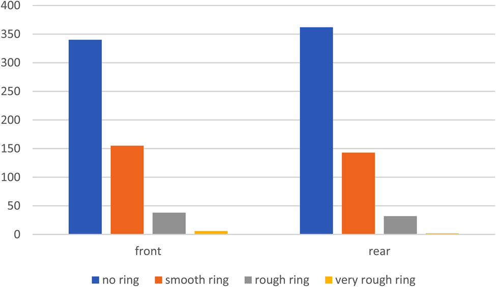
Front teats showed a poorer teat condition than rear teats (Fig. 1). There was a strong association between the teat condition classification of front and rear teats in the cows (r s = 0.79, P = < 0.001).
Assessment of teat condition score (number of front and rear teats) before milking.

The data for colour, swelling at the teat base and teat firmness after milking are shown in Table 1. Rear teats were more often discoloured after milking than front teats (P < 0.05). There were no significant differences between front and rear teats in swelling at the teat base nor in teat firmness (P > 0.05).
Examination after milking of colour changes (1 – normal, 2 – reddened, 3 – bluish), swelling at the teat base (1 – no visible ring, 2 – visible mouthpiece lip mark, 3 – swelling or thickened ring) and teat firmness (1 – soft and no wedge-shaped, 2 – flat or hard)

* Indicates significant difference between front and rear teats (P ≤ 0.05).
Morphology: teat length
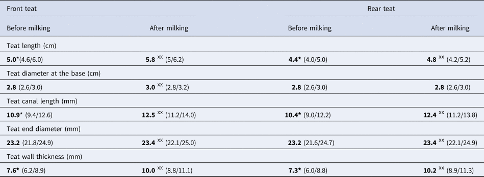
The data for morphological measurements of teat dimensions are presented in Table 2. Before milking front teats were significantly longer than rear teats (P < 0.001). During milking the length of front teats and rear teats increased (P < 0.001). The relative increase of teat length (length after milking/length before milking) was significantly higher (P < 0.001) in the front teats (1.17, 1.07/1.25) in comparison to the rear teats (1.08, 1.00/1.14). The increase of the front teat length during milking was significantly influenced by vacuum level in the mouthpiece chamber during milking and over-milking, but the variables milk yield, teat length, teat end diameter and wall thickness before milking were also included in the model. However, these variables were able to explain only about 22% of the effect (R 2 = 0.215, P = 0.035). For the rear teats only milk yield, teat diameter and teat wall thickness before milking had a significant effect (R 2 = 0.166, P = 0.037) on the changes of the teat length by milking. Variables associated with milking (vacuum levels, milking and over-milking time) were excluded from the model (P > 0.1).
Morphologic teat measurements (teat length, teat diameter) and ultrasonographic teat measurements (teat canal length, teat end diameter, teat wall thickness) of front and rear teats before and after milking, data is presented as median (1st/3rd quartile)

* Indicates significant difference between front and rear teats (P ≤ 0.001).
XX Indicates significant difference between before and after milking (P ≤ 0.001).
Morphology: teat diameter at the base
Teat base increased during milking in the front but not in the rear teats. Since the rear teat diameter was not influenced by milking no model was fitted.
Ultrasonography: teat canal length
Teat dimensions of front and rear teats measured by ultrasonography before and after milking are shown in Table 2. The teat canal length before milking was significantly longer (P < 0.001) at front teats than at rear teats. During milking and over-milking the teat canal length extended significantly at front and rear teats (P < 0.001). The relative increase in length caused by milking was inverse proportional to the teat canal length before milking (r −0.56; P < 0.001). Consequently, the relative increase of teat canal length during milking (length after milking/length before milking) was significantly (P < 0.001) higher at rear teats (1.20, 1.10/1.31) in comparison to the front teats (1.13, 1.05/1.18). For the changes of teat canal length of the front teats the variables vacuum level in the MPC during milking and over-milking, the duration of over-milking but also milk yield, teat diameter and teat wall thickness before milking were included in the best fitting model (R 2 = 0.23, P = 0.029). Teat canal length changes of rear teats were only dependent upon milk yield and the morphological parameters teat end diameter and teat wall thickness before milking (R 2 = 0.21, P = 0.037). No milking associated variables (vacuum levels and duration of milking or over-milking) were included in the model for the rear teats.
Ultrasonography: teat end diameter
There was no difference in teat end diameter between front and rear teats before milking. Teat end diameters increased significantly in front and rear teats (P = 0.001). Changes of teat end diameter of the front teats were influenced by vacuum level in the mouthpiece chamber during milking but also by the morphological parameter teat end diameter and teat wall thickness before milking. The model including these three parameters had a R 2 of 0.27 (P < 0.001). At the rear teats the variables duration of milking, teat end diameter and teat wall thickness before milking (R 2 = 0.24, P = 0.019) were included in the model.
Ultrasonography: teat wall thickness
Before milking the teat wall at front teats was significantly thicker than at rear teats (P = 0.019). Teat wall thickness increased significantly during milking in front and rear teats (P < 0.001). Vacuum level in the MPC during milking and over-milking and duration of milking but also wall thickness and teat diameter before milking as well as days in milk were variables included in the model having an effect on the changes of the front (R 2 = 0.33, P < 0.05) and rear teat wall thickness (R 2 = 0.41, P < 0.001).
Discussion
The main focus of the study was on over-milking time and vacuum level during milking and over-milking and whether these variables may accountable in major part for the differences in milking-induced morphological changes between front and rear teats. To the best of our knowledge this was the first study comparing ultrasound measurements of teat structure from front and rear teats before and after milking and including measurement of vacuum level and over-milking time.
Front teats were over-milked for significantly longer than rear teats, which is similar to the study of Tančin et al. (Reference Tančin, Ipema, Hogewerf and Mačuhová2006) and supports our hypothesis. However, contrary to our expectations, the vacuum levels during milking and especially during over-milking were higher in the rear quarters in comparison to the front quarters. It is reasonable to believe that this is partly due to the differences in teat size between front and rear teats because identical liners were used for all teats. The association between teat size and mouthpiece chamber has been described previously (Rasmussen, Reference Rasmussen1997), although using different measurement equipment. In contrast to the earlier work, our study did not identify an association between lactations status (DIM) and vacuum levels. We did confirm that the teat condition of front teats is poorer than that of the rear teats (Neijenhuis et al., Reference Neijenhuis, Klungel and Hogeveen2001). The morphological and ultrasonographic examination identified differences between front and rear teats before and after milking. Front teats were longer than rear teats but had no differences at the teat diameter at the base. Guarín and Ruegg (Reference Guarín and Ruegg2016) also described longer front teats but also having a wider teat apex.
In the present study the teat canal was longer and teat wall was thicker in front teats. These results are similar to Strapák et al. (Reference Strapák, Strapáková, Rušinová and Szencziová2017) but in contradiction to some others studies (Weiss et al., Reference Weiss, Weinfurtne and Bruckmaier2004; Klein et al., Reference Klein, Flöck, Khol, Franz, Stüger and Baumgartner2005) which did not describe significant differences in teat canal length and teat wall thickness between front and rear teats. We found an increase of teat canal length during milking which is in accordance with other studies (Neijenhuis et al., Reference Neijenhuis, Klungel and Hogeveen2001; Gleeson et al., Reference Gleeson, O'Callaghan and von Rath2002; Paulrud, Reference Paulrud2005). The rear teats showed a larger relative increase of teat canal length, which was also described by Strapàk et al. (Reference Strapák, Strapáková, Rušinová and Szencziová2017). However, contrary to one of our hypotheses the changes of teat canal length, teat diameter, teat wall thickness and teat length of the front teats were influenced only to a limited extent by the milking associated factors: over-milking time, vacuum levels during milking and over-milking. Additionally, these factors were mostly found to be accountable for the changes during milking in front but not in rear teats, which supports our hypothesis that differences in over-milking duration but also in anatomy between front and rear teats are of importance. As was shown by Neijenhuis et al. (Reference Neijenhuis, Barkema, Hogeveen and Noordhuizen2000), the machine-on time is an important factor. It is reasonable to assume that if the cows had been over-milked longer the effects would have been more pronounced and not limited to the front teats. However, we did not detect differences between farms with or without automatic cluster removal. A number of anatomical parameters of the teat also influenced the changes of teat condition caused by milking but their influence was also low. Additionally, we could not detect a significant association between the over-milking time and the appearance of poor teat end condition which is partially contrary to the results of Hillerton et al. (Reference Hillerton, Pankey and Pankey2002) and Besier and Bruckmaier (Reference Besier and Bruckmaier2016). These differences are most likely to be explained by the variations in over-milking time, in milking machinery, cluster types and pulsation and vacuum settings between the various studies (Rasmussen and Madsen, Reference Rasmussen and Madsen2000). However our findings support the idea of teat individual milking and claw removal according to milk flow characteristics (Ipema and Hogewerf, Reference Ipema and Hogewerf2008).
One result of the study is that numerous undetermined factors are influencing the changes of the teat morphology during milking. Although we included various variables in our analyses, factors which had not been measured and were not included in the model obviously had a substantial influence. Such other factors may be the skills of farm worker, type of milking devices, the type and condition of clusters and liners or the pulsation characteristics and cow individual factors. These factors have not been sufficiently recorded in the present study to allow detailed analyses but should be considered in further research projects.
One limitation of the field study was that a limited number of cows per farm could be investigated as we were not supposed to interfere with the milking procedure. Further, we only had the total SCC available, which was not significantly influenced by over-milking time or vacuum level. The result might have been different if quarter based SCC had been available as shown by Guarín and Ruegg (Reference Guarín and Ruegg2016). Additionally, it remains unclear if the exclusion of cows with clinical mastitis from the study may have influenced the results, although as this was generally low it seems unlikely.
To summarize, the study showed that over-milking and vacuum levels influence the teat condition of front quarters. However, under field conditions since the clusters were removed promptly the effect of these parameters on the milking-related changes of the teat condition was limited. We suggest that in farms where long over-milking is frequent, these factors are likely to gain more importance, especially for the front teat condition and somatic cell count. Even if the results of the study suggest that over-milking has a limited effect, the attention of the farmers should be drawn to limiting machine-on time, using correct vacuum and pulsation settings and using different liners for front and rear teats or installing quarter-level milking systems.
Supplementary material
The supplementary material for this article can be found at https://doi.org/10.1017/S0022029921000807.
Acknowledgements
The authors wish to express their particular thanks to the farmers for their collaboration and for allowing us to include their cows in the study during milking. Further thanks are due to Prof. David Logue (Glasgow University) for his help preparing the manuscript.