Infective endocarditis is a bacterial or fungal infection of the endocardium or heart valves. Even with appropriate medical treatment, the infection can lead to catastrophic complications, such as valvular dysfunction, perivalvular extension of infection, formation of an abscess or fistula, or aortic rupture.Reference Bin Abdulhak, Baddour and Erwin1 Although there have been several reports on the complications of this condition in children, very few studies have assessed pseudoaneurysms that developed secondary to infective endocarditis. Here, we report an aortic pseudoaneurysm due to infective endocarditis in a child with trisomy 21 and a history of subaortic stenosis after the repair of aortic coarctation and ventricular septal defect.
Case presentation
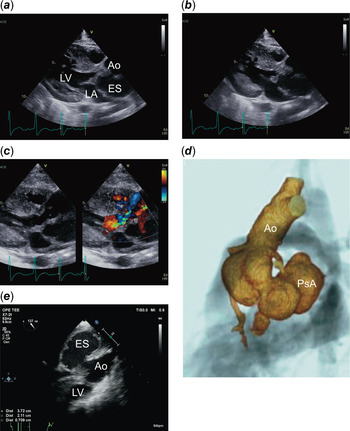
A 7-year-old boy with trisomy 21 was referred to our institute, presenting with a 3-day history of ongoing lethargy and fever. The patient had a history of bicuspid aortic valve and subaortic stenosis following surgery for coarctation of the aorta and ventricular septal defect. Three months prior to this episode, he presented with myocarditis caused by influenza and was hospitalised for 3 months. Immunomodulatory therapies including 2 days of high-dose intravenous immunoglobulin (1 g/kg daily) and high-dose glucocorticoids, followed by 16 days of gradual tapering, were used during hospitalisation. Furthermore, a central venous catheter had been inserted for a month. Physical examination at presentation revealed a heart rate of 100 beats per minute, systolic ejection murmur (Levine II/VI), systolic blood pressure of 96 mmHg, and temperature of 38.4°C. Blood tests revealed an increase in the number of leukocytes (18,520/μl) and elevated serum levels of C-reactive protein (10.8 mg/dl). A chest X-ray revealed thickening of the bronchial wall; radiography findings suggested that bronchitis might have been the cause of fever, and intravenous aminopenicillin (150 mg/kg/day) was initiated. On the following day, methicillin-resistant, coagulase-negative staphylococci were detected in the blood sample that was drawn prior to admission, and the intravenous antibiotic was changed to vancomycin (60 mg/kg/day). Transthoracic echocardiography performed on the 3rd day of hospitalisation showed thickening of the posterior leaflet of the bicuspid aortic valve and trivial aortic regurgitation, suggesting infective endocarditis of the aortic valve. An echolucent space behind the sinus of Valsalva was observed. Abnormal flow between the aorta and echolucent space was detected. The size of the echolucent space did not change during the cardiac cycle (Fig 1a, b, and c). Cardiac CT angiography revealed a space (44.5 × 36.5 × 21.5 mm) between the aortic sinus and the left atrium (Fig 1d). These findings suggested the possibility of a pseudoaneurysm of the sinus of Valsalva caused by infective endocarditis, and surgical treatment was performed to avoid rupture of the pseudoaneurysm.
(a) Transthoracic echocardiogram in the parasternal long-axis view (systolic phase). The figure shows an echolucent space (43.5 × 21.5 mm). (b) Transthoracic echocardiogram in the parasternal long-axis view (diastolic phase). The figure shows a thickened posterior aortic valve leaflet during the diastolic phase. Below the leaflet, there is an echolucent space between the aorta and the left atrium. (c) Transthoracic echocardiogram with colour flow in the parasternal long-axis view. The figure shows an abnormal flow between the aorta and the echolucent space. (d) Cardiac CT angiogram (3D reconstruction). The figure shows that a pseudoaneurysm (44.5 × 36.5 × 28.5 mm) expands below the sinus of Valsalva. (e) Transesophageal echocardiogram. The figure shows an ostium (7.0 mm) between the aorta and echolucent space. Ao=aorta; ES=echolucent space; LA=left atrium; LV=left ventricle; PsA=pseudoaneurysm.

Transesophageal echocardiography performed immediately before surgery showed vegetation on the posterior leaflet of the aortic valve and a perforation (diameter: 7.0 mm) in the echolucent space just above the posterior leaflet (Fig 1e). Because the posterior leaflet was severely eroded, the aortic valve was replaced after debridement using a mechanical prosthetic valve (Open Pivot AP360®□, 16 mm size, Medtronic, Minneapolis, MN, United States of America). The entry hole of the pseudoaneurysm was closed with a glutaraldehyde-treated autologous pericardium after injecting fibrin glue (Bolheal®□, KM Biologics, Kumamoto, Japan) mixed with gentamycin into the pseudoaneurysm. Histological evaluation of the aortic valve revealed infiltration of neutrophils and round cells, suggesting the presence of Staphylococcus spp. Intravenous administration of vancomycin was continued for 8 weeks, with dosage adjusted to maintain the serum vancomycin trough concentrations within a range of 10–15 μg/ml after surgery. Although the pseudoaneurysm remains, confirmed by cardiac CT angiography, it did not increase in size. The patient did not have a relapse of the infection and exhibited no severe complications at discharge.
Discussion
We report the rare case of an aortic root pseudoaneurysm that developed secondary to infective endocarditis in a child with trisomy 21 and a history of surgically repaired CHDs. The reported incidence of infective endocarditis is 1.5–11.6 cases per 100,000 people in 10 countries.Reference Bin Abdulhak, Baddour and Erwin1 In children, the annual incidence rate is reportedly 0.05–0.12 cases per 1000 hospital admissions.Reference Baltimore, Gewitz and Baddour2 Predisposing factors for infective endocarditis include dental infections, surgeries, immunosuppressive therapies, injection drug use, and rheumatic fever. However, a history of CHD is considered the leading risk factor for developing infective endocarditis. One study reported that approximately 80% of children with infective endocarditis had underlying CHD,Reference Coward, Tucker and Darville3 and this result may be related to improved survival of children with CHD. Children with CHD who underwent procedures, including central venous catheter placement and valve or open-heart surgery, had a high risk of developing infective endocarditis.Reference Sun, Lai and Wang4
Perivalvular extension of infection can lead to rare catastrophic complications such as the formation of pseudoaneurysms or fistulous tracts.Reference Baltimore, Gewitz and Baddour2 A pseudoaneurysm can result in haemodynamic deterioration and sudden death when it ruptures; therefore, optimal diagnosis and management are required. Perivalvular complications occurred in 9.8–40% of adult patients with infective endocarditis.Reference Rouzé, Flécher and Revest5 However, in paediatric populations, the incidence of perivalvular complications has not been clarified, and there have been very few reports on pseudoaneurysms at aortic valves caused by infective endocarditis.Reference Barth, Moosdorf, Bauer, Schranz and Akintürk6–Reference Atiyah, Mohsin, Al Faraidi, Al-Hawri, Al Otay and Al Najashi9
The risk factors for infective endocarditis noted in our patient included a history of CHDs, immunosuppressive state, and placement of a central venous catheter; amongst these, stenosis of the subaortic and bicuspid aortic valves may have been related to the formation of the pseudoaneurysm. The narrowing of the aorta caused by these two conditions may have resulted in jets of blood striking the aortic wall. The haemodynamic patterns of the bicuspid aortic valve may have dilated and changed the shear stress of the aortic wall.Reference Mahadevia, Barker and Schnell10 In addition, the jet flow caused by the subaortic stenosis and bicuspid valve may have exacerbated the damage to the infected aortic wall.
The patient luckily survived this life-threatening disease as there was no severe aortic regurgitation, and adhesion tissues from prior cardiac surgeries surrounded his heart.
Conclusion
A pseudoaneurysm of the aorta that develops secondary to infective endocarditis is uncommon in children. Infective endocarditis may lead to the perivalvular extension of infection and formation of a pseudoaneurysm, which results in high mortality and morbidity. Prompt diagnosis and management of infective endocarditis can result in successful outcomes without additional sequelae.
Acknowledgements
The authors express their gratitude to the staff of the Japanese Red Cross Medical Center.
Financial support
This research received no specific grant from any funding agency, commercial, or not-for-profit sectors.
Conflicts of interest
None.
Ethical standards
Written informed consent was obtained from the patient’s family.