Introduction
There is currently an urgent need to establish principles for the assembly of materials with specialized functions/properties at the nanoscale. Construction of nanocomposites with varying architectures and properties can have a wide variety of applications. The development of a theoretical framework for these assemblies is dependent on experimental results, which are needed to establish principles and generalize methodologies.[ Reference Biswas, Bayer, Biris, Wang, Dervishi and Faupel 1 , Reference Xu, Ma, Wang, Xu, Kuang and Kotov 2 ] The assembly of such materials are far-reaching and are usually approached using two methods[ Reference Biswas, Bayer, Biris, Wang, Dervishi and Faupel 1 , Reference Pershina, Sazonov and Filimonov 3 ]: the “top-down” and “bottom-up” methods. In the “top-down” approach nanofabrication tools are employed to create nanoscaled structures with targeted shapes and properties beginning with larger structures and reducing them to the desired scale. One example of this approach is lithography. The “bottom-up” method aims to control material at the molecular or atomic level and build complex assemblies at the nanoscale. This approach relies on directed self-assembly based on mechanisms, which are currently under development.[ Reference Biswas, Bayer, Biris, Wang, Dervishi and Faupel 1 – Reference Tan, Xing and Lu 8 ] The bottom-up approach in principle is more efficient than the top-down approach since it is controlled by thermodynamic properties and not by manipulation. Careful design of building blocks and employment of external stimuli such as magnetic/electric fields, gravity, chemical reactivity, temperature, concentration, pH, reaction time, and light to influence assembly allows for the generation of macroscopic materials with high complexity. The employment of reversible processes to create these materials would be ideal since any errors could easily be corrected. There are virtually an infinite number of structural and morphological types that can be generated using this approach. Nanomaterial morphologies (e.g., shapes and surface structures) and distance-dependent properties (e.g., plasmonic and quantum confinement effects), make nanomaterials ideal for future applications, including catalysis, photonics, environmental detection, biomedical diagnostics, and therapy.[ Reference Xu, Ma, Wang, Xu, Kuang and Kotov 2 , Reference Tan, Xing and Lu 8 , Reference Schroeder, Heller, Winslow, Dahlman, Pratt, Langer, Jacks and Anderson 9 ] Fine control over interactions between nanoparticles (NPs) and composition are critical elements for the construction of these materials. It is additionally anticipated that structures exceeding 100 µm can be produced using nanometer-building blocks, thus making them ideal for incorporation into lithographically produced devices.[ Reference Xu, Ma, Wang, Xu, Kuang and Kotov 2 ]
Pershina et al., in a recent review discussed the utility of DNA for assembly of magnetic nanoparticles (MNPs) for nanobiocomposites and nanobioelectronics.[ Reference Pershina, Sazonov and Filimonov 3 ] They addressed interaction of DNA with metallic MNPs as well as magnetic oxides such as Fe3O4. Interactions of metal ions could occur with different sites of DNA and are dependent on the nature of metal ions. Alkali-metal ions, in general, form bonds with the phosphate groups, whereas transition metal ions bind to phosphate groups and also to DNA bases. In the case of oxide MNPs such as Fe3O4, it was found that the bonding to DNA was through the formation of Fe–O–P bonds.[ Reference Pershina, Sazonov and Filimonov 3 ] One could, therefore, utilize direct grafting of DNA followed by DNA hybridization to assemble a variety of functional materials and composites, including ferrite–ferroelectric core–shell particles such as NiFe2O4–BaTiO3 or CoFe2O4–BaTiO3.[ Reference Bao and Gupta 4 – Reference Tan, Xing and Lu 8 ] In this study, however, the assembly procedure involves first functionalizing DNA and NPs with complementary chemical groups, alkyne and azide groups, respectively, in order to attach DNA to NPs by click reaction. This is followed by the assembly of core–shell NPs by DNA hybridization. This procedure is not specific to the systems studied here and could be employed for assembly of a variety of oxide or metal composites.
Multiferroic nanocomposites
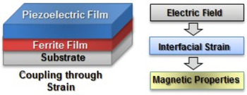
One application of engineered nanomaterials is in the study of multiferroics. A multiferroic is a material that exhibits two or more of the primary ferroic properties (ferromagnetism, ferroelectricity, and ferroelasticity).[ Reference Nan, Bichurin, Dong, Viehland and Srinivasan 10 – Reference Wang, Pan and Chen 15 ] A composite made of a ferromagnetic and ferroelectric material allows for the coupling between the electric and magnetic order parameters (Fig. 1) and represents an engineered multiferroic. The magnetoelectric (ME) coupling is mediated by mechanical forces. When an ac magnetic field is applied to a ferromagnetic material, it oscillates (in size) due to magnetostriction. When this mechanical deformation is transferred to a ferroelectric/piezoelectric material (e.g., PZT and quartz crystal) it responds by producing an electric field due to the piezoelectric effect. Thus, the mechanical strain on these materials produces a magnetic field–electric field conversion. Generation of these composites provides the opportunity for studies on the physics of ME coupling, and have enormous potential for the design of novel miniature functional devices.[ Reference Sun and Srinivasan 11 ] Such devices include multiple-state logic memories or electrically written magnetic memories, spintronic non-volatile memories, magnetic/electric field sensors, transformers, gyrators, resonators, phase shifters, filters, tunable microwave and optical devices, and antennas.[ Reference Nan, Bichurin, Dong, Viehland and Srinivasan 10 , Reference Sun and Srinivasan 11 ]
Electric field control of electromagnetic properties of a ferrite–piezoelectric material. The control is through mechanical forces.

Chemical assembly
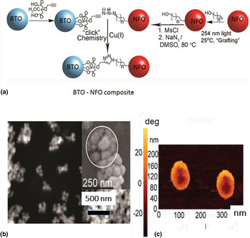
Our studies on ferromagnetic–ferroelectric composites revealed a giant low-frequency ME effect and enhancement at electromechanical and ferromagnetic resonance. We also developed theoretical models for the ME phenomena.[ Reference Nan, Bichurin, Dong, Viehland and Srinivasan 10 ] The strain-mediated ME coupling in core–shell nanocomposites assemblies of ferroelectric (barium titanate, BTO) and ferromagnetic (nickel ferrite, NFO) materials is expected to be very strong due to a surface-to-volume ratio that is a factor of 103–106 higher than for bulk and thick/film composites. Core–shell NPs of 10–400 nm NFO and 50–600 nm BTO were assembled by attaching complementary chemical groups, azide and alkyne, to particles and then assembling them by the click reaction as shown in Fig. 2.
(top) Synthesis of BTO and NFO core–shell NPs by chemical self-assembly: particles were functionalized with complementary coupling groups (propargyl and azide groups) and then assembled in the presence of copper catalyst. (bottom left) SEM of core–shell particle. (bottom left, inset) SEM of collection of core–shell particles. (bottom right) MFM phase image showing the core–shell structure.[ Reference Sreenivasulu, Popov, Chavez, Hamilton, Lehto and Srinivasan 16 – Reference Srinivasan, Sreenivasulu, Benoit, Petrov and Chavez 20 ]

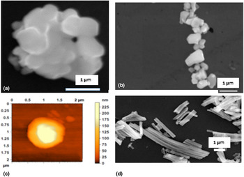
Studies by scanning electron microscopy (SEM), transmission electron microscopy (TEM), and scanning microwave microscopy (SMM) at 1–26 GHz showed the core–shell structures with well-defined interfaces (Fig. 3).[ Reference Sreenivasulu, Popov, Chavez, Hamilton, Lehto and Srinivasan 16 – Reference Srinivasan, Sreenivasulu, Benoit, Petrov and Chavez 20 ] Magnetic and ferroelectric characterization revealed ferromagnetic ordering for NFO particles and ferroelectric nature for BTO. We have generalized the method for attaching the groups to the NPs so that any organic group can be attached. This technique is very effective in creating covalent attachments between NFO and BTO NPs and is not limited by the diameters for these particles. A shortcoming, however, is that the attachments are non-dynamic (cannot be reorganized once assembled) and create nanocomposites that are limited in size, which may be due to low coupling efficiency for the click reaction.
(a) SEM image showing a DNA-assembled particle of 600 nm BTO core–200 nm NFO shell. (b) SEM image of an array of core–shell particles assembled under a magnetic field. (c) SMM topography image of the particle. (d) SEM of bundle of ferrite-PZT core–shell nanowires assembled with DNA.

DNA-assisted assembly of multiferroic nanocomposites
The use of DNA in the NP self-assembly process has proven to be one of the most versatile methods to date for creating nanostructures of varying structures and compositions.[ Reference Tan, Xing and Lu 8 , Reference Guo, Hu and Deng 21 , Reference Ma, Yang, Wei and Yuan 22 ] To accomplish this, DNA is conjugated to nanomaterial/particles via modified functional groups on the custom-synthesized DNA strand. Such groups include sulfhydryl, amine, carboxylate, azide, alkyne, and others located at either the 5′ or 3′ end.[ Reference Tan, Xing and Lu 8 , Reference Ravan, Kashanian, Sanadgol, Badoei-Dalfard and Karami 23 – Reference Kumar, Hwang, Kumar and Nam 25 ] Methods for covalently attaching single-stranded DNA (ssDNA) to NP surfaces have included gold–thiolate interaction,[ Reference Banchelli, Nappini, Montis, Bonini, Canton, Berti and Baglioni 26 – Reference Pita, Abad, Vaz-Dominguez, Briones, Mateo-Marti, Martin-Gago, Morales Mdel and Fernandez 28 ] Michael addition,[ Reference Kitamura, Nakai, Kohda, Furuta-Okamoto and Iwata 29 ] disulfide bond formation,[ Reference Shen, Wang, Yang and Jiang 30 – Reference Santra, Yapec, Theodoropoulou, Dobson, Hebard and Tan 32 ] phosphate–amino linkage via carbodiimide activation,[ Reference Zhu, Zhang, He and Fang 33 ] amide bond formation,[ Reference Medley, Smith, Wigman and Chetwyn 34 – Reference Yin, Li, Liu, Ren, Yang and Qu 38 ] Schiff-base formation,[ Reference Fuentes, Mateo, Rodriguez, Casqueiro, Tercero, Riese, Fernandez-Lafuente and Guisan 39 ] glutaraldehyde-assisted cross-linking,[ Reference Amagliani, Omiccioli, Campo, Bruce, Brandi and Magnani 40 – Reference Nie, Wang, Li and Chen 43 ] and cross-linking using compounds containing sulfhydryl and amino-(N-hydroxysuccinimide) groups.[ Reference Josephson, Perez and Weissleder 44 – Reference Osterberg, Rizzi, Zardan Gomez de la Torre, Stromberg, Stromme, Svedlindh and Hansen 47 ] The commercial availability of custom-synthesized DNA/RNA containing up to 100 bp with any desired sequence and several 5′ or 3′ organic functional groups offers a great opportunity to explore the effect of sequence and method the attachment. Employment of DNA to assemble NP via hybridization into superlattices has been an example of particle-to-materials fabrication.[ Reference Park, Lytton-Jean, Lee, Weigand, Schatz and Mirkin 48 – Reference Lu, Yager, Zhang, Xin and Gang 50 ] With biologic assemblies many length-scales are crossed using a structural hierarchy. Recently a class of intercalators, [Ru(dipyrido[2,3-a:3′,2′-c]phenazine) (N−N)2]2+-synthesized with DNA was found to increase the bond strength between DNA and NPs.[ Reference Seo, Wang, Shade, Rouge, Brown and Mirkin 51 ] Although work with the employment of DNA as a linker has been reported, essentially no work has been published using RNA.
The Cu(I)-catalyzed azide–alkyne cycloaddition (CuAAC) “click” reaction has also been employed to achieve attachment of DNA to NPs.[ Reference Cutler, Zheng, Xu, Giljohann and Mirkin 52 – Reference Kaittanis, Boukhriss, Santra, Naser and Perez 54 ] Recent work by Mirkin and co-workers has demonstrated that alkyne-functionalized ssDNA can be bound to NPs via CuAAC “click” chemistry yielding NPs with high-density monolayers of DNA on the surface.[ Reference Cutler, Zheng, Xu, Giljohann and Mirkin 52 ] In their work, they attach single-stranded oligomeric DNA to superparamagnetic iron oxide nanoparticles as a proof-of-concept study. In our work, 3-buten-1-ol was grafted to the surfaces of 600 nm BTO and 200 nm NFO using 254 nm light under anaerobic conditions. These groups were subsequently converted to alkyl–azide groups (Fig. 2 top) and alkyne-functionalized DNA (custom synthesized by Keck Laboratories, Inc.) was attached via the click reaction.[ Reference Sreenivasulu, Lochbiler, Panda, Srinivasan and Chavez 55 ]
RNA-assisted assembly of multiferroic nanocomposites
RNA-based nanotechnology is a newly emerging field.[ Reference Shukla, Haque, Tor, Wilhelmsson, Toulme, Isambert, Guo, Rossi, Tenenbaum and Shapiro 56 – Reference Carter, Dolska, Owczarek, Ackerson, Eaton and Feldheim 59 ] There has been an increasing interest in RNA for the assembly of nanostructures over the past few years as indicated by the increase in publications. Compared with DNA, RNA possesses higher thermodynamic stability, a higher variety of base-pairing alternatives, and programmable tertiary structure formation (helices, stems, loops, hairpins, bulges, and pseudoknots).[ Reference Shukla, Haque, Tor, Wilhelmsson, Toulme, Isambert, Guo, Rossi, Tenenbaum and Shapiro 56 ] The known degradation of RNA from RNase enzymes has previously caused scientists to avoid employing RNA in nanotechnology. This has resulted in disinterest in this field. Assuming that this remains a problem with oligomeric RNA, one could use alternative strategies to generate degradation-resistant RNA.[ Reference Liu, Guo, Cinier, Shlyakhtenko, Shu, Chen, Shen and Guo 60 ] Improvements in stability could come from using modified bases (i.e., 5-Br-U, 5-I-U), modified phosphate linkages (i.e., phosphothiolate, boranophosphate), peptide nucleic acids (i.e., PNA), or C2′ modifications (i.e., 2′-fluorine, 2′-O-methyl, or 2′-O-amine).[ Reference Shukla, Haque, Tor, Wilhelmsson, Toulme, Isambert, Guo, Rossi, Tenenbaum and Shapiro 56 , Reference Sharma, Haque, Pi, Shlyakhtenko, Evers and Guo 58 ] Using computer-designed oligonucleotide RNA, Jaeger and co-workers were able to generate three-dimensional (3D) nanoscaffolds that assembled in vitro.[ Reference Afonin, Bindewald, Yaghoubian, Voss, Jacovetty, Shapiro and Jaeger 61 ] Other 3D structures have been assembled using similar techniques.[ Reference Grabow, Zakrevsky, Afonin, Chworos, Shapiro and Jaeger 62 ] Due to the great diversity of base-pair and folding interactions with RNA, computer programs have been developed to help predict 3D structures.[ Reference Markham and Zuker 63 , Reference Matveeva, Kang, Spiridonov, Saetrom, Nemtsov, Ogurtsov, Nechipurenko and Shabalina 64 ] NP formation of modified RNA via metal (i.e., Fe and Co) complexation has also been reported.[ Reference Carter, Dolska, Owczarek, Ackerson, Eaton and Feldheim 59 ]
Formation of ordered structures by magnetic fields
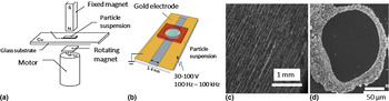
One of the key objectives of our research is to assemble nanomaterials into superstructures, which are predicted to exhibit strong ME coupling at low frequencies and resonance ME effects over a wide frequency range of interest. The procedure involves heating the nanomaterials in a solution to 80 °C for dehybridization, followed by cooling under a field so that DNA-assisted assembly and field-directed assembly occur simultaneously. The set ups as in Fig. 4 are useful for assembly under magnetic and electric fields.[ Reference Wang and Ko 65 , Reference Smoukov, Gangwal, Marquez and Velev 66 ] The uniformity, position, and patterns can be controlled with positioning or patterning of magnets on a substrate or the frequency and amplitude of applied voltage. Our efforts toward H-assembly of superstructures have been successful. Figure 4 shows SEM images of 2.5 × 2.5 mm2 array and a ring of DNA-assembled core–shell particle obtained with uniform and rotating fields, respectively.
(a) A setup for H-assisted assembly. Binding site is a substrate with patterned Ni/permalloy bottom and assists in assembly of desired structure for core–shell or coaxial tubes/wires.[ Reference Wang and Ko 65 ] (b) E-field-assisted assembly set up.[ Reference Smoukov, Gangwal, Marquez and Velev 66 ] (c) SEM image showing a 2.5 × 2.5 mm2 2D superstructure of 600 nm BTO and 200 nm NFO core–shell particles obtained in a uniform H-field. (d) A ring of the core–shell particles assembled in a rotating magnetic field.

Magnetic, ferroelectric, and multiferroic properties of assembled composites
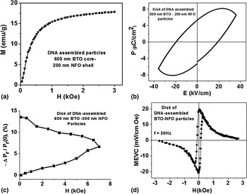
It is essential to characterize the DNA-assembled composites and superstructures in terms of ferroic order parameters and ME coupling strength.[ Reference Sreenivasulu, Lochbiler, Panda, Srinivasan and Chavez 55 , Reference Zhang, Dai, Chow, Sun, Lo and Chan 67 , Reference Zhang, Dai, Lu and Chan 68 ] The ferromagnetic properties of the composite was studied by measurements of room-temperature magnetization M as a function of the static magnetic field H. Figure 5(a) shows such date for the core–shell composite and a ferromagnetic character is clearly evident from the data. The magnetization shows saturation value of 17.5 emu/g for H ~ 2.5 kOe, which is about 60% of the value expected for the bulk NFO-only sample. The ferroelectric nature of the composites was confirmed with polarization P versus electric field E measurements with a (Radiant Technology) ferroelectric tester. The data in Fig. 5(b) indicate remnant polarization of 4 mC/m2 and a saturation value of ~8 mC/m2, values that are somewhat smaller than values reported for the bulk or thick film BTO.
(a) Room-temperature magnetization as a function of static magnetic field for as-assembled BTO–NFO composite. (b) Polarization P versus electric field E data for a disc of DNA-assembled BTO–NFO core–shell particles. (c) Fraction change in the remnant polarization P r versus H obtained from data as in Fig. 5(b) when the sample is subjected to an in-plane static magnetic field. (d) Low-frequency MEVC versus H for a sintered disc of DNA-assembled BTO–NFO particles.[ Reference Sreenivasulu, Lochbiler, Panda, Srinivasan and Chavez 55 ]

Magneto-electric characterization of the composites could be done by measuring the sample response to: (i) an applied magnetic field (termed direct-ME effect) and (ii) an applied electric field (converse ME effect). Here we provide results on the direct-ME effect by two different techniques: static magnetic field H-induced polarization P in the sample and low-frequency ME voltage coefficient. Measurements were first carried out on pressed pellets of as-assembled core–shell composites. Figure 5(c) shows the results on direct-ME effect (DME) by static magnetic field H-induced polarization through P versus E measurements in a disc of DNA-assembled sample. The samples were subjected to a uniform in-plane magnetic field. A decrease in P due to H was measured. The fractional change in the remnant polarization ΔP r/P r (H = 0) = [P r(H)–P(H = 0)]/P r (H = 0) versus H measured from P versus E for H = 0–7 kOe are shown. As H is increased from 0 to 7 kOe, ΔP r/P r increases with H to ~7% at H = 7 kOe. When H is decreased back to zero, a hysteresis is seen in ΔP r/P r versus H with the magnitude ΔP r/P r increasing to 13% for H = 0. Thus, the results in Fig. 5(c) provide clear evidence for magnetostriction-induced change in polarization for the composite.[ Reference Sreenivasulu, Lochbiler, Panda, Srinivasan and Chavez 55 ]
We also studied the nature of DME by low-frequency ME voltage coefficient (MEVC) measurements on sintered discs of DNA-assembled sample. The samples were first poled in E = 1 kV/cm at room temperature and then subjected to a dc bias magnetic field H and an ac field of H ac = 1 Oe at 30 Hz. The ac and dc magnetic fields were parallel to each other and applied parallel to the disc plane. The ME voltage induced across the disc thickness was measured as a function of H. Figure 5(d) shows MEVC versus H data for the DNA-assembled sample. The MEVC shows a zero-bias (H = 0) value of 20 mV/cm Oe and is indicative of a built-in magnetic field arising from dipole–dipole interactions between NFO particles in the core–shell structures. With increase in H, MEVC decreases and the coupling vanishes at H = 2 kOe due to saturation of magnetostriction resulting in zero value for the piezomagnetic coupling q. A hysteresis is also seen in MEVC versus H data. When H is reversed the MEVC becomes negative (a 180° phase difference with H ac). For comparison we measured MEVC on thin discs of the composites made by mixing NFO and BTO particles. The MEVC was much smaller than for DNA-assembled samples. For our DNA-assembled nanocomposites, we observed strong ME coupling in as-assembled composites, which is evident from polarization induced by magnetic field H and MEVC results in Fig. 5. We found that annealing the samples at high temperatures removes the organic components and enhances the interaction of the components yielding excellent ME coupling evident by low-frequency ME voltage coefficient and H-induced polarization.[ Reference Sreenivasulu, Lochbiler, Panda, Srinivasan and Chavez 55 ]
Composites with a ferrite core and BTO or lead zirconate titanate (PZT) shell can also be synthesized by a two-step hydrothermal and annealing process or a surfactant-assisted thermal method.[ Reference Liu, Zhao, Huang, Zhao and Zhou 69 , Reference Raidongia, Nag, Sundaresan and Rao 70 ] Several systems, including Fe3O4/PbTiO3, CoFe2O4/BaTiO3, and CoFe2O4/Pb(Zr,Ti)O3 were synthesized. The synthesis technique facilitated control over size of the core and thickness of shell.[ Reference Liu, Zhao, Huang, Zhao and Zhou 69 ] Measurements of ME coupling strength by magnetodielectric effect was reported for CoFe2O4/BaTiO3. The dielectric constant at 134 K decreased by 1.7% for an applied field of 10 kOe.[ Reference Raidongia, Nag, Sundaresan and Rao 70 ] Thus, one infers that the ME coupling in the composite is much weaker than the measured coupling strength in the DNA-assembled core–shell particles (Fig. 5). It is quite clear that assembling the nanocomposites using the DNA-assisted method followed by sintering affords materials with superior ME coupling strengths.[ Reference Sreenivasulu, Lochbiler, Panda, Srinivasan and Chavez 55 ]
Conclusions and outlook
Most of the work involving DNA-attachment to NPs has focused on noble metals such as gold and thiol-tethered DNA. The attachment of programmable nucleic acid oligomers to metal oxide NPs is new and provides them with smart properties enabling them to seek out interactions via DNA/RNA–DNA/RNA hybridization. Incorporation of these modifications to multiferroic precursors is a newly emerging field and may hold promise in the design of material structure and properties at the nanoscale.
Acknowledgements
We gratefully acknowledge help by Drs. Sreenivasulu Gollapudi and Manashi Panda in sample synthesis and characterization. The research was supported by a grant from the Army Research Office (grant no: W911NF1210545). The efforts were also supported in part by grants from the National Science Foundation (grant numbers DMR-0902701, ECCS-1040304, ECCS-1337716, CHE-0748607, and CHE-0821487).