INTRODUCTION
The family Acrocirridae was erected by Banse (Reference Banse1969), who removed the genera Acrocirrus Grube, 1873 and Macrochaeta Grube, 1850 from the Cirratulidae, because of the presence of frontal palps, arrangement of branchiae, and shape of noto- and neurochaetae. Several genera have been transferred from Flabelligeridae to Acrocirridae based on the unique spinous nature of the notopodial chaetae (Hartman, Reference Hartman1971; Orensanz, Reference Orensanz1974; Averintsev, Reference Averintsev1980; Salazar-Vallejo et al., Reference Salazar-Vallejo, Gillet and Carrera-Parra2007). The family now consists of 9 genera and about 37 nominal species, but at least two other pelagic genera are still undescribed (Osborn & Rouse, Reference Osborn and Rouse2010).
The genus Acrocirrus is most diverse in the Pacific Ocean and only the type species Acrocirrus frontifilis (Grube, 1860) has been described from the Mediterranean but is reported to be widely distributed in the Indian and Atlantic Oceans (Banse, Reference Banse1969; Wolf, Reference Wolf, Uebelacker and Johnson1984). Banse (Reference Banse1969) recognized three distinct groups of Acrocirrus species: (1) species with compound chaetae throughout and restricted to the North Pacific Ocean (A. columbianus Banse, Reference Banse1978; A. crassifilis Moore, 1923; A. muroranensis Okuda, 1934; A. occipitalis Banse, Reference Banse1978; A. uchidai Okuda, 1934; and A. validus von Marenzeller, 1879); (2) species with modified chaetiger 11 (segment 13) and equipped with a heavy neuropodial acicular hook (A. aciculigerus Kudenov, Reference Kudenov1976; A. frontifilis (Grube, 1860); A. heterochaetus Annenkova, 1934; A. incisa Kudenov, 1975; and A. okotensis Imajima, 1963); and (3) species with simple hooks in at least some abdominal chaetigers (A. okotensis Imajima, 1963 and A. trisectus Banse, Reference Banse1969).
We describe here Acrocirrus bansei sp. nov. from Coconut Island, Kaneohe Bay, Oahu, the first acrocirrid species reported from the Hawaiian Islands and distinct from the North Pacific species group. An updated key to all recognized species of the genus is given.
MATERIALS AND METHODS
Specimens were collected from a seawater table that contained fine coral sand collected from a protected reef flat at the Hawaii Institute of Marine Biology on Coconut Island, Kaneohe Bay, Oahu in June 1983. The worms were fixed with 10% buffered formalin and preserved in 70% ethanol.
Line drawings were made with the aid of a camera lucida attached to a light microscope. Two specimens were prepared for scanning electron microscopy (SEM) by dehydration through a series of increasing concentration of ethanol ending with 2 changes of absolute ethanol followed by critical point drying (in a SAMDRI-795). After that, specimens were mounted on stubs and coated with gold/palladium. SEM observations were carried out using the Hitachi S-4800 at the Biological Electron Microscopy Facility (BEMF), University of Hawaii at Manoa.
Type material of Acrocirrus bansei sp. nov. are deposited in the British Museum of Natural History, UK (BMNH), Bernice Pauahi Bishop Museum, Hawaii, USA (BPBM), and United States National Museum of Natural History, Smithsonian Institution, Washington, DC, USA (USNM).
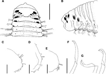
Acrocirrus bansei sp. nov. (A) Anterior end in dorsal view, holotype (USNM 1154139); (B) anterior end in lateral view, holotype (USNM 1154139); (C) parapodium from chaetiger 9; (D) parapodium from chaetiger 11; (E) parapodium from chaetiger 54; (F) thoracic neurochaeta, lateral view; (G) abdominal neurochaeta, lateral view. Scale bars: A and B, 0.5 mm; C, D and E, 0.25 mm; F and G, 50 µm.

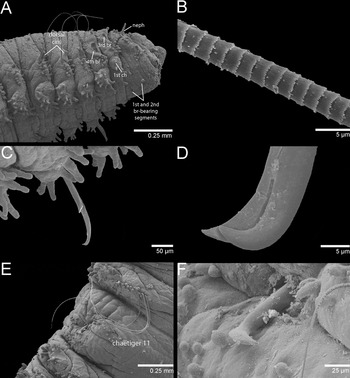
Scanning electron microscopy of Acrocirrus bansei sp. nov. showing (paratypes USNM 1154140-41): (A) anterior end in lateral view; (B) middle region of notochaeta; (C) neuropodia and thoracic neurochaeta; (D) tip of thoracic neurochaeta; (E) modified chaetiger 11; (F) aciculum from chaetiger 11. br, branchia; ch, chaetiger; neph, nephridiopore.

TYPE MATERIAL
Holotype: Coconut Island, Kaneohe Bay, Oahu, Hawaii, USA, 21°26′0.92″N 157°47′13.0″W, shallow subtidal, 10 June 1983, coll. J. H. Bailey-Brock (USNM 1154139).
Paratypes: same locality, date and collector as holotype, (3, BMNH 2011.10-12; 4, BPBM R3436; 4, mounted on stubs, USNM 1154140-41).
COMPARATIVE MATERIAL EXAMINED
Acrocirrus frontifilis (Grube, 1860), North Atlantic Ocean, off Morocco (Fauvel, Reference Fauvel1936, as Macrochaeta clavicornis and redetermined by Banse, Reference Banse1969 as A. frontifilis), BMNH, No. 1928.4.26.719/20, coll. P. Fauvel.
DESCRIPTION
Holotype 27 mm long, 1.2 mm wide with 95 chaetigers. Paratypes ranging from 14–25 mm long, 0.5–1 mm wide for about 58–92 chaetigers. Holotype with body rounded dorsally, ventrally flat, wider anteriorly, tapering to narrow posterior. Dorsum with two longitudinal rows of rounded papillae and others smaller rounded papillae sparsely distributed; most papillae concentrated on parapodia. Holotype dark brown with posterior 15 chaetigers paler shade of yellow; paratypes yellow brown to dark brown with posterior chaetigers paler shades of yellow and brown.
Prostomium blunt with a mid-terminal crest and transverse line across dorsal surface; 2 pairs of eyes in holotype, 2 or 3 pairs of eyes in paratypes, one large and postero-lateral, other pair very small and postero-centrally located, third pair (when present) postero-lateral and same size as the second pair (Figures 1A, B & 2A). Palps inserted on peristomial basis and as long as fifteen chaetigers. Peristomium lies behind and below prostomium. First branchial segment fused ventrally with the second branchial segment (Figure 1B). Four pairs of branchiae as long as 10 segments with rounded and flattened papillae surrounding the posterior half of branchial scar; first branchiae-bearing segment with a papilla antero-dorsal to each branchia; second branchiae-bearing segment with a conspicuous papilla (nephridiopore) below each branchia (Figure 2A); dorsal portion of third branchiae-bearing segment extended anteriorly over anterior segments reaching prostomium; a pair of distinct papillae present anteriorly (Figures 1A, B & 2A).
Neuropodia a rounded lobe with rows of digitate papillae (Figures 1D & 2C); neurochaetae from segment 3 (third branchiae-bearing segment), 4–5 compound falcigers throughout, blades become shorter posteriorly (Figures 1F, G & 2C, D); cutting edges of neurochaetae facing rearward through segment 12 (chaetiger 10), thereafter facing forward. Segment 13 (chaetiger 11) biannulated with parapodia reduced to low notopodial lobes, compound falcigers replaced by a heavy acicular hook (Figure 2E, F). Notochaetae from segment 5 (chaetiger 3) with notopodial cirri and 5–11 short papillae ventrally (Figures 1C, D & 2A, B); 2 (rarely 3) spinous capillaries anteriorly reducing to 1 on mid- and posterior chaetigers; notopodial cirri absent after chaetigers 27–34 (Figure 1E).
Pygidium with anal aperture placed terminally and surrounded by minute papillae.
REMARKS
The new species belongs to a group of Acrocirrus species in which chaetiger 11 is modified and equipped with a heavy neuropodial acicular hook (Banse, Reference Banse1969). This group includes Acrocirrus incisa, A. frontifilis, A. heterochaetous and A. okotensis. Among them, Acrocirrus bansei sp. nov. is most similar to A. frontifilis by the presence of notopodial cirri, which are not present in any other Acrocirrus species. Acrocirrus bansei sp. nov. differs from A. frontifilis because notopodial cirri are present in all segments of the voucher material for A. frontifilis but absent on posterior chaetigers of A. bansei sp. nov., by the presence of several distinct rounded papillae surrounding the posterior half of the branchial groove, and presence of frontal papillae on the third branchiae-bearing segment in A. bansei sp. nov.
We believe that some undescribed Acrocirrus species might have been pooled together into the A. frontifilis name by the presence of the notopodial cirri. Wolf (Reference Wolf, Uebelacker and Johnson1984) records A. frontifilis from the Gulf of Mexico based on the presence of the notopodial cirri, chaetal distribution and placement of papillae on the parapodia. However, this is also a common characteristic of the species described here, which differ from A. frontifilis as mentioned above.
ETYMOLOGY
This species is named after Dr Karl Banse due to his recognition of acrocirrids as a separate family from the cirratulids, and his description and redescription of several taxa in the genus Acrocirrus.
BIOLOGY
One specimen was found regenerating the posterior end.
DISTRIBUTION
This species is only known from shallow subtidal waters of Coconut Island, Kaneohe Bay, Oahu, Hawaii.
KEY TO SPECIES OF ACROCIRRUS
(modified from Kudenov, Reference Kudenov1976; Banse, Reference Banse1978)
1. Segment 13 with acicular hooks………2
— Segment 13 without acicular hooks………7
2. Three to six acicula per parapodium of segment 13; acicula not distally enlarged………A. aciculigerus Kudenov, 1976
— One aciculum per parapodium of segment 13; acicula distally enlarged and beaked………3
3. Abdominal neurochaetae as simple falcigers………A. okotensis Imajima, 1963
— Abdominal neurochaetae as composite falcigers………4
4. Notopodial cirri present………5
— Notopodial cirri absent………6
5. Notopodial cirri present throughout; third branchiae-bearing segment without frontal papillae………A. frontifilis (Grube, 1860)
— Notopodial cirri absent after chaetigers 27–34; third branchiae-bearing segment with frontal papillae………A. bansei sp. nov.
6. Parapodial lobes incised; noto- and neurochaetae from third branchiae-bearing segment………A. incisa Kudenov, 1975
— Parapodial lobes entire; notochaetae from third branchiae-bearing segment and neurochaetae from chaetiger 2 or chaetiger 3………A. heterochaetus Annenkova, 1934
7. Abdominal neurochaetae as simple falcigers………A. trisectus Banse, 1969
— Abdominal neurochaetae as composite falcigers………8
8. First branchiae-bearing segment dorsally not visible; parapodia with one noto- and neurochaeta………A. occipitalis Banse, 1978
— First branchiae-bearing segment dorsally visible (at least partly); parapodia with more than one noto- and neurochaeta………9
9. Neurochaetae from fourth branchiae-bearing segment………A. crassifilis Moore, 1923
— Neurochaetae from third branchiae-bearing segment………10
10. Neurochaetae from chaetiger 2………A. uchidai Okuda, 1934
— Neurochaetae from chaetiger 3………11
11. Usually one notochaeta and two neurochaetae per parapodium………A. columbianus Banse, 1978
— Several noto- and neurochaetae per parapodium………12
12. Upper face of prostomium smooth………A. muroranensis Okuda, 1934
— Upper face of prostomium ridged………A. validus von Marenzeller, 1879
ACKNOWLEDGEMENTS
This is contributed paper WRRC-CP-2012-03 of the Water Resources Research Center, University of Hawai'i at Mānoa, Honolulu. Drs R.E. Brock and R. Fitzhardinge assisted in the field. Laboratory facilities were provided by the Hawaii Institute of Marine Biology at Coconut Island. We thank Tina M. Carvalho (BEMF) for her assistance with SEM procedure and Dr Emma Sherlock (BMNH) for kindly loaning voucher material of A. frontifilis from Morocco. Dr Emma Sherlock (BMNH), Dr Linda Ward (USNM) and Holly Bolick (BPBM) coordinated the deposition of type specimens. We also thank Dr Karl Banse and two anonymous referees for their constructive criticism of the manuscript.