Introduction
Many free-living animals, including molluscs, fishes, seabirds, and marine mammals, inhabit the intertidal zone (Japanese Association of Benthology Reference Benthology2020). Trematode parasites have been frequently detected from such intertidal animals (Kuris et al. Reference Kuris, Hechinger, Shaw, Whitney, Aguirre-Macedo, Boch, Dobson, Dunham, Fredensborg, Huspeni, Lorda, Mababa, Mancini, Mora, Pickering, Talhouk, Torchin and Lafferty2008; Madhavi and Bray Reference Madhavi and Bray2018; Yamaguti Reference Yamaguti1975). Many trematodes are reported to use two or three hosts in their life cycles (Olsen Reference Olsen1986; Yamaguti Reference Yamaguti1975). In most trematode species, they have larval (miracidia, cercaria, and metacercaria), parthenital (sporocysts and rediae), and marital stages (sexual adults or marita) (Littlewood and Bray Reference Littlewood and Bray2014). Molluscs are the first intermediate hosts for sporocysts or rediae that generate cercariae by asexual proliferation. Cercariae released from molluscs develop into metacercariae in the second intermediate hosts. The metacercariae ingested by the definitive vertebrate hosts and develop into maritae, which release eggs into the environment with host feces. Additionally, predators of the second intermediate hosts might become paratenic hosts, which are potential sources of infection for the definitive hosts. For some trematode species, cercariae directly invade the definitive host to develop into maritae or develop into infective encysted-metacercariae in the environment and invade orally the definitive host. Among the life cycles of trematodes, molluscs act as the first intermediate hosts for most species and thus determine whether the life cycle runs successfully (Olsen Reference Olsen1986; Yamaguti Reference Yamaguti1975). However, the details of the life cycles of most trematode species remain to be investigated. This is because few morphological diagnostic characteristics among the larval and parthenital stages other than maritae and extensive morphological differences between these and marital stages make morphological identification of them difficult (Dawes Reference Dawes1956; Ito 1962). Although illuminating complete life cycles is important to address ecological and evolutionary questions (e.g., Kuris et al. Reference Kuris, Hechinger, Shaw, Whitney, Aguirre-Macedo, Boch, Dobson, Dunham, Fredensborg, Huspeni, Lorda, Mababa, Mancini, Mora, Pickering, Talhouk, Torchin and Lafferty2008; Louvard et al. Reference Louvard, Yong, Cutmore and Cribb2024; Miura et al. Reference Miura, Kuris, Torchin, Hechinger and Chiba2006; Nakao et al. Reference Nakao, Waki, Sasaki, Anders, Koga and Asakawa2017; Niewiadomska and Pojmanska Reference Niewiadomska and Pojmanska2011; Poulin and Cribb Reference Poulin and Cribb2002), for most trematodes only the definitive hosts have been known.
In recent years, DNA barcodes have been utilised to test the conspecificity of the trematode larvae, parthenitae, and maritae (e.g., Cribb et al. Reference Cribb, Cutmore, Wee, Browne, Morales and Pitt2024; Izumi et al. Reference Izumi, Akiyama, Suzumura and Ogawa2021; Nakao et al. Reference Nakao, Waki, Sasaki, Anders, Koga and Asakawa2017; Sasaki et al. Reference Sasaki, Miura and Nakao2022; Seo et al. Reference Seo, Ansai, Sase, Saito, Takano, Kojima and Waki2024). In previous studies, suitable barcoding fragments that could enable species identification have been thought to be obtained from the nuclear 28S ribosomal RNA (rDNA), the second internal transcribed spacer region (ITS2 rDNA), and mitochondrial cytochrome c oxidase subunit 1 (COI) regions, and these regions were often used in combination (e.g., Cribb et al. Reference Cribb, Cutmore, Wee, Browne, Morales and Pitt2024; Seo et al. Reference Seo, Ansai, Sase, Saito, Takano, Kojima and Waki2024). 28S rDNA is also suitable for conducting the phylogenetic analysis of broad trematode lineages (e.g., Louvard et al. Reference Louvard, Yong, Cutmore and Cribb2024; Martin et al. Reference Martin, Downie and Cribb2020; Waki et al. Reference Waki, Hoso, Nitta, Seo and Urabe2024). Identifications of larval or parthenital stages will reveal the life cycles of the trematoodes; however, compared to the number of species described based on maritae, larvae, parthenitae, and life cycles have been revealed for few trematode species. This can be attributable to the lack of a DNA survey of maritae for comparisons, and some sequences of larval and parthenital trematodes deposited on the online DDBJ/EMBL/GenBank databases (ISNDC) have indeed been identified only at the genus or family level (e.g., Kakui Reference Kakui2022; Louvard et al. Reference Louvard, Yong, Cutmore and Cribb2024; Miura et al. Reference Miura, Kuris, Torchin, Hechinger, Dunham and Chiba2005; Nakao and Sasaki Reference Nakao and Sasaki2021). However, identifications of the genus, family, and higher taxonomic groups might also enable the prediction of their life cycles based on the host information of their relatives.
In Japan, various larval and parthenital trematodes have been detected from mollusks, especially intertidal gastropods (Ito 1962; Miura et al. Reference Miura, Kuris, Torchin, Hechinger, Dunham and Chiba2005; Sasaki et al. Reference Sasaki, Miura and Nakao2022); however, these trematodes have rarely been identified at the species or genus levels, and their life cycles have seldom been discussed. Therefore, we investigated trematodes from intertidal gastropods in Japan and used DNA barcoding to elucidate or predict their life cycles. The study highlights the significance of gastropods as intermediate hosts of trematodes.
Materials and methods
Sample collections
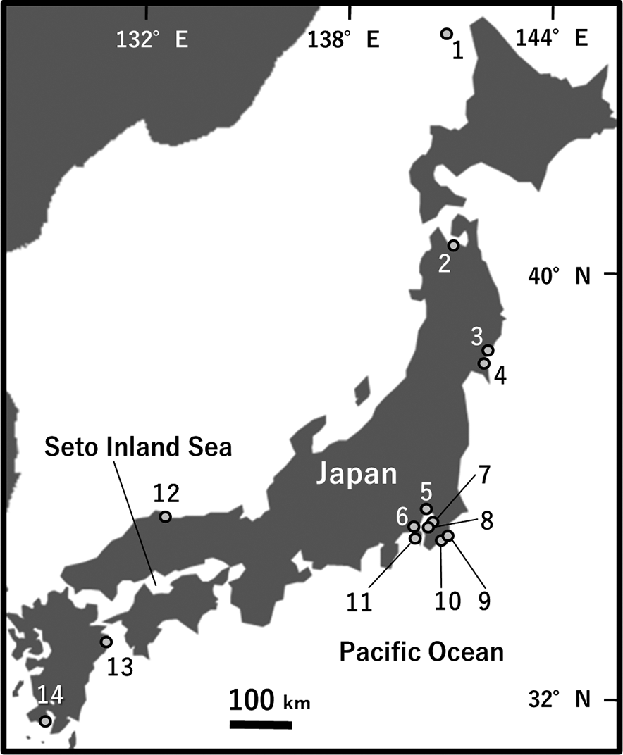
Field sampling of gastropods was carried out from the intertidal areas at 14 localities in Japan, from 2019 to 2024 (Figure 1, Tables 1 and 2). Public access and the presence of gastropods were the criteria used for the selection of the collection sites. The collected gastropods were transported to the laboratory and identified based on their morphological characteristics by referring to Okutani (Reference Okutani2017). Their shells were individually crushed with a hammer, and the internal organs were dissected under a stereomicroscope in 1% NaCl solution. When sporocysts or rediae were detected, they were kept in 70 or 99% ethanol for subsequent molecular analyses and morphological observations, whereas metacercariae were fixed in 70 or 99% ethanol after mounting between a glass slide and a coverslip.
Sampling sites of the snail hosts in Japan. The locality numbers correspond to those provided in Table 1.

Samples of gastropods tested for infection with trematodes. Site numbers correspond to those given in Figure 1. Pref.: Prefecture

Identification of larval and parthenital stages of trematode species from gastropods. Site numbers correspond to those given in Figure 1. Pref., Prefecture; Trematode, the family and, if there is such assumption, genus or species of a trematode; Host, the species of the host; Stage, developmental stage in the host; re, rediae; sp, sporocyst; mc, metacercariae; pmc, parthenogenetic metacercariae; Site, the locality where infected host was sampled, numbers as in Table 1 and Figure 1; 28S rDNA and COI, ISNDC accession numbers for the corresponding newly obtained sequences; References, articles which contained key information to support identification of material in this study

* Species of which maritae were detected from vertebrate hosts in this study.
Maritae and metacercariae from vertebrate hosts were also collected, when the opportunity arose, and used for molecular comparisons with the samples from gastropods (Table 3). The specimens of these maritae were fixed and kept in 70% ethanol between glass slides and were subsequently utilised for morphological and molecular analyses. Metacercariae were kept in 70% ethanol and were used for molecular identification.
Identification of maritae and metacercariae of trematodes from vertebrates sampled in this study. Pref., Prefecture; Stage, developmental stage in the host; ma, marita; mc, metacercariae; 28S rDNA and COI, INSDC accession numbers for the corresponding newly obtained sequences; References, articles which contained key information to support identification of material in this study

Morphological observations
The rediae and cercariae specimens kept in ethanol were observed under a light microscope (BX53, Olympus) after mounting on slides with Hoyer’s medium. The cercariae were observed ventrally. A camera lucida (U-DA, Olympus) attached to the microscope was used for making line drawings when mature specimens were obtained.
Maritae and metacercariae were observed using permanent mounts of ethanol-preserved samples. The samples were observed under a light microscope after staining with acetocarmine or Heidenhein’s iron-hematoxylin, dehydrating in an ethanol series, clearing in xylene or creosote, and mounting on slides with Canada balsam. Permanent mounts of some selected maritae and metacercariae were prepared after cutting a piece of tissue from each specimen for molecular analyses. Line drawings were made as described above. Whole worms of metacercariae from the flounder Liopsetta pinnifasciata (Kner, 1870) were used for DNA extraction; hence, permanent mounts were not prepared.
The preserved specimens of sporocysts and rediae and permanent mounts of metacercariae and maritae were deposited in the Meguro Parasitological Museum (MPM) or Minamisanriku Nature Center (MNC).
DNA barcoding and phylogenetic analyses
Alkaline lysates of small tissue pieces of sporocysts, rediae, metacercariae, and maritae were individually prepared as templates for the polymerase chain reaction (PCR) in accordance with the method described by Nakao et al. (Reference Nakao, Waki, Sasaki, Anders, Koga and Asakawa2017). DNA templates for the metacercariae of Stictodora lari Yamaguti, Reference Yamaguti1939 and Proctoeces sp. 2 (see Results) were obtained by using their whole bodies. The partial fragments of the 28S rRNA and COI regions were amplified by the following primer sets: 28S rRNA: dig12 and 1500R (Tkach et al. Reference Tkach, Kudlai and Kostadinova2016), or LSU5 (Littlewood et al. Reference Littlewood, Curini-Galletti and Herniou2000) and 1600R (Williams et al. Reference Williams, Reid and Littlewood2003); COI: JB3 (Bowles and McManus, Reference Bowles and McManus1993) and CO1-R trema (Miura et al. Reference Miura, Kuris, Torchin, Hechinger, Dunham and Chiba2005), JB3 and another reverse primer JB4.5 (Bowles et al. Reference Bowles, Blair and McManus1992), or Dig_cox1Fa and Dig_cox1R (Wee et al. Reference Wee, Cribb, Bray and Cutmore2017). These primers were also used for sequencing. Whenever necessary, the internal sequencing primers used for 28S rDNA were as follows: ECD2S (reverse, Williams and Ozawa Reference Williams and Ozawa2006), D3m (reverse, Takano and Kano Reference Takano and Kano2014), and 900F (forward, Lockyer et al. Reference Lockyer, Olson, Østergaard, Rollinson, Johnston, Attwood, Southgate, Horak, Snyder, Le, Agatsuma, DP, Carmichael, Naem and Littlewood2003). A commercial service (Eurofins Genomics Inc., Tokyo, Japan) performed the Sanger sequencing of the PCR amplicons. The BioEdit sequence alignment editor (Hall Reference Hall1999) was used to prepare the alignment datasets of 28S rDNA and COI.
The systematic positions of the larvae and parthenitae at the species or higher taxonomic levels were determined by comparing the obtained 28S rDNA and COI sequences with the published data using the Basic Local Alignment Search Tool (BLAST) against the DDBJ/EMBL/GenBank databases as follows. We identified a set of most similar sequences and the related species/genera to each of our sequences with BLAST. We also compared our larval and parthenital sequences with those of our trematodes from vertebrates. As for COI, if the closely related sequences (> 80% similarity) were not found in our vertebrate trematodes and in the databases, COI sequences were not used for species identification. The sequences of our samples and closely related ones found in the databases were used to make an alignment data set using BioEdit. Then, intraspecific divergence values for their overlapping regions (670–1267 sites for 28S rDNA; 317–574 sites for COI) were computed based on the p-distance, using the genetic software MEGA X (Kumar et al. Reference Kumar, Stecher, Li, Knyaz and Tamura2018). The divergence values of related species were used as thresholds to decide whether it was reasonable to assume conspecificity of our trematodes from molluscs versus those from the online databases and our trematodes from vertebrate hosts. If the divergence values between our larval and parthenital samples and species-identified trematodes were less than or subequal to the threshold intraspecific distance value of the related species/genera, they were judged to be conspecific. If not, their higher taxonomic group (genus or family) was examined by phylogenetic analyses using 28S rDNA sequences in MEGA X. The maximum likelihood (ML) method was used for reconstructing the phylogenetic trees under the Kimura 2-parameter (K2P) + G or Hasegawa–Kishino–Yano (HKY) + G models selected by MEGA X. Nodal support was evaluated with 1,000 bootstrap replicates.
Results
Seventeen gastropod species (682 individuals in total) were sampled from 14 localities (Figure 1, Table 1). Among these, infection with sporocysts, rediae, and metacercariae (including parthenogenetic metacercariae) was observed in six species (47 individuals, Figure 2). Identification of these specimens was facilitated by samples from one bird and four fish individuals. Below, we first provide a description of material from these vertebrate hosts, followed by the main data from the gastropod hosts.
Trematodes detected from intertidal gastropods and fish. Arrows and asterisks represent trematodes. (a) Soft tissues of host snail Batillaria attramentaria infected with rediae of Philophthalmus hechingeri. (b) Cercariae of Stictodora lari detected from the host gastropod Batillaria attramentaria. (c) Cercariae of Maculifer pacificus detected from the host gastropod Monodonta confusa. (d) A metacercaria of Proctoeces sp. 2 in the host gastropod Nipponacmea fuscoviridis. e) Metacercariae of Stictodora lari from the host flounder Liopsetta pinnifasciata.

Trematodes from vertebrates
Trematodes in vertebrate hosts in this study were morphologically or molecularly identified as following.
Suborder Gymnophallata Pérez-Ponce de León & Hernández-Mena, 2019
Family Fellodistomidae Nicoll, 1909
Genus Proctoeces Odhner, 1911
Proctoeces sp. 1
The marita from the Japanese black seabream Acanthopagrus schlegelii (Bleeker, 1854) was broken, making it difficult to identify its taxonomic position morphologically, but the marita was judged to be a member of the genus Proctoeces – namely, Proctoeces sp. 1 – using a 28S rDNA tree as discussed later. Morphological characters were not provided for this damaged specimen.
Sampling locality and date: Hiroshima Prefecture, Japan (October 26, 2020)
Vertebrate host: The Japanese black seabream Acanthopagrus schlegelii (Bleeker, 1854)
Deposition: MPM Coll. No. 25314
DNA marker: 28S rDNA, LC856695
Suborder Opisthorchiata La Rue, 1957
Family Heterophyidae Leiper, 1909
Genus Stictodora Looss, 1899
Stictodora lari Yamaguti, 1939
The maritae from the slaty-backed gull Larus schistisagus Stejneger, 1884 were morphologically identified as a member of the genus Stictodora (Figure 3a), based on their characteristics of elongated and pyriform bodies, two testes, small gonotyls, and rounded ventral suckers with many spines (Bray et al. Reference Bray, Gibson and Jones2008). Moreover, the following morphological characteristics exhibited by these maritae led to their identification as Stictodora lari: long prepharynx subequal to that of esophagus, seminal receptable between ovary and testis, and vitelline posterior to anterior margins of testes (Yamaguti Reference Yamaguti1939). Measurements of the maritae are given in Table 4. Twenty-four species have been classified in the genus Stictodora, including nonmarine species (WoRMS, 2024). Among these, Stictodora cablei Dronen, Blend & Gardner, 2007, Stictodora lari, and Stictodora morosovi Leonov, Reference Leonov1957 have the seminal receptable between ovary and testis, and vitelline posterior to anterior margins of testes (Dronen et al. Reference Dronen, Blend, Gardner and Jiménez2007; Leonov Reference Leonov1957). Of these, Stictodora lari is the only species where the length of the prepharynx and esophagus are subequal. Stictodora cablei has a prepharynx much longer than its esophagus, and Stictodora morosovi has an esophagus much longer than its prepharynx. Three metacercariae from the flounder Liopsetta pinnifasciata were also identified as Stictodora lari by the present molecular analysis (Figure 2e). The morphological characters of the metacercariae have not been provided, as whole worms were used for DNA extraction.
Maritae sampled in this study. (a) Stictodora lari. (b) Maculifer pacificus. Abbreviations: E, esophagus; C, caecum; Cp, cirrus pouch; Eg, egg; O, ovary; Os, oral sucker; Ph, pharynx; Po, parenchymatous organ; Por, Postoral ring; Pp, pars prostatica; Pr, Prepharynx; Sr, seminal receptacle; T, Testis; U, uterus; V, vitellarium; Vs, ventral sucker.

Comparison of morphological measurements of Stictodora species in different references. All measurements are in μm unless indicated otherwise

– Not described in the citation
* Diameter
** Sizes of two testes
Sampling localities and dates: Abashiri City, Hokkaido Prefecture, Japan (November 10, 2017, maritae); Hokkaido Prefecture, Japan (October 18, 2018, metacercariae)
Vertebrate hosts: The flounder Liopsetta pinnifasciata (Kner, 1870) (metacercariae); the slaty-backed gull Larus schistisagus Stejneger, 1884 (maritae)
Deposition: MPM Coll. No. 25317, maritae in L. schistisagus
DNA markers: 28S rDNA, LC856703; COI, LC856728–LC856730
Suborder Xiphidiata Olson, Cribb, Tkach, Bray & Littlewood, 2003
Family Opecoelidae Ozaki, 1925
Genus Maculifer Nicoll, 1915
Maculifer pacificus Yamaguti, 1938
The maritae from the panther puffer Takifugu pardalis (Temminck & Schlegel, 1850) and one from the pare puffer Takifugu vermicularis (Temminck & Schlegel, 1850) were assigned to the genus Maculifer based on their elongate body, ventral sucker on anterior two-third of the body, and well-observable post oral ring (Figure 3b; Jones et al. Reference Jones, Bray and Gibson2005; Martin et al. Reference Martin, Ribu, Cutmore and Cribb2018). All maritae were identified as Maculifer pacificus based on the following combination of morphological characters: testes situated diagonal, anterior margin of posterior testis is posterior to mid-level of anterior testis, egg length smaller than 67 μm (Martin et al. Reference Martin, Ribu, Cutmore and Cribb2018). Measurements of the maritae are given in Table 5. Eight species have been known in the genus Maculifer (Martin et al. Reference Martin, Ribu, Cutmore and Cribb2018). Among them, Maculifer pacificus and Maculifer japonicus Layman, 1931 have testes situated diagonal, and anterior margin of posterior testes are posterior to mid-level of anterior testes (Martin et al. Reference Martin, Ribu, Cutmore and Cribb2018; Yamaguti Reference Yamaguti1938). These two species can be distinguished by the length of eggs (< 75 μm in Maculifer pacificus vs > 75 μm in Maculifer japonicus).
Comparison of morphological measurements of Maculifer species in different references. All measurements are in μm unless indicated otherwise

– Not described in the citation
* Sizes of two testes
** Diameter
*** Width
Sampling localities and dates: Imabari City, Ehime Prefecture, Japan (February 26, 2019, from Takifugu vermicularis); Takehara City, Hiroshima Prefecture, Japan (March 16, 2017, feom T. pardalis)
Vertebrate hosts: The panther puffer Takifugu pardalis (Temminck & Schlegel, 1850); the pare puffer Takifugu vermicularis (Temminck & Schlegel, 1850)
Depositions: MPM Coll. No. 25323, three maritae in T. pardalis; MPM Coll. No. 25322, marita in T. vermicularis
DNA markers: 28S rDNA, LC856724; COI, LC856736–LC856738
Trematodes from gastropods: identification, molecular analyses, and life cycle information
Based on molecular analyses using 28S rDNA (855–1620 bp) and COI (368–940 bp) sequences, the trematodes from the gastropods were divided into 14 species of nine families. In the descriptions, all measurements are presented as means, with ranges in parentheses, and are in micrometres unless otherwise stated.
Suborder Echinostomata La Rue, 1926
Family Philophthalmidae Looss, 1899
Rediae with immature cercariae of a single species, Philophthalmus hechingeri, were detected from one individual of Batillaria attramentaria at Site 5 throughout the survey (Figure 4a, Table 1).
Larval and parthenital trematodes from intertidal gastropods. (a) Redia of Philophthalmus hechingeri. (b) Metacercaria of Proctoeces sp. 1. (c) Parthenogenetic metacercaria of Gymnophallidae gen. sp. 1. (d) Cercaria of Prodistomum Type 4. (e, f) Cercaria and redia of Stictodora lari. (g, h) Cercaria and redia of Cryptocotyle lingua. (i, j) Ventral and lateral faces of cercaria of Maculifer pacificus.

Genus Philophthalmus Looss, 1899
Philophthalmus hechingeri Sasaki, Miura & Nakao, 2022
Based on the comparison of the 28S rDNA sequence obtained and those deposited in the DDBJ/EMBL/GenBank databases (LC474511 and LC623767) (Sasaki et al. Reference Sasaki, Miura and Nakao2022; Sato et al. Reference Sato, Sasaki, Nabeta, Tomioka, Uga and Nakao2019), our sample was identified as P. hechingeri (Supplementary Table S1, Supplementary File S1). The COI fragment could not be amplified, possibly because of the degradation of the samples under poor storage conditions. Morphological descriptions were not provided because only immature cercariae were sampled.
Sampling locality and date: Ichikawa City, Chiba Prefecture, Japan (April 20–28, 2022)
Hosts of marita: Not recorded naturally. The Japanese quail Coturnix japonica Temminck & Schlegel, 1849 (experimental infection, Sasaki et al. Reference Sasaki, Miura and Nakao2022); human eye (accidental infection, Sasaki et al. Reference Sasaki, Miura and Nakao2022)
Host of metacercaria: Metacercaria of this genus are generally not expected to form within a host, but rather in the external environment (West Reference West1961).
Host of redia: Batillaria attramentaria (G. B. Sowerby II, 1855) (Sasaki et al. Reference Sasaki, Miura and Nakao2022; this study)
Synonym: Philophthalmus sp. (Sato et al. Reference Sato, Sasaki, Nabeta, Tomioka, Uga and Nakao2019)
Deposition: MPM Coll. No. 25348, rediae in B. attramentaria at Site 5
DNA marker: 28S rDNA, LC856693
Suborder Gymnophallata Pérez-Ponce de León & Hernández-Mena, 2019
Family Fellodistomidae Nicoll, 1909
Molecular analysis using 28S rDNA sequences have indicated that five metacercariae from four gastropod species belong to the genus Proctoeces (Figures 4b, 5, Table 1), and our material might represent three species as evident from the intraspecific distance values (Supplementary Table S2, Supplementary File S2). The comparison of the COI sequences among the three species was not possible since the amplification of the COI fragment was successful from only one of the three putative species.
Maximum-likelihood tree of Proctoeces inferred from partial sequences of nuclear 28S ribosomal DNA (732 sites), with Lintonium spp. (MZ687081 and MZ687083) as outgroup taxa. Bootstrap values above 50% are shown. Database accession numbers are shown after species names. Asterisks represent newly obtained sequences (LC856694−LC856699). Scale bar indicates number of substitutions per site.

Genus Proctoeces Odhner, 1911
Proctoeces cf. major
Based on comparison between our materials and the previously published sequences of maritae of Proctoeces cf. major parasitizing the East Asian common octopus Octopus sinensis A. d’Orbigny, 1834 in Japan (LC618022–618028; Izumi et al. Reference Izumi, Akiyama, Suzumura and Ogawa2021), three metacercariae from Purpuradusta gracilis, Monodonta confuse, and Trochus sacellum at Sites 9, 10, and 12, respectively, were identified as Proctoeces cf. major. Amplification of the COI fragment did not succeed for any metacercariae.
Description of metacercaria: Body elongate oval, 1239 in length and 449 in width. Oral sucker oblong, 164 in length and 224 in width. Prepharynx short, 36 in length. Pharynx muscular, 131 in length and 113 in width. Esophagus short, 49 in length. Ceca long and terminating near posterior end of body. Ventral sucker oblong, 314 in length and 373 in width. Testes rounded. Anterior testis 121 in length and 107 in width. Posterior testis 166 in length and 94 in width. Ovary anterior to posterior testes, 72 in length and 66 in width.
Sampling localities and dates: Katsuura City, Chiba Prefecture, Japan (June 24, 2024, metacercaria in Purpuradusta gracilis); Yonago City, Tottori Prefecture, Japan (October 10, 2020, metacercaria in Monodonta confusa); Kamogawa City, Chiba Prefecture, Japan (July 7, 2024, metacercaria in Trochus sacellum)
Host of marita: The East Asian common octopus Octopus sinensis A. d’Orbigny, 1834 (Izumi et al. 2002)
Hosts of metacercaria: Monodonta confusa Tapparone Canefri, 1874 (this study); Purpuradusta gracilis (Gaskoin, 1849) (this study); Trochus sacellum R. A. Philippi, 1855 (this study)
Host of sporocyst: Not recorded
Synonym: Not provided
Deposition: MPM Coll. No. 25313, metacercaria in P. gracilis at Site 9
DNA markers: 28S rDNA, LC856697–LC856699
Proctoeces sp. 1
One metacercaria from Monodonta confusa at Site 13 was possibly conspecific with an marita from the Japanese black seabream Acanthopagrus schlegelii sampled in this study, according to the similarity of 28S rDNA sequences (Supplementary Table S2, Supplementary File S2). Amplification of the COI fragment was not successful. Proctoeces maculatus was detected from Acanthopagrus schlegelii in the same region (Yamaguti 1934), but Proctoeces sp. 1 and P. maculatus may be not conspecific because sequences of ours and of the morphologically identified P. maculatus (e.g., OR724717, Vernmaak et al. 2023) did not form a single clade in the 28S rDNA tree (Figure 5).
Description of metacercaria (Figure 4b): Morphological characters were similar to those of Proctoeces cf. major. Body 2012 in length and 801 in width. Oral sucker rounded, 228 in length. Prepharynx short, 48 in length. Pharynx muscular, 132 in length and 164 in width. Esophagus long, 183 in length. Ceca long and terminating near posterior end of body. Ventral sucker rounded, 275 in length and 265 in width. Testes rounded. Anterior testis 108 in length and width. Posterior testis 159 in length and 144 in width. Ovary between two testes, 87 in length and 89 in width.
Sampling locality and date: Saeki City, Oita Prefecture, Japan (September 17, 2019)
Host of marita: The Japanese black bream Acanthopagrus schlegelii (Bleeker, 1854) (this study)
Host of metacercaria: Monodonta confusa Tapparone Canefri, 1874 (this study)
Host of sporocyst: Not recorded
Synonym: Not provided
Deposition: MPM Coll. No. 25315, metacercaria in M. confusa at Site 13
DNA marker: 28S rDNA, LC856694
Proctoeces sp. 2
Species of a metacercaria collected from Nipponacmea fuscoviridis at Site 2 (Figure 2d) was not identified based on our analyses (Supplementary Table S2, Supplementary File S2). This metacercaria formed a robust clade (bootstrap percentage, BP = 91%) with some Proctoeces species in the 28S rDNA tree (Figure 5), thereby leading to it being labeled as Proctoeces sp. 2. A COI fragment of the metacercaria was also determined, but sequences of the closely related species of Proctoeces sp. 2 were not found in the online databases. Morphological descriptions and depositions were not provided because the whole worm was used for DNA extraction.
Sampling locality and date: Aomori City, Aomori Prefecture, Japan (June 3, 2023)
Host of marita: Not recorded
Host of metacercaria: Nipponacmea fuscoviridis (Teramachi, 1949) (this study)
Host of sporocyst: Not recorded
Synonym: Not provided
DNA markers: 28S rDNA, LC856696; COI, LC856725
Family Gymnophallidae Odhner, 1905
Parthenogenetic metacercariae from one individual of Nipponacmea fuscoviridis at Site 2 (Figure 4c, Table 1) were molecularly assigned to this family.
Genus Parvatrema Cable, 1953
Parvatrema sp. 1
The parthenogenetic metacercariae were thought to be a member of the genus Parvatrema and labeled as Parvatrema sp. 1 (Figure 4c, Table 1). No similar sequence was found for 28S rDNA and COI fragment of this species in the online databases (Supplementary Table S3, Supplementary File S3). A close genetic relationship and the monophyly was predicted between this and the Parvatrema species including Parvatrema margaritense (e.g., OR571574, Galaktionov et al. Reference Galaktionov, Gonchar, Postanogova, Miroliubov and Bodrov2024) (BP = 99%) based on 28S rDNA sequences (Figure 6). Our sequence was also nested in a larger clade of the genus Parvatrema species including Parvatrema cf. donacis (e.g., MN384712, Hill-Spanik et al. Reference Hill-Spanik, Sams, Connors, Bricker and de Buron2021) (Figure 6).
Maximum-likelihood tree of gymnophallid trematodes inferred from partial sequences of nuclear 28S ribosomal DNA (775 sites), with Steringophorus thulini (AJ405297 and AJ405298) as outgroup taxa. Bootstrap values above 50% are shown. Database accession numbers are shown after species names. Asterisks represent a newly obtained sequence (LC856700). Scale bar indicates number of substitutions per site. PM: parthenogenetic metacercaria.

Description (based on three parthenogenetic metacercariae): Body oval, 649 (616–691) in length and 523 (480–584) in width. Oral sucker on anterior terminal of body, 62 (50–73) in length and 90 (78–101) in width. Prepharynx indistinct. Pharynx muscular, 51 (43–66) in length and 53 (49–60) in width. Esophagus short. Ventral sucker posterior to caecum bifurcation. Ventral sucker small, 49 (47–51) in length and 43 (42–44) in width.
Sampling locality and date: Aomori City, Aomori Prefecture, Japan (May 31, 2023)
Host of marita: Not recorded
Host of metacercaria: Nipponacmea fuscoviridis (Teramachi, 1949) (this study)
Host of sporocyst: Not recorded
Synonym: Not provided
Deposition: MPM Coll. No. 25316, parthenogenetic metacercariae in N. fuscoviridis at Site 2
DNA markers: 28S rDNA, LC856700; COI, LC856726
Suborder Lepocreadiata Olson, Cribb, Tkach, Bray & Littlewood, 2003
Family Lepocreadiidae Odhner, 1905
Rediae with mature cercariae from Purpuradusta gracilis at Site 10 (Figure 4d, Table 1) were identified as a member of the lepocreadiid genus Prodistomum.
Genus Prodistomum Linton, 1910
Prodistomum Type 4
Based on molecular analyses using 28S rDNA and COI sequences, rediae with mature cercariae from two individuals of Purpuradusta gracilis were identified as ‘Prodistomum Type 4’ reported as maritae from the Chub mackerel Scomber japonicus Houttuyn, 1782 from the Pacific coast of Japan (Cribb et al. Reference Cribb, Cutmore, Wee, Browne, Morales and Pitt2024) (Supplementary Tables S4 and S5, Supplementary File S4 and S5).
Description (based on three cercariae): Body elongated, 383 (289–489) in length and 151 (140–162) in width. Oral sucker on anterior margin of body, 52 (50–54) in length and 51 (42–57) in width. Prepharynx long, 52 (43–69) in length. Pharynx rounded, 18 (14–24) in length and 18 (15–20) in width. Ventral sucker middle of body, 46 (41–51) in length and 43 (39–48) in width. Testes well-observed and rounded. Anterior testis 39 (36–41) in length and 36 (32–42) in width. Posterior testis 34 (28–40) in length and 38 (35–41) in width. Ovary indistinct. Excretory vesicle I-shaped. Tail longer than body, 614 (595–632) in length and 57 (52–62) in width. Tail with prominent setae along lateral sides of its whole length.
Sampling locality and date: Kamogawa City, Chiba Prefecture, Japan (July 7, 2024)
Host of marita: The Chub mackerel Scomber japonicus Houttuyn, 1782 (Cribb et al. Reference Cribb, Cutmore, Wee, Browne, Morales and Pitt2024)
Host of metacercaria: Not recorded
Host of redia: Purpuradusta gracilis (Gaskoin, 1849) (this study)
Synonym: Not provided
Deposition: MPM Coll. No. 25349, cercariae in P. gracilis at Site 10
DNA markers: 28S rDNA, LC856701 and LC856702; COI, LC864039
Suborder Opisthorchiata La Rue, 1957
Family Heterophyidae Leiper, 1909
Three individuals of Batillaria attramentaria (one from Sites 7, 8, and 11) hosted rediae of this family (Figure 4e, f, Table 1). Based on the sequences of 28S rDNA and COI, redia from each of the three molluscs were found to be different species, as evident from the intraspecific distance values. Morphological and phylogenetic analyses revealed that the three species belonged to the genus Stictodora. The amplification of the 28S rDNA fragments was unsuccessful for the rediae of one species, Stictodora lari.
Genus Stictodora Looss, 1899
Stictodora lari Yamaguti, 1939
Three metacercariae from the flounder Liopsetta pinnifasciata and four maritae from the slaty-backed gull Larus schistisagus (newly obtained in this study; see above) were used for molecular comparisons to identify the species and life cycle of the rediae from the host gastropod at Site 7. Intraspecific distance values of COI sequences based on our worms (rediae, metacercariae, and a marita) and those from the online databases for the family indicated that our worms belong to a single species (Supplementary Table S6, Supplementary File S6). Conspecificity was also suggested between Stictodora lari and ‘Cercaria batillariae lineage HL6’ reported from Batillaria attramentaria widely distributed on the Japanese coast (Miura et al. Reference Miura, Kuris, Torchin, Hechinger, Dunham and Chiba2005) by the comparison of COI sequences.
Maritae of Stictodora lari were also reported from the black-tailed gull Larus crassirostris in Japan (Yamaguti Reference Yamaguti1939), and its metacercariae were reported from tissues under the skin of the yellowfin goby Acanthogobius flavimanus in Korean waters (Chai et al. Reference Chai, Park, Hong, Choi and Lee1989). In addition, the host flounders sampled in this study were generally piscivorous (Tomiyama Reference Tomiyama2021). Thus, the flounder should be a paratenic host that fed on the second intermediate hosts, based on the metacercariae that were detected from its body cavity in the present study. In addition, many seabirds and fishes distributing outside Japan have been reported to be the definitive and second intermediate hosts of this species, respectively (Bearup Reference Bearup1961; Nekrasov et al. Reference Nekrasov, Pronin, Sanzhieva and Timoshenko1999).
Description (based on five cercariae, Figure 4e): Body elongate oval, 210 (156–237) in length and 81 (70–86) in width. Oral sucker nearly anterior margin of body, 34 (31–38) in length and 33 (29–36) in width. Pharynx and caecum indistinct. Ventral sucker also indistinct. Eyespots anterior one-firth of body. Excretory vesicle V-shaped. Tail long, 425 (391–478) in length and 32 (26–35) in width
Sampling locality and date: Kisarazu City, Chiba Prefecture, Japan (July 5, 2019)
Hosts of marita: The European herring gull Larus argentatus Pontoppidan, 1763 (Nekrasov et al. Reference Nekrasov, Pronin, Sanzhieva and Timoshenko1999); the black-tailed gull Larus crassirostris Vieillot, 1818 (Yamaguti Reference Yamaguti1939); the silver gull Larus novaehollandiae Stephens, 1826 (as Chroicocephalus novaehollandiae (Stephens, 1826), Bearup, Reference Bearup1961); the slaty-backed gull Larus schistisagus Stejneger, 1884 (this study)
Hosts of metacercaria: The yellowfin goby Acanthogobius flavimanus (Temminck & Schlegel, 1845) (Chai et al. Reference Chai, Park, Hong, Choi and Lee1989); Gambusia sp. (experimental infection, Bearup Reference Bearup1961); Lebistes sp. (experimental infection, Bearup Reference Bearup1961); the flounder Liopsetta pinnifasciata (Kner, 1870) (this study)
Hosts of redia: Batillaria attramentaria (G. B. Sowerby II, 1855) (Miura et al. Reference Miura, Kuris, Torchin, Hechinger, Dunham and Chiba2005; this study); Batillaria australis (Quoy & Gaimard, 1834) (as Velacumantus australis (Quoy & Gaimard, 1834), Bearup, Reference Bearup1961)
Synonym: Cercaria batillariae lineage HL6 (Miura et al. Reference Miura, Kuris, Torchin, Hechinger, Dunham and Chiba2005)
Deposition: MPM Coll. No. 25350, rediae in B. attramentaria at Site 7
DNA marker: COI, LC856727
Stictodora sp. 1
No closely related sequence, that could be identified at the species levels, was found for the 28S rDNA and COI fragments obtained from the rediae sampled at Site 11 (Supplementary Table S6, S7, Supplementary File S6, S7). These rediae formed a strongly supported clade with other members of the genus Stictodora (BP = 100%), in the 28S rDNA tree (Figure 7). Therefore, this taxon was considered to be an unidentified species of the genus, Stictodora sp. 1. Comparison of the COI sequences revealed that Stictodora sp. 1 corresponds to ‘Cercaria batillariae lineage HL 5’ reported from Batillaria attramentaria found on the Pacific coast of Japan (Miura et al. Reference Miura, Kuris, Torchin, Hechinger, Dunham and Chiba2005). Descriptions were not provided because only immature cercariae were sampled in this study.
Maximum-likelihood tree of heterophyid trematodes inferred from partial sequences of nuclear 28S ribosomal DNA (843 sites), with Opisthorchis spp. (MF099790 and PP439313) as outgroup taxa. Bootstrap values above 50% are shown. Database accession numbers are shown after species names. Asterisks represent newly obtained sequences (LC856703−856705). Scale bar indicates number of substitutions per site.

Sampling locality and date: Yokosuka City, Kanagawa Prefecture, Japan (December 8, 2020)
Host of marita: Not recorded
Host of metacercaria: Not recorded
Host of redia: Batillaria attramentaria (G. B. Sowerby II, 1855) (Miura et al. Reference Miura, Kuris, Torchin, Hechinger, Dunham and Chiba2005; this study)
Synonym: Cercaria batillariae lineage HL5 (Miura et al. Reference Miura, Kuris, Torchin, Hechinger, Dunham and Chiba2005)
Deposition: MPM Coll. No. 25351, rediae in B. attramentaria at Site 13
DNA markers: 28S rDNA, LC856704; COI, LC856731
Stictodora sp. 2
A similar situation was observed for the rediae sampled at Site 8. Partial sequences of 28S rDNA and COI were not sufficient for identification at the species level (Supplementary Table S6, S7, Supplementary File S6, S7). In the 28S rDNA tree, this species was nested within the strongly supported clade of the genus Stictodora (BP = 100%) (Figure 7) and was therefore labeled as Stictodora sp. 2. Stictodora sp. 2 corresponds to ‘Cercaria batillariae lineage HL4’ identified in the Batillaria attramentaria on the Pacific coast and the Seto Inland Sea of Japan (Miura et al. Reference Miura, Kuris, Torchin, Hechinger, Dunham and Chiba2005).
Description (based on three cercariae): Morphological characteristics similar to cercariae of Stictodora lari. Body 289 (273–301) in length and 102 (89–116) in width. Oral sucker 39 (36–43) in length and 40 (38–45) in width. Tail long, 485 (286–580) in length and 40 (37–42) in width.
Sampling locality and date: Futtsu City, Chiba Prefecture, Japan (July 5, 2019)
Host of marita: Not recorded
Host of metacercaria: Not recorded
Host of redia: Batillaria attramentaria (G. B. Sowerby II, 1855) (Miura et al. Reference Miura, Kuris, Torchin, Hechinger, Dunham and Chiba2005; this study)
Synonym: Cercaria batillariae lineage HL4 (Miura et al. Reference Miura, Kuris, Torchin, Hechinger, Dunham and Chiba2005)
Deposition: MPM Coll. No. 25352, rediae in B. attramentaria at Site 8
DNA markers: 28S rDNA, LC856705; COI, LC856732
Family Opisthorchiidae Looss, 1899
Rediae with mature cercariae of this family were isolated from an individual of Littorina brevicula at Site 4 (Figure 4g, h, Table 1) and were thought to be Cryptocotyle lingua. Amplification of the COI fragment was not successful.
Genus Cryptocotyle Lühe, 1899
Cryptocotyle lingua (Creplin, 1825)
Our rediae can be identified as Cryptocotyle lingua (Supplementary Table S8, Supplementary File S8) since their 28S rDNA sequences are identical to those of cercariae and rediae of C. lingua from western Russia (White Sea) and Germany (MW361240, AY222228) in an overlapping site (Olson et al. Reference Olson, Cribb, Tkach, Bray and Littlewood2003; Tolstenkov et al. Reference Tolstenkov, Chatzigeorgiou and Gorbushin2023). Maritae of this trematode have been collected from the herring gull Larus argentatus in Japan (Yamaguti Reference Yamaguti1939). The definitive hosts of this trematode have been reported to be various birds and mammals worldwide (Morozov Reference Morozov and Skriabin1952; Olsen Reference Olsen1986; Stunkard Reference Stunkard1930; Yamaguti Reference Yamaguti1975; Yoshimura Reference Yoshimura1965). The second intermediate host was not recorded from Japan. In addition, metacercariae have not been detected in any fish species worldwide that have Japan as part of their distribution.
Description (based on three cercariae): Body elongated oval, 240 (222–270) in length and 105 (100–113) in width. Oral sucker nearly anterior margin of body, 47 (44–49) in length and 52 (47–59) in width. Pharynx indistinct, 14 in length and 17 (16–17) in width when visible. Caeca indistinct. Ventral sucker anterior one-third or fourth of body, 18 (16–20) in length and 19 (19–19) in width. Eyespots at level of ventral sucker. Excretory vesicle V-shaped. Tail long, 353 (324–382) in length and 51 (49–52) in width.
Sampling locality and date: Minamisanriku Town, Miyagi Prefecture, Japan (July 26, 2021)
Hosts of marita: The domestic dog Canis lupus familiaris Linnaeus, 1758 (experimental infection, Willey & Stunkard Reference Willey and Stunkard1942; as Canis familiaris Linnaeus, 1758, Morozov Reference Morozov and Skriabin1952); the domesticated red fox Vulpes vulpes Linnaeus, 1758 (Morozov Reference Morozov and Skriabin1952); the arctic fox Vulpes lagopus (Linnaeus, 1758) (Morozov Reference Morozov and Skriabin1952); the harbor seal Phoca vitulina Linnaeus, 1758 (Morozov Reference Morozov and Skriabin1952); Larus sp. (as ‘herring gull’, Køie Reference Køie1977); the European herring gull Larus argentatus Pontoppidan, 1763 (Morozov Reference Morozov and Skriabin1952); the lesser black-backed gull Larus fuscus Linnaeus, 1758 (Morozov Reference Morozov and Skriabin1952); the great black-backed gull Larus marinus Linnaeus, 1758 (Morozov Reference Morozov and Skriabin1952); the black-headed gull Larus ridibundus Linnaeus, 1766 (Morozov Reference Morozov and Skriabin1952); the night heron Nycticorax nycticorax (Linnaeus, 1758) (Morozov Reference Morozov and Skriabin1952); the black-legged kittiwake Rissa tridactyla (Linnaeus, 1758) (Morozov Reference Morozov and Skriabin1952); the razorbill Alca torda Linnaeus, 1758 (Morozov Reference Morozov and Skriabin1952); the roseate tern Sterna dougalli Montagu, 1813 (Morozov Reference Morozov and Skriabin1952); the common tern Sterna hirundo Linnaeus, 1758 (Morozov Reference Morozov and Skriabin1952)
Hosts of metacercaria: The Atlantic cod Gadus morhua Linnaeus, 1758 (as Gadus callarias Linnaeus, 1758, Morozov, Reference Morozov and Skriabin1952; Duflot et al. Reference Duflot, Gay, Midelet, Kania and Buchmann2021); the ballan wrasse Labrus bergylta Ascanius, 1767 (Yamaguti Reference Yamaguti1975); the tautog Tautoga onitis (Linnaeus, 1758) (Stunkard Reference Stunkard1930); the whiting Merlangius merlangus (Linnaeus, 1758) (Duflot et al. Reference Duflot, Gay, Midelet, Kania and Buchmann2021); the shorthorn sculpin Myoxocephalus scorpius (Linnaeus, 1758) (as Cottus scorpius Linnaeus, 1758, Morozov Reference Morozov and Skriabin1952); the European plaice Pleuronectes platessa Linnaeus, 1758 (Morozov Reference Morozov and Skriabin1952); the European flounder Platichthys flesus (Linnaeus, 1758) (Morozov Reference Morozov and Skriabin1952); the two-spotted goby Pomatoschistus flavescens (Fabricius, 1779) (as Gobius ruthensparri Euphrasen, 1786, Yamaguti Reference Yamaguti1975); the sand goby Pomatoschistus minutus (Pallas, 1770) (as Gobius minutus Pallas, 1770, Morozov Reference Morozov and Skriabin1952); the cunner Tautogolabrus adspersus (Walbaum, 1792) (Stunkard Reference Stunkard1930)
Hosts of redia: Littorina brevicula (R. A. Philippi, 1844) (this study); Peringia ulvae (Pennant, 1777) (Morozov, Reference Morozov and Skriabin1952); Littorina littorea (Linnaeus, 1758) (Køie Reference Køie1977; Olsen Reference Olsen1986; Olson et al. Reference Olson, Cribb, Tkach, Bray and Littlewood2003; Tolstenkov et al. Reference Tolstenkov, Chatzigeorgiou and Gorbushin2023; Yamaguti Reference Yamaguti1975)
Synonyms: Cercaria lophocerca de Filippi, 1857; Cryptocotyle americana Ciurea, 1924; Cryptocotyle macrorhinis (MacCallum, 1916) Dermocystis ctenolabri Stafford, 1905; Distoma lingua Creplin, 1825; Distomum macrorhinis MacCallum, 1916; Hallum caninum Wigdor, 1918; Tocotrema lingua (Creplin, 1825)
Deposition: MNC-OT-02848, rediae in L. brevicula at Site 4
DNA marker: 28S rDNA, LC856706
Suborder Pronocephalata Skrjabin, 1955
Family Notocotylidae Lühe, 1909
A sporocyst from an individual of Littorina brevicula at Site 1 was labeled as Notocotylidae gen. sp. 1.
Notocotylidae gen. sp. 1
We obtained 28S rDNA and COI sequences of the sporocyst, but both were not sufficient for identification at the species level (Supplementary Table S9, Supplementary File S9). In addition, its generic position could not be determined from the phylogenetic analysis based on 28S rDNA, although this sporocyst was assumed to belong to the family Notocotylidae (Figure 8). This worm was immature and has thus not been illustrated. We did not deposit specimens because whole worm was used for molecular analyses.
Maximum-likelihood tree of notocotylid trematodes inferred from partial sequences of nuclear 28S ribosomal DNA (896 sites), with Nudacotyle undicola (MF538578 and MN745944) as outgroup taxa. Bootstrap values above 50% are shown. Database accession numbers are shown after species names. Asterisks represent a newly obtained sequence (LC856707). Scale bar indicates number of substitutions per site.

Sampling locality and date: Rishiri Island, Hokkaido Prefecture, Japan (October 21, 2020)
Host of marita: Not recorded
Host of metacercaria: Not recorded
Host of sporocyst: Littorina brevicula (R. A. Philippi, 1844) (this study)
Synonym: Not provided
DNA markers: 28S rDNA, LC856707, COI, LC856733
Suborder Xiphidiata Olson, Cribb, Tkach, Bray & Littlewood, 2003
Family Microphallidae Ward, 1901
Six immature sporocysts of this family were sampled from two gastropod species from Sites 3, 7, and 11. Based on molecular analyses using 28S rDNA sequences, the five sporocysts in Batillaria attramentaria and one in Littorina brevicula were judged to be different species, as evident from the intraspecific distance values. Amplification of the COI fragment was successful only for the former species.
Microphallidae gen. sp. 1
The five sporocysts from Batillaria attramentaria at Sites 7 and 11 were labeled as Microphallidae gen. sp. 1. 28S rDNA and COI data did not suggest which species or genus these specimens belong to (Supplementary Table S10, Supplementary File S10; Figure 9). Descriptions were not provided because only immature sporocysts were sampled in this study.
Maximum-likelihood tree of microphallid trematodes inferred from partial sequences of nuclear 28S ribosomal DNA (1063 sites), with Parabascus lepidotus (ON036091 and ON036092) as outgroup taxa. Bootstrap values above 50% are shown. Database accession numbers are shown after species names. Asterisks represent newly obtained sequences (LC856708−LC856713). Scale bar indicates number of substitutions per site.

Sampling localities and dates: Kisarazu City, Chiba Prefecture, Japan (July 5, 2019); Yokosuka City, Kanagawa Prefecture, Japan (December 8, 2020)
Host of marita: Not recorded
Host of metacercaria: Not recorded
Host of sporocyst: Batillaria attramentaria (G. B. Sowerby II, 1855) (this study)
Synonym: Not provided
Deposition: MPM Coll. No. 25353, sporocyst in B. attramentaria at Site 11
DNA markers: 28S rDNA, LC856708–LC856712; COI, LC856734 and LC856735
Genus Maritrema Nicoll, 1907
Maritrema sp. 1
The phylogenetic analysis based on 28S rDNA sequences (Figure 9) indicated that sporocysts from Littorina brevicula at Site 3 formed a strongly supported clade (BP = 100%) with species of the genus Maritrema, so they were labeled as Maritrema sp. 1. The particular species could not be identified (Supplementary Table S10).
Sampling locality and date: Kesennuma City, Miyagi Prefecture, Japan (August 19, 2019)
Host of marita: Not recorded
Host of metacercaria: Not recorded
Host of sporocyst: Littorina brevicula (R. A. Philippi, 1844) (this study)
Synonym: Not provided
Deposition: MPM Coll. No. 25354, sporocyst in L. brevicula at Site 3
DNA marker: 28S rDNA, LC856713
Family Opecoelidae Ozaki, 1925
Sporocysts with cercariae in Batillaria attramentaria and Monodonta confusa at Sites 4, 6, and 14 were determined to be a single species belonging to the family Opecoelidae (Figures 2c, 4i, j, Table 1).
Genus Maculifer Nicoll, 1915
Maculifer pacificus Yamaguti, 1938
Intraspecific distance values of 28S rDNA and COI sequences for the family indicated that the sporocysts and the maritae of Maculifer pacificus in the puffers (the panther puffer Takifugu pardalis and the pare puffer Takifugu vermicularis) belong to a single species (Supplementary Table S11, 12, Supplementary File S11, S12). In addition to these two host puffers, maritae of Maculifer pacificus were also detected from other tetraodontid fishes in Japan (Machida and Kuramochi Reference Machida and Kuramochi1999; Ogawa and Inoue Reference Ogawa and Inouye1997; Yamaguti Reference Yamaguti1938; Yamaguti Reference Yamaguti1959). Our 28S rDNA sequences were found to closely resemble that of Opistholebes amplicoelus Nicoll, 1915 of the subfamily Opistholebetinae Fukui, 1929 (98.70% similarity, MH933875) (Kakui Reference Kakui2022, Martin et al. Reference Martin, Ribu, Cutmore and Cribb2018, Reference Martin, Downie and Cribb2020), suggesting that Maculifer pacificus belongs to this subfamily.
Description (based on five cercariae): Body elongated, 303 (297–310) in length and 91 (78–106) in width. Oral sucker on anterior margin of body, 60 (53–68) in length and 56 (53–59) in width. Stylet in anterior of oral sucker, length 10 (9–12). Prepharynx long, 49 (42–60) in length. Pharynx 21 (20–23) in length and 21 (18–22) in width. Ventral sucker posterior one-third of body, 65 (64–66) in length and 66 (54–74) in width. Excretory vesicle clearly observed, reaching middle of ventral sucker. Tail short, 45 (41–51) in length and 54 (53–56) in width.
Sampling localities and dates: Minamisanriku Town, Miyagi Prefecture, Japan (July 26 and August 21, 2021, in Monodonta confusa Tapparone Canefri, 1874); Yokohama City, Kanagawa Prefecture, Japan (July 20, 2020, in Batillaria attramentaria (G. B. Sowerby II, 1855)); Ibusuki City, Kagoshima Prefecture, Japan (November 13, 2020, in M. confusa)
Hosts of marita: the half-smooth golden pufferfish Lagocephalus spadiceus (Richardson, 1845) (as Spheroides spadiceus (Richardson, 1845), Yamaguti Reference Yamaguti1938); Takifugu niphobles (Jordan & Snyder, 1901) (as Spheroides niphobles Jordan & Snyder, 1901, Yamaguti Reference Yamaguti1938); the panther puffer Takifugu pardalis (Temminck & Schlegel, 1850) (as Spheroides pardalis (Temminck & Schlegel, 1850), Yamaguti Reference Yamaguti1959; Machida & Kuramochi Reference Machida and Kuramochi1999; this study); the tiger puffer Takifugu rubripes (Temminck & Schlegel, 1850) (Ogawa & Inoue Reference Ogawa and Inouye1997); the pare puffer Takifugu vermicularis (Temminck & Schlegel, 1850) (as Spheroides vermicularis Jordan & Snyder, 1901, Yamaguti Reference Yamaguti1938; this study)
Host of metacercaria: Not recorded
Hosts of sporocyst: Batillaria attramentaria (G. B. Sowerby II, 1855) (this study); Monodonta confusa Tapparone Canefri, 1874 (this study)
Synonym: not provided.
Deposition: MNC-OT-02849 and MPM Coll. No. 25355, sporocyst in M. confusa at Site 4 and 14, respectively
DNA markers: 28S rDNA, LC856714–LC856723; COI, LC856739–LC856743
Discussion
In this study, the life cycles of several trematode species, inhabiting Japanese coastal areas, were partially clarified based on molecular analyses and host records in previous studies. Some of the trematodes from the intertidal gastropods could not be identified in this study because DNA barcodes of identified maritae have not been obtained. The candidates are trematode species that have been reported from Japan and belong to the same group to our unidentified larvae and parthenitae. Comprehensive sampling and investigation of both vertebrate and invertebrate animals in the future would enable the detection of maritae and other stages.
Although DNA sequence dataset alone cannot always guarantee correct identification, especially in case only 28S rDNA sequences of larval and parthenital stages were obtained (e.g., Cribb et al. Reference Cribb, Cutmore, Wee, Browne, Morales and Pitt2024), our identifications provide adequate results and discussions for trematode lifecycles because closely related species tend to use similar hosts in their lifecycles (Olsen Reference Olsen1986; Yamaguti Reference Yamaguti1975). In this study, the unknown hosts of larval, parthenital, and marital stages for several trematode species were able to be predicted based on the host group of their related trematode species as follows (Figure 10):
Determined and hypothetical routes of trematode transmission related to the intertidal gastropods surveyed in the present study in the coastal regions in Japan. Small and capital alphabets represent the host molluscs and other marine host animals, respectively. a: Littorina brevicula. b: Nipponacmea fuscoviridis, c: Batillaria attramentaria. d: Monodonta confusa. e: Trochus sacellum. f: Purpuradusta gracilis, g: Unknown mollusks act as the first intermediate host. A: Seabird of Charadriiformes. B: Seabird of Anseriformes. C: Unknown mammal acts as the definitive host. D: Unknown crustacean acts as the second intermediate host. E: Acanthogobius flavimanus. F: Liopsetta pinnifasciata. G: Acanthopagrus schlegelii. H: Scomber japonicus. I: Unknown fish acts as the second intermediate or definitive hosts. J: Unknown jellyfish acts as the second intermediate host. K: Octopus sinensis. L: Takifugu pardalis and Takifugu vermicularis. M: Unknown sea urchins and gastropods act as the second intermediate host. Arrows indicate trematode transmissions. Red arrows represent cercariae infections in the second intermediate hosts. Yellow arrows represent infection to the definitive hosts without the second intermediate host. Dark arrows indicate the metacercariae infections to definitive hosts. White arrow indicates a metacercariae infection to a paratenic host.

Suborder Echinostomata
The maritae of Philophthalmus hechingeri (Philophthalmidae) were not reported from nature. The report from human eye in Japan (Sato et al. Reference Sato, Sasaki, Nabeta, Tomioka, Uga and Nakao2019) was probably accidental. Possible definitive hosts of Philophthalmus hechingeri are Charadriiformes seabirds, similar to other members of the family Philophthalmidae (Sasaki et al. Reference Sasaki, Miura and Nakao2022). Cercariae of this family are known to develop into infective encysted-metacercariae upon striking organic or nonorganic objects (West Reference West1961), such as foods of seabirds, suggesting the absence of a second intermediate host for Philophthalmus hechingeri.
Suborder Gymnophallata
Three putative Proctoeces species (Fellodistomidae) were reported in this study at the metacercarial stage, and their first intermediate hosts are unknown. Perhaps some mussels harbor sporocysts of these species, similar to other Proctoeces members (Yamaguti Reference Yamaguti1975). For Proctoeces sp. 2, the definitive host remained unknown, and it could be fishes or octopuses (Izumi et al. Reference Izumi, Akiyama, Suzumura and Ogawa2021; Oliva et al. Reference Oliva, Valdivia, Cárdenas, Muñoz, Escribano and George-Nascimento2018; Wee et al. Reference Wee, Cribb, Bray and Cutmore2017; Yamaguti Reference Yamaguti1975). Alternatively, maritae could develop right in the second intermediate host (Yamaguti Reference Yamaguti1975); thus, Proctoeces sp. 2 possibly have the potential of developing into maritae in gastropod hosts.
The definitive host of Parvatrema sp. 1 (Gymnophallidae) was unclear in this study; however, as is the case with several other members of its family, seabirds of Charadriiformes and Anseriformes can act as the definitive host (Gibson et al. Reference Gibson, Jones and Bray2002; Olson et al. Reference Olson, Cribb, Tkach, Bray and Littlewood2003; Yamaguti Reference Yamaguti1975). Its first intermediate host is also not known, but sporocysts of this family have been reported from various bivalves (Shimura et al. Reference Shimura, Yoshinaga and Wakabayashi1982; Yamaguti Reference Yamaguti1975).
Suborder Lepocreadiata
The second intermediate host of Prodistomum Type 4 (Lepocreadiidae) was not found. Metacercariae of some Prodistomum species have been detected in gelatinous zooplanktons (Cribb et al. Reference Cribb, Cutmore, Wee, Browne, Morales and Pitt2024). Moreover, Scomber fishes, including the definitive host Scomber japonicus, have been reported to feed on jellyfishes (Lamb et al. Reference Lamb, Hunter, Pinnegar, van der Kooij, Creer and Taylor2019). Therefore, gelatinous zooplanktons, such as jellyfishes, are thought to be the second intermediate hosts of Prodistomum Type 4.
Suborder Opisthorchiata
Metacercariae and maritae of Stictodora sp. 1 and Stictodora sp. 2 (Heterophyidae) were not found; however, seabirds (Charadriiformes and Anseriformes) and fishes are most probably the definitive and second intermediate hosts, respectively, similar to other members of the genus (Dronen et al. Reference Dronen, Blend, Gardner and Jiménez2007; Yamaguti Reference Yamaguti1939, Reference Yamaguti1975). No second intermediate host for Cryptocotyle lingua (Opisthorchiidae) has been reported in Japan including in the present study; however, the second intermediate host has been reported worldwide to be gadid, gobiid, labrid, and pleuronectid fish species (Duflot et al. Reference Duflot, Gay, Midelet, Kania and Buchmann2021; Morozov Reference Morozov and Skriabin1952; Stunkard Reference Stunkard1930). In addition, fishes of Atlantic Ocean, the ballan wrasse Labrus bergylta (Labridae) and the two-spotted goby Pomatoschistus flavescens (as Gobius ruthensparri, Gobiidae), have been experimentally infected with the cercariae, which developed into encysted metacercariae in these hosts (Yamaguti Reference Yamaguti1975). Although fish species recorded as the second intermediate hosts of Cryptocotyle lingua are not distributed in Japan, fishes of the same group that were reported in Japan may act as the hosts of this trematode.
Suborder Pronocephalata
Potential definitive hosts of Notocotylidae gen. sp. 1 include seabirds (Anseriformes and Charadriiformes) and marine mammals (sea lions and whales), which also serve as the definitive hosts of other confamilial members (Carvajal et al. Reference Carvajal, Durán and George-Nascimento1983; Malatesta et al. Reference Malatesta, Frati, Cerioni, Agrimi and Di Guardo1998; Yamaguti Reference Yamaguti1975). Notocotylidae gen. sp. 1. should lack a second intermediate host since the cercariae of this family develop to the infective encysted metacercarial stage in the external environment (Olsen Reference Olsen1986; Yamaguti Reference Yamaguti1975).
Suborder Xiphidiata
We did not find their definitive and second intermediate hosts of Microphallidae gen. sp. 1 and Maritrema sp. 1 (Microphallidae). Seabirds of Anseriformes and Charadriiformes or mammals were the likely definitive host of these species, and crustaceans could have been the second intermediate hosts, similar to other microphallid trematodes (Martorelli et al. Reference Martorelli, Fredensborg, Mouritsen and Poulin2004; Tkach et al. Reference Tkach, Littlewood, Olson, Kinsella and Swiderski2003; Yamaguti Reference Yamaguti1975). For Maculifer pacificus (Opecielidae), the second intermediate hosts were unknown. Gastropods or sea urchins are possible second intermediate hosts of Maculifer pacificus, similar to other members of subfamily Opistholebetinae (Jousson et al. Reference Jousson, Bartoli and Pawlowski1999; Martin et al. Reference Martin, Downie and Cribb2020).
The substrates used by the host gastropods in the sampling habitat were recorded in this study (Table 1). Our records were consistent with the habitats, the favorable depths and sediments, for these gastropod species reported in previous reports and monographs (Table 6) (Habe and Kosuge Reference Habe and Kosuge1967; Iijima Reference Iijima2001; Ohgushi Reference Ohgushi1956; Okutani Reference Okutani2017). The major habitat for Littorina brevicula, which served as the intermediate host of avian and mammal trematodes (Table 6), is the upper intertidal zone on rocky shores (Ohgushi Reference Ohgushi1956). This may because trematode eggs were released with the feces of definitive hosts, birds, and mammals in the upper intertidal zone, compared with in the middle and low intertidal zones. The other infected gastropod species except Batillaria attramentaria inhabited lower zones of the rocky shore and tended to act as the first and second intermediate hosts of fish and octopus trematodes (Table 6). These gastropod species might frequently come in contact with fecal eggs from fishes and octopuses, as well as with cercariae from the first intermediate hosts. Lastly, Batillaria attramentaria on intertidal muddy and sandy shores are frequently infected with sporocysts and rediae of avian trematodes (Table 6). This may because muddy and sandy shores are important feeding habitats for seabirds (Weller Reference Weller1999), which release feces with trematode eggs frequently.
Habitats of gastropods sampled in the present study and their infected trematodes

* Definitive host is unknown but predicted in the present and/or past studies.
In addition, a possible phylogenetic constraint for the host use was found in this study. Gastropods of subclasses Patellogastropoda and Vetigastropoda were commonly used as the second intermediate hosts of trematodes of the suborder Gymnophallata (Table 6), while gastropods of Caenogastropoda were generally used as the first intermediate hosts by most trematodes of the other suborders. Such host-use patterns would be illuminated at various taxonomic levels by further trematode surveys. At the species level, sporocyst of Maculifer pacificus were detected from two species of Gastropoda belonging to different subclasses. In the land snail trematodes of Brachylaima, sporocysts have been detected from different infraorder snail species (Waki et al. Reference Waki, Nakao, Sasaki, Ikezawa, Inoue, Ohari, Kameda, Asada, Furusawa and Miyazaki2022), suggesting host specificity of sporocysts was very low for some trematodes species.
Supplementary material
The supplementary material for this article can be found at http://doi.org/10.1017/S0022149X25100485.
Data availability
The sequence data analysed during the current study are available in the GenBank repository, LC856693–LC856743. The other datasets analysed during the current study are available from the corresponding author on reasonable request.
Acknowledgments
We greatly appreciate Fumi Kawano and Kenji Okoshi for helping our research. We would also like to express our gratitude to Aiko Iwasaki and Shumpei Morita for their assistance in collecting the parasites from the limpets. We are especially grateful to two anonymous reviewers for their invaluable comments.
Author contribution
Eriko Ansai and Tsukasa Waki wrote the main manuscript text. Eriko Ansai, Mizuki Sasaki, Hakuyu Sekine, Kazuyuki Munakata, Tetsuya Sase, Masato Nitta, Shota Suzuki, Takuzo Abe, Hiroaki Fukumori, Yuto Nakatsubata, and Masahiro Suzuki sampled the hosts and the trematodes. Eriko Ansai, Mizuki Sasaki, Hakuyu Sekine, Kazuyuki Munakata, Tetsuya Sase, Masato Nitta, and Tsukasa Waki identified the worms. Eriko Ansai, Mizuki Sasaki, Hakuyu Sekine, Kazuyuki Munakata, Tetsuya Sase, Yuto Nakatsubara, Tsuyoshi Takano, Hiroaki Fukumori, and Tsukasa Waki analysed the molecular datasets. Eriko Ansai and Tsukasa Waki prepared the figures, tables, and online resources. All authors reviewed the manuscript.
Financial support
This study was partially supported by the Research Institute of Marine Invertebrates, Japan (Ko-04).
Competing interests
The authors declare none.
Ethical standard
No approval of research ethics committees was required to accomplish the goals of this study because experimental work was conducted with unregulated species (fish and snails) and a dead bird.