Introduction
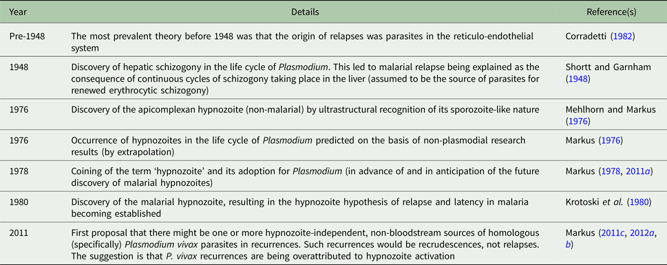
The malaria parasite Plasmodium vivax, like P. malariae, P. ovale curtisi and P. o. wallikeri, exhibits the phenomenon of ‘persistent parasitism’ (Sutherland, Reference Sutherland2016), which is increasingly being encountered during the course of epidemiological investigations (Betson et al. Reference Betson, Clifford, Stanton, Kabatereine and Stothard2018). One of the forms in which P. vivax persists is the ‘hypnozoite’ (Markus, Reference Markus1978, Reference Markus2011a). This hepatic stage is conventionally thought to be the source of relapse in P. vivax malaria (Markus, Reference Markus2011b, Reference Markus and Mehlhorn2016a), and the indirect evidence for the validity of the hypothesis is compelling, despite the theory remaining parasitologically unproven in humans. As regards hypnozoite-independent recurrences, it was concluded approximately 7 years ago (Table 1) that there might be more to P. vivax recurrences biologically than is readily apparent (Markus, Reference Markus2011c, Reference Markus2012a,Reference Markusb). Additional suggestions made since then are covered in this paper. Concepts involved are summarized below and attention is called to contrasting styles of discourse.
Relapse in malaria: main related events and hypotheses

‘Recurrence’ means that the cause of renewed clinical illness or peripheral blood parasitaemia is a recrudescence, a relapse, or a reinfection. In this article, the last-mentioned cause is excluded by the description ‘non-reinfection recurrence’. A ‘recrudescence’ has a merozoite source, whereas the origin of a ‘relapse’ is hypnozoite activation. For further details regarding these malaria-related definitions, see Box 1 in Markus (Reference Markus2015) and Box 1 in Markus (Reference Markus2017). The combined information in the two cited publications is a comprehensive terminological explanation that includes judgemental comments.
Evolution of the hypnozoite theory of relapse
The character of discourse on the hypnozoite hypothesis of relapse and latency in malaria has changed over time. The factual way in which parasitologists talk today about what hypnozoites allegedly do was also my attitude up until a few years ago (Markus, Reference Markus2011b). By contrast, malariologists in the 1980s did not always rationalize so rigidly, particularly those who carried out hypnozoite-associated studies. Knowing more about malarial recurrence than their colleagues and being cognizant of past mistakes (Shortt and Garnham, Reference Shortt and Garnham1948; Pays, Reference Pays2012), they sometimes seemed reluctant to state firmly that relapses are hypnozoite-mediated. One is hard-pressed to find this earlier type of qualified statement in today's publications. The scientific wait-and-see attitude has all but disappeared, the greater caution evident in some of the old literature having been discarded. Current, almost exclusively factual statements about hitherto unproven aspects of malarial relapse are, however, not necessarily wrong. On the contrary, the dogma that hypnozoites are the source of relapse in P. vivax and P. ovale sensu lato malaria is almost certainly correct, at least for P. vivax (P. ovale falls outside the scope of this article). Nevertheless, Richter et al. (Reference Richter, Franken, Holtfreter, Walter, Labisch and Mehlhorn2016), when writing open-mindedly about their idea of graded dormancy, expressed surprise at ‘… how rapidly the scientific world switched to the hypnozoite concept’. Schmidt (Reference Schmidt1986) was similarly puzzled by the ‘… promptness with which the hypnozoite concept was accepted …’. The current dogma is reminiscent of an earlier (but probably erroneous) conviction that prevailed between 1948 and 1980 about the origin of relapse (Table 1). That concept was usually worded as a fact. Textbooks and journal articles would emphatically state that sequential cycles of schizogony take place in the liver during P. vivax malaria. In regard to malarial relapse, we find a clash between what has been accepted historically and what has seemingly been correct. This prompted Corradetti (Reference Corradetti1982) to remark that he considered it ‘… safer to doubt any theory until it has been clearly demonstrated’. He was referring primarily, but not solely, to pre-1948 relapse dogma (Table 1), which also appeared ‘factually’ in textbooks and elsewhere. Of relevance to the elucidation of the biology of Plasmodium in general is the observation by Pays (Reference Pays2012) that ‘… deference towards scientific orthodoxy and a degree of reluctance to question and to criticise can delay advances in knowledge’.
As mentioned above, relapse in malaria was between 1948 and 1980 (Table 1) explained in terms of repeated cycles of hepatic schizogony (Shortt and Garnham, Reference Shortt and Garnham1948). In writing about the concept, Desowitz (Reference Desowitz2000) noted that the arguments ‘… are now fading with the years, as the graduate students and associates … (of Shortt and Garnham) also now fade into history’. This is true for the post-1980 hypnozoite concept of relapse (Table 1) as well, which in the view of most malariologists has replaced the previous theory. Note from the illustrative early (mainly 1980s) quotations below how the hypnozoite hypothesis became transmogrified into a ‘fact’, this being virtually the only way the concept is referred to by authors today. As I have already indicated, tentative or qualified phrasing on this topic is almost completely absent from present-day researchers’ publications.
The earlier discourse style enabled hypnozoite researchers to avoid ‘putting their heads on any blocks’, so to speak. Bray and Garnham (Reference Bray and Garnham1982) used the words ‘believe’ and ‘belief’ when talking about the hypnozoite concept of relapse. Furthermore, they cautiously wrote that sporozoites of particular (so-called ‘non-relapsing’) primate plasmodial species ‘… are thought not to differentiate into hypnozoites’ (italics added). Bray (Reference Bray1984) began a statement tentatively: ‘If one accepts the hypnozoite theory of relapse and long-term delayed primaries …’. Garnham (Reference Garnham1988) also hedged his discourse: ‘… the hypnozoite and its probable course in the life cycle …’; and ‘… on the assumption that the presence of hypnozoites is responsible for latency’. In the following year, Krotoski (Reference Krotoski1989) cautioned: ‘It must be emphasized … that additional work … is necessary in order to establish the (hypnozoite) concept fully …’. Soon after the discovery by Mehlhorn and Markus (Reference Mehlhorn and Markus1976) of the apicomplexan (but non-malarial) hypnozoite (Table 1), Krotoski et al. (Reference Krotoski, Krotoski, Garnham, Bray, Killick-Kendrick, Draper, Targett and Guy1980) reported the serendipitous recognition in hepatic tissue of malarial hypnozoites (Table 1). This stage was first seen on 13 June 1979 by Dr David Martin, who was working in Krotoski's laboratory. Writing a decade later, Mons and Sinden (Reference Mons and Sinden1990) offered a scientifically flawless opinion similar to that of Krotoski (Reference Krotoski1989), quoted above. They said that the hypnozoite is widely accepted as the source of true relapses but warned that ‘… more research is needed to prove beyond doubt that this parasite is not a re-invaded merozoite, but a dormant, sporozoite-derived parasite’. No relevant life cycle work took place during the next couple of decades. Investigations of the recommended type (Krotoski, Reference Krotoski1989; Mons and Sinden, Reference Mons and Sinden1990) have been carried out only very recently, and provide strong support for the validity of the hypnozoite theory of malarial relapse (Mikolajczak et al. Reference Mikolajczak, Vaughan, Kangwanrangsan, Roobsoong, Fishbaugher, Yimamnuaychok, Rezakhani, Lakshmanan, Singh, Kaushansky, Camargo, Baldwin, Lindner, Adams, Sattabongkot and Kappe2015; Soulard et al. Reference Soulard, Bosson-Vanga, Lorthiois, Roucher, Franetich, Zanghi, Bordessoulles, Tefit, Thellier, Morosan, Le Naour, Capron, Suemizu, Snounou, Moreno-Sabater and Mazier2015; Cubi et al. Reference Cubi, Vembar, Biton, Franetich, Bordessoulles, Sossau, Zanghi, Bosson-Vanga, Benard, Moreno, Dereuddre-Bosquet, Le Grand, Scherf and Mazier2017; Voorberg-van der Wel et al. Reference Voorberg-van der Wel, Roma, Gupta, Schuierer, Nigsch, Carbone, Zeeman, Lee, Hofman, Faber, Knehr, Pasini, Kinzel, Bifani, Bonamy, Bouwmeester, Kocken and Diagana2017). Publications that contain the results of important biological and related research since 2013 on P. cynomolgi are listed by Voorberg-van der Wel et al. (Reference Voorberg-van der Wel, Roma, Gupta, Schuierer, Nigsch, Carbone, Zeeman, Lee, Hofman, Faber, Knehr, Pasini, Kinzel, Bifani, Bonamy, Bouwmeester, Kocken and Diagana2017).
Hypnozoite-independent speculations
To explore the presumed hypnozoite basis of relapse, Imwong et al. (Reference Imwong, Boel, Pagornrat, Pimanpanarak, McGready, Day, Nosten and White2012) undertook a study that had the primary objective of distinguishing between single and separate parasite inoculations. They found that the first recurrences in very young children are frequently homologous (i.e. genetically closely related). Epidemiologically, it is indeed considered that a predominance of homologous recurrences early in life suggests bites by only one or a few infective mosquitoes (Koepfli and Mueller, Reference Koepfli and Mueller2017). By contrast, more heterogeneity in this respect is thought to reflect greater hypnozoite diversity, resulting from multiple infective mosquito bites. To add to this likely scenario, we might ask whether the ubiquity of genotypic homology in recurrent P. vivax malaria in individuals of various ages indicates that there can be, apart from a presumed hypnozoite origin, another non-circulating source or sources of homologous, relapse-like P. vivax recurrences (Table 1). If so, these would be recrudescences, not relapses.
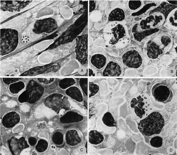
Research has validated the question by revealing that our understanding of the survival of plasmodial sporozoites and merozoites in the mammalian skin and lymphatic system is incomplete (Landau et al. Reference Landau, Chabaud, Mora-Silvera, Coquelin, Boulard, Rénia and Snounou1999; Gueirard et al. Reference Gueirard, Tavares, Thiberge, Bernex, Ishino, Milon, Franke-Fayard, Janse, Ménard and Amino2010; Wykes et al. Reference Wykes, Kay, Manderson, Liu, Brown, Richard, Wipasa, Jiang, Jones, Janse, Waters, Pierce, Miller, Stow and Good2011; Voza et al. Reference Voza, Miller, Kappe and Sinnis2012; Wykes and Horne-Debets, Reference Wykes and Horne-Debets2012; Ménard et al. Reference Ménard, Tavares, Cockburn, Markus, Zavala and Amino2013). Since 2010, there has accordingly been some speculation as to whether quiescent P. vivax sporozoites or extra-erythrocytic merozoites might occur in dermal or lymphatic sites (Fig. 1) and be associated with recurrences (Gueirard et al. Reference Gueirard, Tavares, Thiberge, Bernex, Ishino, Milon, Franke-Fayard, Janse, Ménard and Amino2010; Markus, Reference Markus2011c, Reference Markus2012a,Reference Markusb, Reference Markus2016b; Ménard et al. Reference Ménard, Tavares, Cockburn, Markus, Zavala and Amino2013). Furthermore, it should be noted that erythrocytes are not the only cells in peripheral blood from humans and non-human primates in which forms of Plasmodium (that are not necessarily reproductively active) have been seen (Landau et al. Reference Landau, Chabaud, Mora-Silvera, Coquelin, Boulard, Rénia and Snounou1999; Wykes and Horne-Debets, Reference Wykes and Horne-Debets2012; Markus, Reference Markus2017). There is also the question I raised (Markus, Reference Markus2015) of whether drug-induced, temporary blood-stage-parasite dormancy can sometimes be correlated with P. vivax recrudescences – an aspect of the recurrence puzzle that is covered in the penultimate section of this paper. However, more obvious possible erythrocytic-stage sources that might be associated with homologous instances of recurrent P. vivax malaria are bone marrow and the spleen, a subject dealt with in one of the sections below.
‘Merophores’ of Plasmodium vinckei petteri in murine splenic impression smears (from Landau et al. Reference Landau, Chabaud, Mora-Silvera, Coquelin, Boulard, Rénia and Snounou1999). Stain: Giemsa-colophonium. Merophores can possibly be associated with rodent malarial recrudescences. We need to determine whether such parasites that are not destroyed intracellularly occur in the life cycles of any primate plasmodial species. (A) Merophore sack inside a macrophage, day 7 post-inoculation (p.i.). At this time during the course of a murine malarial infection, the average size of macrophages in the spleen is approximately 23 µm. (B) Merophore leucocyte (polymorphonuclear cell), 8 days p.i. (C) Merophore leucocyte (macrophage), 8 days p.i. (D) Merophore leucocyte (polymorphonuclear cell), 8 days p.i. This historical plate was originally published at a magnification of ×1750 (no scale bar provided). Reproduced with the permission of the Société Française de Parasitologie, obtained via Jean-Lou Justine, current Editor-in-Chief of the journal Parasite.

Significance of blood-stage-induced malarial infections
Simian P. cynomolgi malaria is regarded as a disease model for human P. vivax malaria (Waters et al. Reference Waters, Higgins and McCutchan1993; Fonseca et al. Reference Fonseca, Joyner, MaHPIC Consortium, Galinski and Voit2017; Joyner et al. Reference Joyner, MaHPIC Consortium, Wood, Moreno, Garcia and Galinski2017), and recrudescences are often a feature of untreated, blood-stage-initiated P. cynomolgi malaria (Corradetti, Reference Corradetti1966). It follows that recurrent P. cynomolgi parasitaemia in sporozoite-infected monkeys might occasionally, or perhaps even frequently, have the same non-hypnozoite origin or origins (whatever it or they may be) as recrudescences in monkeys infected by inoculation of erythrocytic parasites (Markus, Reference Markus2017). Prevailing hypnozoite-related dogma obfuscates this probability. Thus, recurrences in sporozoite-transmitted P. cynomolgi malaria might regularly have a hypnozoite-independent origin, like that of recrudescences of erythrocytic-stage-induced P. malariae infections in humans, which frequently take place in the long term (Corradetti, Reference Corradetti1966). Such P. malariae recurrences cannot be relapses (even if hypnozoites do occur in the life cycle of P. malariae) because the evidence is that hypnozoites are derived from sporozoites, which are not present in post-passage, P. malariae blood-stage inocula. When extrapolated to P. cynomolgi’s sister species P. vivax, these comparative arguments support my contention that some homologous P. vivax recurrences in naturally acquired human infections are erroneously being ascribed to hypnozoite activation (Table 1).
As for hypnozoite formation, it must be emphasized that, as stated above, it is thought to be only sporozoite-induced (not blood-stage-associated) infections that result herein; malarial hypnozoites apparently being directly sporozoite-derived (Markus, Reference Markus2012b). This was the original extrapolative interpretation from the results of my non-malarial, laboratory-based apicomplexan biological investigations (Table 1; Markus, Reference Markus1975, Reference Markus1976), as acknowledged by Garnham (Reference Garnham1985), and which strongly influenced the thinking of malarial hypnozoite researchers around 1980 (R. Killick-Kendrick, personal communication). By this time, I was no longer at Imperial College London, where much of the malarial and other research took place.
In summary, non-reinfection, recurrent P. vivax malaria has a bimodal origin, the parasite source being either merozoites (even if only in the bloodstream) or, presumably, hypnozoites. Homologous (as opposed to heterologous) recurrences are highly suggestive of a merozoite origin, although not exclusively so. The point I am making here is that many authors are probably overattributing homologous P. vivax malarial recurrences to the activation of hypnozoites. Close genetic relatedness of the parasites in a P. vivax recurrence to those that were present in peripheral blood during the initial bout of parasitaemia could be an indication that the recurrence is a recrudescence (merozoite origin) and not, in fact, necessarily a relapse (hypnozoite origin). One newly recognized, possible parasite source that might initiate or contribute to homologous, hypnozoite-independent P. vivax recurrences is bone marrow (Malleret et al. Reference Malleret, Li, Zhang, Tan, Suwanarusk, Claser, Cho, Koh, Chu, Pukrittayakamee, Ng, Ginhoux, Ng, Lim, Nosten, Snounou, Rénia and Russell2015; Fonseca et al. Reference Fonseca, Joyner, MaHPIC Consortium, Galinski and Voit2017; Markus, Reference Markus2017), as discussed below.
Erythrocytic parasites in bone marrow and the spleen
The idea that human bone marrow may harbour plasmodial parasites goes back more than 125 years (Franken et al. Reference Franken, Bruijns-Pötschke, Richter, Mehlhorn and Labisch2017). This aside, the recorded occurrence of P. vivax in bone marrow was discussed by Baird (Reference Baird2013) and Barber et al. (Reference Barber, William, Grigg, Parameswaran, Piera, Price, Yeo and Anstey2015) in regard to malarial severity. Furthermore, bone marrow has been mentioned in the context of recrudescence of P. falciparum malaria (Shanks, Reference Shanks2015), a subject related to the bone marrow-associated concept summarized here for P. vivax. Erythrocytic forms of P. vivax are not seen exclusively in cells in vivo that are obviously reticulocytes. They can also be present in what seem morphologically to be normocytes, but which could be reticulocytes with parasite-induced loss of their reticular material (Lim et al. Reference Lim, Pereira, Shaw-Saliba, Mascarenhas, Maki, Chery, Gomes, Rathod and Duraisingh2016; Malleret et al. Reference Malleret, Rénia and Russell2017). Reports of the occurrence of P. vivax in red blood cells of whatever ages in bone marrow (which is reticulocyte-rich) and the spleen have appeared from time to time in the literature. For example, see Siqueira et al. (Reference Siqueira, Magalhães, Melo, Ferrer, Castillo, Martin-Jaular, Fernández-Becerra, Ordi, Martinez, Lacerda and del Portillo2012), Baird (Reference Baird2013), Barber et al. (Reference Barber, William, Grigg, Parameswaran, Piera, Price, Yeo and Anstey2015), Malleret et al. (Reference Malleret, Li, Zhang, Tan, Suwanarusk, Claser, Cho, Koh, Chu, Pukrittayakamee, Ng, Ginhoux, Ng, Lim, Nosten, Snounou, Rénia and Russell2015) and Baro et al. (Reference Baro, Deroost, Raiol, Brito, Almeida, de Menezes-Neto, Figueiredo, Alencar, Leitão, Val, Monteiro, Oliveira, del Pilar Armengol, Fernández-Becerra, Lacerda and del Portillo2017). Asexual multiplication of P. vivax must surely take place in the bone marrow, especially considering the low peripheral parasitaemias that are characteristic of P. vivax malaria.
It is generally assumed that P. vivax malarial recrudescences are the result of asexual reproduction of circulating stages. But is the bone marrow a source of parasites in recrudescent P. vivax malaria (Malleret et al. Reference Malleret, Li, Zhang, Tan, Suwanarusk, Claser, Cho, Koh, Chu, Pukrittayakamee, Ng, Ginhoux, Ng, Lim, Nosten, Snounou, Rénia and Russell2015)? The question as to whether P. vivax parasites in bone marrow are ever the origin of or contribute to homologous (specifically) recurrent peripheral parasitaemia was raised for the first time recently (Markus, Reference Markus2017). Life cycle research on Plasmodium cynomolgi needs to be done in relation to bone marrow, as commented by Fonseca et al. (Reference Fonseca, Joyner, MaHPIC Consortium, Galinski and Voit2017) and Markus (Reference Markus2017). It will, in addition, be important to ascertain whether P. vivax’s erythrocytic forms can be present in the bone marrow of appropriately humanized mice. Plasmodium vivax has been seen in human bone marrow when it has not been detected microscopically in the bloodstream (Ru et al. Reference Ru, Mao, Zhang, Pang, Zhao, Liu and Wickramasinghe2009; Malleret et al. Reference Malleret, Li, Zhang, Tan, Suwanarusk, Claser, Cho, Koh, Chu, Pukrittayakamee, Ng, Ginhoux, Ng, Lim, Nosten, Snounou, Rénia and Russell2015). We need to know more about this, using molecular diagnosis (Gruenberg et al. Reference Gruenberg, Moniz, Hofmann, Wampfler, Koepfli, Mueller, Monteiro, Lacerda, de Melo, Kuehn, Siqueira and Felger2018), because the occurrence of P. vivax in bone marrow probably has implications for the eradication of malaria (Malleret et al. Reference Malleret, Li, Zhang, Tan, Suwanarusk, Claser, Cho, Koh, Chu, Pukrittayakamee, Ng, Ginhoux, Ng, Lim, Nosten, Snounou, Rénia and Russell2015; Markus, Reference Markus2017).
When examining primate or murine organs/tissues such as bone marrow or the spleen for stages of Plasmodium, Giemsa-stained impression smears should be used (Markus, Reference Markus1975; Mehlhorn and Markus, Reference Mehlhorn and Markus1976; Landau et al. Reference Landau, Chabaud, Mora-Silvera, Coquelin, Boulard, Rénia and Snounou1999), not only haematoxylin and eosin-stained sections. When apicomplexan parasites are found in the former, interpretation of their nature is easier, as compared with the use of the latter. Giemsa is helpful for staining Plasmodium and related coccidian parasites in tissue sections as well (Markus et al. Reference Markus, Killick-Kendrick and Garnham1974).
Lastly, whether erythrocytic parasites in the spleen can multiply or are always destroyed is at present unknown for both P. vivax trophozoites and splenic P. falciparum ring stages (Safeukui et al. Reference Safeukui, Correas, Brousse, Hirt, Deplaine, Mulé, Lesurtel, Goasguen, Sauvanet, Couvelard, Kerneis, Khun, Vigan-Womas, Ottone, Molina, Tréluyer, Mercereau-Puijalon, Milon, David and Buffet2008; Fig. 2 in Markus, Reference Markus2016b). Is the spleen a source of recrudescent malarial parasites?
Drug-associated considerations in recurrence
It is well known that both blood schizontocides and the supposedly radically curative drug primaquine are not always effective for P. vivax malaria (e.g. Campo et al. Reference Campo, Vandal, Wesche and Burrows2015; Carmona-Fonseca, Reference Carmona-Fonseca2015; Zuluaga-Idarraga et al. Reference Zuluaga-Idarraga, Perez and Aguirre-Acevedo2015; Commons et al. Reference Commons, Thriemer, Humphreys, Suay, Sibley, Guerin and Price2017). Recurrences can therefore be a consequence of failure to clear all parasites. Drug resistance might be the problem in many instances. However, in the absence of systems for P. vivax that enable reproducible testing of the effects of compounds (van Schalkwyk et al. Reference van Schalkwyk, Moon, Blasco and Sutherland2017), little is actually known about P. vivax drug resistance (Chaorattanakawee et al. Reference Chaorattanakawee, Lon, Chann, Thay, Kong, You, Sundrakes, Thamnurak, Chattrakarn, Praditpol, Yingyuen, Wojnarski, Huy, Spring, Walsh, Patel, Lin, Juliano, Lanteri and Saunders2017). More pharmacogenetic studies would probably facilitate the evaluation of anti-P. vivax drug efficacy (Milner et al. Reference Milner, Berman, Caridha, Dickson, Hickman, Lee, Marcsisin, Read, Roncal, Vesely, Xie, Zhang, Zhang and Li2016; Silvino et al. Reference Silvino, Costa, de Araújo, Ascher, Pires, Fontes, Carvalho, de Brito and Sousa2016; Baird et al. Reference Baird, Battle and Howes2018; Pewkliang et al. Reference Pewkliang, Rungin, Lerdpanyangam, Duangmanee, Kanjanasirirat, Suthivanich, Sa-ngiamsuntorn, Borwornpinyo, Sattabongkot, Patrapuvich and Hongeng2018).
Such considerations aside, primaquine is known to be merozoitocidal and gametocytocidal, in addition to being, apparently, hypnozoitocidal. There was early direct microscopic in vivo parasitological evidence for the last-mentioned effect (Garnham, Reference Garnham1988), based on some findings for P. cynomolgi in rhesus monkeys Macaca mulatta. To complicate interpretational matters further in respect of treated individual P. vivax-infected humans (as opposed to presumed reduction by primaquine of the collective hypnozoite load in a whole group of patients), it has yet to be determined whether primaquine will kill parasites with equal efficacy in all parts of the body where they might occur (Markus, Reference Markus2015). The need for such information is indicated by the fact that whereas primaquine eliminates rodent plasmodial stages in the liver, it is either less effective or ineffective against the same murine parasites when they are present dermally (Gueirard et al. Reference Gueirard, Tavares, Thiberge, Bernex, Ishino, Milon, Franke-Fayard, Janse, Ménard and Amino2010; Voza et al. Reference Voza, Miller, Kappe and Sinnis2012). There is yet another drug factor that we should take into account in regard to recurrences. I have recently debated whether relapse-like clinical and parasitaemic recrudescences can sometimes follow temporary, drug-induced, P. vivax erythrocytic-stage quiescence (Markus, Reference Markus2015), something that might occur in vitro (Thomson-Luque et al. Reference Thomson-Luque, Shaw-Saliba, Kocken and Pasini2017). The phenomenon has been clearly shown ex vivo for P. falciparum in experiments that are ongoing (Gray et al. Reference Gray, Gresty, Chen, Zhang, Gutteridge, Peatey, Chavchich, Waters and Cheng2016; Dembélé et al. Reference Dembélé, Ang, Chavchich, Bonamy, Selva, Yi-Xiu Lim, Bodenreider, Yeung, Nosten, Russell, Edstein, Straimer, Fidock, Diagana and Bifani2017; Zhang et al. Reference Zhang, Gallego-Delgado, Fernández-Arias, Waters, Rodriguez, Tsuji, Wek, Nussenzweig and Sullivan2017).
Moreover, the efficacy of drugs for clearing P. vivax from bone marrow, haemopoietic or otherwise, might prove to be important if bone marrow is a significant reservoir site for the parasites. It is worth noting that Joyner et al. (Reference Joyner, MaHPIC Consortium, Wood, Moreno, Garcia and Galinski2017) recorded the presence of haemozoin in bone marrow of a rhesus monkey with P. cynomolgi malaria, but they made no mention of the presence of parasites there (or elsewhere). However, the animal had received subcurative antimalarial treatment 48 h earlier to reduce parasitaemia. If P. cynomolgi is normally readily detectable in the bone marrow of infected, untreated rhesus monkeys, the absence of P. cynomolgi parasites in this instance could indicate that rapid clearance of stages from bone marrow by means of drugs is possible. Future research will undoubtedly shed light on the matter.
An outstanding question is whether or not recrudescences of P. vivax malaria can result from interplay between immunity and any drugs, such as the range of compounds for which Pukrittayakamee et al. (Reference Pukrittayakamee, Chantra, Simpson, Vanijanonta, Clemens, Looareesuwan and White2000) reported therapeutic responses. In relation to the current malaria eradication research agenda (Rabinovich et al. Reference Rabinovich, Drakeley, Djimde, Hall, Hay, Hemmingway, Kaslow, Noor, Okumu, Steketee, Tanner, Wells, Whittaker, Winzeler, Wirth, Whitfield and Alonso2017), it is a subject that should receive attention. This type of interaction has been found to affect total clearance or otherwise of P. yoelii parasites by artesunate (Claser et al. Reference Claser, Chang, Russell and Rénia2017), so follow-up is needed. Interpretation for P. vivax malaria might not always be straightforward. Boyd and Kitchen (Reference Boyd and Kitchen1944) found that P. vivax recurrences took place in treated artificial infections that had been sporozoite-induced, but usually not when the initial infection was allowed to run its course and become self-terminating. Intuitively, this situation is perhaps not what one would have expected.
Concluding remarks
The uncertainties to which I have drawn attention make the confident categorization of individual homologous P. vivax recurrences as hypnozoite-associated relapses difficult to impossible, irrespective of whether they are short-term or long-term recurrences. A long-term homologous P. vivax recurrence in a naturally infected person is not necessarily a relapse, even if the patient has been treated with both primaquine and a blood schizontocide such as chloroquine (considering the frequency of drug failure). Pending greater understanding of recurrent P. vivax malaria, an out-of-the-box (in terms of hypnozoite-related dogma) range of non-hypnozoite explanations for homologous, relapse-like P. vivax recurrences should be considered – along the lines of, for example, the analytical approach adopted for a patient with recurrent P. malariae infection (Rutledge et al. Reference Rutledge, Marr, Huang, Auburn, Marfurt, Sanders, White, Berriman, Newbold, Anstey, Otto and Price2017). It is considered that selective whole-genome amplification might be helpful for future characterization of P. vivax recurrences (Cowell et al. Reference Cowell, Loy, Sundararaman, Valdivia, Fisch, Lescano, Baldeviano, Durand, Gerbasi, Sutherland, Nolder, Vinetz, Hahn and Winzeler2017), determination of the origin of which is particularly problematic in endemic areas (Popovici et al. Reference Popovici, Friedrich, Kim, Bin, Run, Lek, Cannon, Ménard and Serre2018). Research topics to address some of the issues surrounding recurrent P. vivax malaria have been suggested (Markus, Reference Markus2012b, Reference Markus2015, Reference Markus2016b, Reference Markus2017).
Finally, elucidation of the mechanisms of activation (hypnozoites) or reactivation (merozoites) of hidden P. vivax parasites could be important in the practical context of attempted malaria eradication. In respect of the induction of hypnozoite latency, one possibility is that phosphorylation of the α subunit of eukaryotic translation initiation factor 2 is involved (Holmes et al. Reference Holmes, da Silva Augusto, Zhang, Wek and Sullivan2017). If reactivated quiescent merozoites do facilitate transmission of P. vivax malaria, then they are collectively (like hypnozoites, probably) an obstacle to eradication of the disease. Whatever the parasite source in a P. vivax recurrence, renewed peripheral parasitaemia will normally be accompanied by the formation of gametocytes which, if ingested by mosquitoes, will frequently lead to ongoing transmission of malaria.
Financial support
Funding was received from the Wits Research Institute for Malaria as well as the National Research Foundation (NRF) in South Africa. Any finding, conclusion, recommendation or opinion expressed here is that of the author, and the NRF does not accept any responsibility in this regard.
Conflict of interest
The author declares no conflicts of interest.