Introduction
The name Sphinctrina tigillaris was introduced by Berkeley & Broome (Reference Berkeley and Broome1865a ) for a short-stiped minute British calicioid fungus with 1-septate spores “·00015–·0003 long”. The species was given the English name “Rafter Sphinctrina” by Cooke (Reference Cooke1871), having originally been described from a wooden beam in a church, but appears to have been subsequently overlooked by lichenologists and other mycologists. It is not mentioned in any of the standard catalogues of British lichens or other fungi, nor was it considered in the monograph of the European species of Sphinctrina (Löfgren & Tibell Reference Löfgren and Tibell1979). This situation may well be due to confusion with the nomenclaturally unrelated and familiar calicioid lichen now known as Cyphelium tigillare (Ach.) Ach. 1815, based on the name Lichen tigillaris Ach. 1799 (Tibell Reference Tibell1971). Berkeley & Broome's original material in the fungarium of the Royal Botanic Gardens Kew (K) has now been re-examined, and this contribution addresses the correct application of this long-forgotten name.
Methods
Historical collections of Sphinctrina tigillaris in K were sectioned using a Reichert-Austria OME sliding microtome and mounted in lactic acid and lactofuchsin as semi-permanent slides. These and squash mounts in water were examined on a Leica DM LB2 compound microscope. Micrographs were obtained with camera attachment DFC320 on this microscope and on a Leica MZ16 stereomicroscope using Leica Application Suite (LAS) software, version V3.8. High resolution images of the three vouchers found on the type sheet are available through JSTOR Plant Science via the following web links: http://plants.jstor.org/specimen/k-m000179042?s=t for lectotype, http://plants.jstor.org/specimen/k-m000153753?s=t for isolectotype, and http://plants.jstor.org/specimen/k-m000153754?s=t for syntype.
Taxonomy
Chaenothecopsis tigillaris (Berk. & Broome) D. Hawksw. comb. nov.
MycoBank No.: MB808957
Basionym: Sphinctrina tigillaris Berk. & Broome, Ann. Mag. Nat. Hist., ser. 3 15: 450 (1865).
Synonym: Calicium tigillare (Berk. & Broome) Sacc., Syll. Fung. 8: 836 (1889); nom. illegit. (Art. 53.1) [non Calicium tigillare (Ach.) Pers. 1811].
Type: Great Britain, England, Northamptonshire, King's Cliffe church, on Perenniporia meridionalis on a wooden beam, 1864, M. J. Berkeley [?] [K(M) 179042—lectotype designated here (ex-hb. Berkeley), MycoBank No. MBT178073; K(M) 153753—isolectotype (ex-hb. Broome)].
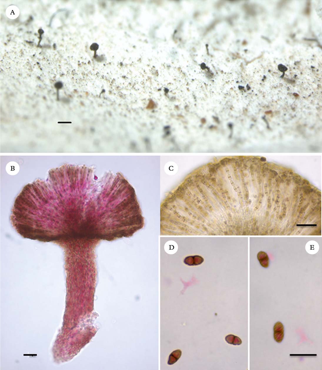
(Fig. 1)
Chaenothecopsis tigillaris. A, habit showing stalked ascomata on Perenniporia meridionalis; B, microtome section of ascoma mounted in lactofucsin; C, hymenial tissues in lactic acid, showing the ascospores uniseriately arranged in asci; D & E, uniseptate ascospores, showing spiral ornamentation. A & C [isolectotype, K(M) 153753]; B, D & E [lectotype, K(M) 179042]. Scales: A=200 µm; B & C=20 µm; D & E=10 µm.
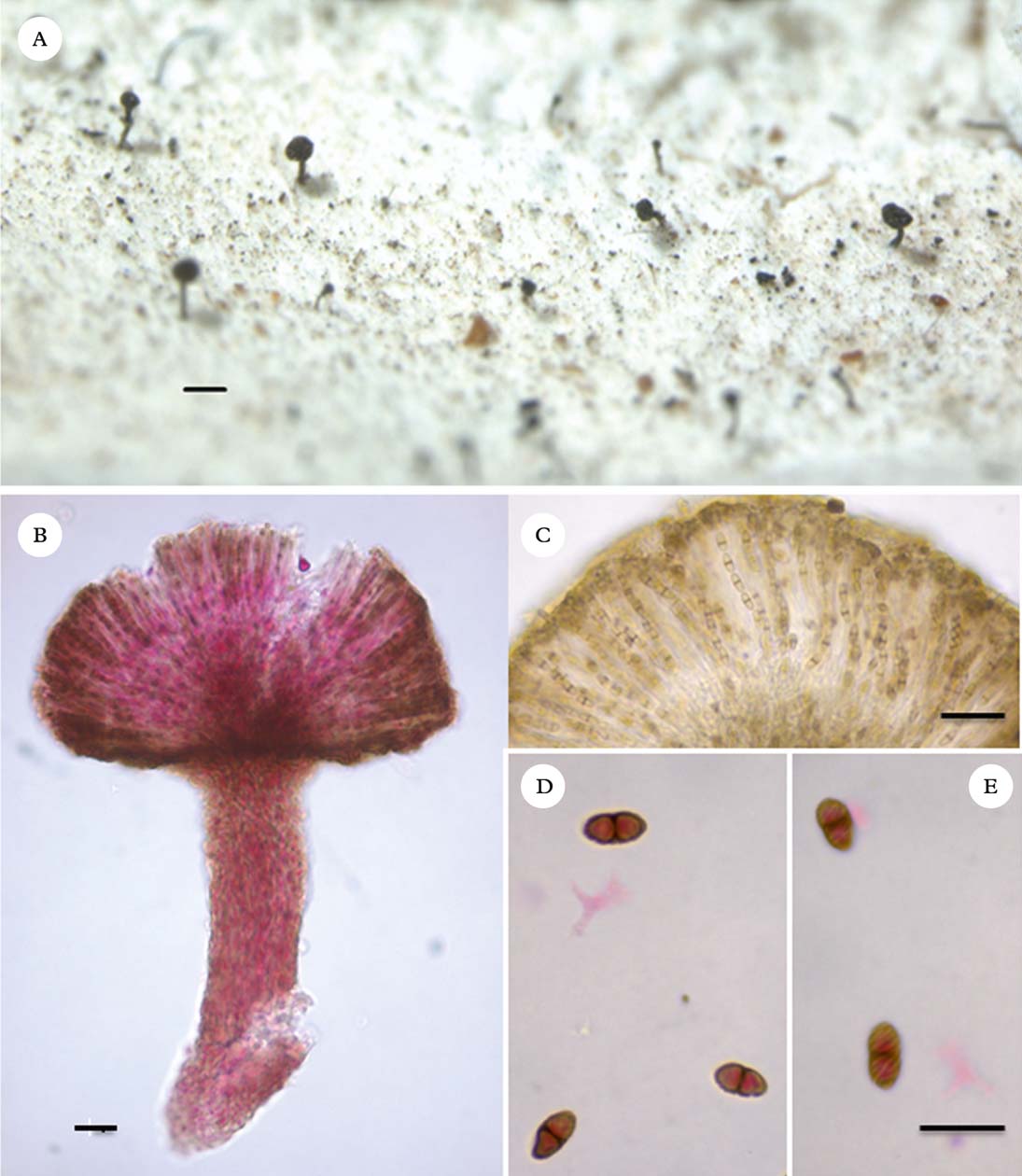
Ascomata scattered over the hymenophore surface of the host basidiomata, arising singly, calicioid, black, somewhat glossy, mainly 150–200 µm tall. Stipe 35–50 µm thick below the capitulum, composed of compacted brownish hyphae forming a dense intertwined irregularly periclinally orientated textura intricata, becoming paler towards the base, hyphae smooth-walled, somewhat torulose, uneven in thickness, 2·0–2·5 µm wide, ±homogeneous in structure; KOH−. Capitulum expanding abruptly from the stipe, hemispherical, surface convex, ±smooth, not forming a mazaedium, 195–210 µm diam. Hypothecium dark brown, formed by a continuation of the stipal hyphae expanding radially and periclinally, textura intricata, c. 50 µm in height. Exciple dark brown, 4·0–7·5 µm thick, 2–3 cells thick, composed of short-torulose hyphae to somewhat irregularly angular isodiametrical cells, individually mainly 3·5–4·5 µm diam. Hymenium hyaline, but appearing brownish from the ascospores, 65–75(–80) µm tall. Epithecium continuous, persistent, brown, 4·5–6·5 µm thick, formed of adhering almost pseudoparenchymatous cells, individually 3·0–4·5 µm diam., with somewhat unevenly thickened walls, formed from terminal branches of interascal filaments. Asci persistent, narrowly cylindrical, unitunicate in structure, 45–52×2·5–3·5 µm, 8-spored. Ascospores uniseriately arranged in the ascus, narrowly ellipsoid, pale brown, 1-transseptate, the septum slightly darker brown, appearing smooth, but with some spiral verruculose ornamentation evident at the highest magnifications (>×1000), 4·5–6·0(–7·0)×(1·5–)2·0–2·5(–3·0) µm.
Typification. The original diagnosis refers to two collections, “on an old Polyporus from a beam in King's Cliffe Church, and on wood at Batheaston, C.E. Broome.” There are two specimens under the name Sphinctrina tigillaris from King's Cliffe on a polypore in K, one from hb. Berkeley and one from hb. Broome; the latter appears to be a piece physically cut from the first. The larger of the two is selected as lectotype here; it is the isolectotype, however, that has measurement annotations in Berkeley's hand. In addition, two packets from King's Cliffe were found in the basidiomycete section of the fungarium filed under Perenniporia medulla-panis [K(M) 75884]; one from hb. Berkeley and one from hb. Cooke. These two specimens were originally determined by Berkeley as Polyporus subfuscoflavidus (as “subfuscus-flavidus”), redetermined as P. medulla-panis by J. Lowe in 1960, and now redetermined as P. meridionalis. It is likely that this material, at least in part, was documented by Berkeley & Broome (Reference Berkeley and Broome1865b ) as being found “on oak planks in the roof of King's Cliffe Church”. One set of specimens from hb. Berkeley was also found to support the Chaenothecopsis; that specimen is undated (see below) but could be either a second isolectotype or a later collection from the same church. Given the close association between these calicioid and pored species, both of which were documented as British in 1865, it seems curious that a complete name for the polypore is not amongst the annotations accompanying the original C. tigillaris collections.
There is a single specimen from Broome's herbarium on wood on the same sheet as the isolectotype, but with no locality, date, or indication of the collector and which is annotated “=Calicium tympanellum” [i.e. the species now known as Cyphelium inquinans (Keissler Reference Keissler1938)]. While it is possible this is the specimen from Batheaston, that cannot be established without doubt, and it is not congeneric with the collection from the polypore (see below). As Berkeley retained only the fungus on the polypore in his collection, and that collection was made the year prior to the publication of the name, this is the most appropriate material for designation as the lectotype.
Additional material. Great Britain: England: V. C. 32, Northamptonshire: King's Cliffe church, on Perenniporia meridionalis [as Polyporus subfuscus-flavidus], s.d., ex hb. Berkeley (K(M) 75884, sub P. meridionalis).
Key to the calicioid fungi known on polypores
Of the six species keyed out here, only Chaenotheca brunneola, Chaenothecopsis caespitosa, and C. tigillaris are known from the British Isles.
-
1 Ascomata not producing a mazaedium, ascospores 1-septate, ellipsoid; not lichenized ... 2
Ascomata producing a distinct mazaedium, ascospores non-septate, spherical or cuboid; lichenized ... 5
-
2(1) Ascomata arising singly ... 3
Ascomata arising in dense caespitose tufts, stalks branched and anastomosed, ascospores smooth, 9–14×3·0–4·5 µm ... Chaenothecopsis caespitosa
-
3(2) Ascospores<10 µm in length ... 4
Ascospores (10–)12–15(–20)×3–4 µm ... Phaeocalicium polyporaeum
-
4(3) Ascospores 4·5–6·0(–7·0)×(1·5–)2·0–2·5(–3·0) µm ... Chaenothecopsis tigillaris
Ascospores 3–5×2·5–3·0 µm ... Chaenothecopsis polyporae
-
5(1) Ascospores spherical, walls smooth or with irregular fissures, (2·0–)3·5–4·5(–5·5) µm diam; stalk black, not pruinose; algal partner Dictyochloropsis ... Chaenotheca brunneola
Ascospores spherical to cuboid, walls irregularly thickened, 3–4 µm diam.; stalk and capitulum with a yellow-green pruina; algal partner Stichococcus ... Chaenotheca brachypoda
Discussion
Titov (Reference Titov2006) treated an impressive 58 species in Chaenothecopsis, many of which were illustrated in colour and with scanning electron micrographs (SEM) of the ascospores; none agreed with C. tigillaris. The absence of a lichenized thallus and the rather small ascospores are suggestive of Mycocalicium (Tibell Reference Tibell1999), but the spiral ornamentation on the ascospores precludes a placement in that genus. The asci of the type material of Sphinctrina tigillaris were not in a sufficiently good state to determine the details of the ascus tip structure to confirm the placement in Chaenothecopsis; in that genus, the ascus tips should be elongated with a long and narrow central channel, while no such channel is evident in the ascus tips of Mycocalicium and Phaeocalicium species (Schmidt Reference Schmidt1970; Tibell Reference Tibell1975; Hawksworth Reference Hawksworth1980).
We presume that C. tigillaris is a saprobe rather than a pathogen of Perenniporia meridionalis as infected parts of the host basidome continue to generate basidiospores.
The third specimen under the name Sphinctrina tigillaris in Kew, which came from the Broome herbarium [K(M) 153754], could be a syntype but lacks information as to the locality and date. This specimen occurred directly on wood, with no indication of any polypore being present and a thin immersed lichenized thallus is evident in microtome sections. The ascomata are mazaedial, arise from a short stipe up to c. 200 µm tall, and expand to a capitulum of c. 250 µm diam.; some capitula have a faint reddish brown pruina on the lower parts. The ascospores are brown, 1-septate, broadly ellipsoid, verruculose with a spiral ornamentation (at over ×1000), and mainly 7–9×3·0–4·5 µm. This specimen is a species of Calicium, and not Cyphelium inquinans as the later annotation on the label (see above) suggests. It actually belongs to the widespread Calicium salicinum Pers. 1794, a species most commonly found on wood, which is described in detail by Tibell (Reference Tibell1999).
Chaenothecopsis tigillaris is not the only calicioid fungus to be reported from poroid basidiomata, as indicated in the key above:
(1) Chanenotheca brachypoda, a lichen-forming species in which the ascomata produce a mazaedium, is primarily associated with rotting wood and bark, but is also reported from “old polypores” in Scandinavia (Tibell Reference Tibell1999). Rikkinen (Reference Rikkinen1995) mentions it from dead basidiomes of polypores “on rotten birch”, and he informs us that the fungus typically extends from open cankers onto the hymenial surfaces of Fomes fomentarius and Piptoporus betulinus (J. Rikkinen, pers. comm.). The report of “C. stemonea f. albocinerea” on unnamed species in central Finland by Koskinen (Reference Koskinen1955) could also belong here, but the application of that name is uncertain (Tibell Reference Tibell1980).
(2) Chaenotheca brunneola, also a lichen-forming species in which the ascomata produce a mazaedium, was mentioned by Tibell as sometimes growing on “Polyporus sp.” (Tibell Reference Tibell1980), and occasionally on Trametes versicolor (Tibell Reference Tibell1981). It is described and illustrated in detail by Tibell (Reference Tibell1980), who records the non-septate ascospores as spherical and “(2·2)3·4–[4·0]-4·6(5·4) µm”.
(3) Chaenothecopsis caespitosa, described from a decaying indeterminate polypore and subsequently found on rotten wood of Taxus, has complex compacted branched ascomata 2–4 mm tall, and also larger ascospores, measuring 9–14×3·0–4·5 µm (Hawksworth Reference Hawksworth1980; Titov Reference Titov2006).
(4) “Chaenothecopsis polyporae” is a designation given to a calicioid fungus found on an unidentified tree-inhabiting polypore in Canada. It was mentioned as “Chaenothecopsis sp.” and stated to have ascospores 3–5×2·5–3·5 µm by Goward (Reference Goward1999). Goward had, however, used “Chaenothecopsis polyporae” on a packet of this fungus which he sent to Titov for study (T. Goward, in litt.). Titov (Reference Titov2006) provided a brief description of “Chaenothecopsis polyporae Goward (ined.)”, but he did not validate the designation. On the basis of the slightly smaller size given for the ascospores, Goward's fungus could be close to, or even be identical with, Chaenothecopsis tigillaris. However, we have not studied any of his material and no other measurements apart from the ascospore size range were available; it is also not known if that fungus had similar spore ornamentation.
(5) Phaeocalicium polyporaeum occurs on the upper surface of Trichaptum biforme basidiomata. It has been considered to grow on Trametes (syn. Coriolus) species (Keissler Reference Keissler1938; Hawksworth Reference Hawksworth1980; Tibell Reference Tibell1981), a genus in a different order, and Trichaptum biforme was added as an additional host by Tibell (Reference Tibell1981), who also distributed material growing on it (Tibell Reference Tibell1989). However, the hosts of all six collections in K (including Lojka's type from Hungary) were re-examined and all found to represent T. biforme. This fungus is larger, with delicate stipes to almost 1 mm in height, and 0–1-septate often indistinctly septate ascospores measuring (10–)11–15(–20)×(2·5–)3·0–4·0(–5·0) µm (Hawksworth Reference Hawksworth1980; Tibell Reference Tibell1981; Titov Reference Titov2006). Peck (Reference Peck1876) reported C. tigillaris (as Sphinctrina) on Polyporus abietinus (now assigned to Trichaptum) from Albany and Buffalo in New York State and noted that “the spores in our specimens are ·0003–·0006 long”. Based on spore and host data alone, we suggest these specimens are more likely to represent either P. polyporaeum or an undescribed taxon.
The host polypore of Chaenothecopsis tigillaris was determined as Perenniporia meridionalis Decock & Stalpers Reference Decock and Stalpers2006 and is of particular interest. That fungus favours dead Quercus wood in warmer forests of central and southern Europe although, interestingly, it is also recorded on construction timber in more northerly locations (Decock & Stalpers Reference Decock and Stalpers2006). This polypore had not previously been reported from the UK, but has only recently been formally distinguished from P. medulla-panis (Jacq.) Donk 1967 by the larger pores and basidiospores, and the differing reaction of the wider vegetative hyphae to Melzer's reagent (Decock & Stalpers Reference Decock and Stalpers2006). That polypore also occurs on dead Quercus wood, but has a much more northerly distribution in Europe. On a re-examination of the UK collections filed as P. medulla-panis in K (all from England), the five that belonged to Perenniporia were redetermined as P. meridionalis. This resulted in a shift in the English status of the former species from “Extinct” (Evans et al. Reference Evans, Henrici and Ing2006) to “Not English” (Ainsworth Reference Ainsworth2014). Furthermore, no authentic British or Irish vouchers have been traced, and so P. medulla-panis is scheduled for exclusion from the British and Irish Basidiomycota checklist (http://www.basidiochecklist.info/).
We have not located any collections of Chaenothecopsis tigillaris made since the original in 1864, but the fungus clearly should be searched for on any Perenniporia specimens encountered, particularly those on building timbers or other worked wood. The five English specimens redetermined as Perenniporia meridionalis (see above) were, with one exception recorded on oak timber in 1935, collected in the 19th century. Although mostly lacking ecological data, these basidiomata and associated wood, where present, had characters consistent with similarly anthropogenic habitats. As this fungus is evidently thermophilous elsewhere in Europe (Decock & Stalpers Reference Decock and Stalpers2006), it is possible that both it and its calicioid associate are not British natives and were inadvertently introduced, perhaps repeatedly, with imported timber. In the absence of any compelling evidence that P. meridionalis has ever established a viable British population in the outdoor environment, it qualifies for inclusion as an alien in the checklist (http://www.basidiochecklist.info/) and would therefore remain as “Not Evaluated” (NE) in an IUCN-compliant regional conservation assessment (IUCN 2014).
We are indebted to Amanda Waterfield for first drawing this material to our attention while curating material in the Royal Botanic Gardens Kew. Trevor Goward kindly provided background information on the material studied by the late Alexander N. Titov, Jouko Rikkinen generously shared his unpublished observations on Chaenotheca brachypoda, and Leif Tibell indicated that he had not encountered this fungus before. This contribution was prepared while DLH was in receipt of funding from the Spanish Ministerio de Ciencia e Innovación project CGL2011-25003.