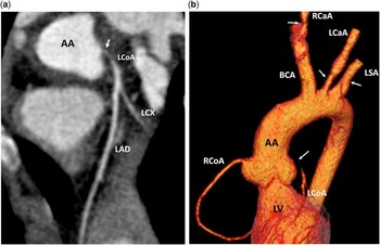
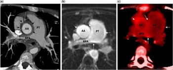
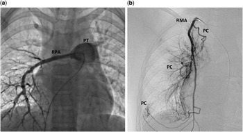
An 11-year-old girl with no medical history or cardiovascular disease risk factor was referred to our hospital for chest pain with elevated troponin T (0.4 μg/L, n<0.03) and inflammatory syndrome with chronic anaemia (fibrinogen: 5.4 g/L, C reactive protein: 68 mg/L, platelets: 408 g/L, and haemoglobin: 10.3 g/dl). Her blood pressure was normal (100/70) with no difference between upper and lower limbs. Electrocardiogram showed sinus rhythm with ST segment depression and negative T wave in the anterolateral leads (V1–V5, D1, AVL). Echocardiography showed a non-dilated left ventricle with normal global contractility (ejection fraction 67%) but with antero-septal and anterior hypokinaesia. Moderate functional mitral regurgitation was noted. The left anterior descending artery had thickened walls and its flow was retrograde. Coronary angiography revealed sub-occlusion of the proximal left coronary artery, with retrograde flow originating from collaterals of the right coronary artery. (Supplementary videos 1 and 2=angio N° 71 and N° 91). Multidetector CT angiography showed that the left coronary ostium stenosis was caused by wall thickening of the ascending aorta (Figs 1a and b, 2a). CT angiography also revealed severe bilateral stenosis of the carotid arteries (80% on the right and 50% on the left), mild bilateral subclavian artery stenosis (20%) (Fig 1b), and right pulmonary artery stenosis (Figs 2a and 3a), strongly suggestive of severe Takayasu arteritis. Collateral vessels from the right internal mammary artery contributed to right pulmonary flow (Fig 3b). Late Gadolinium enhancement on MRI, as well as uptake on 18-fluorodeoxyglucose positron-emission tomography, both located in the same areas of the ascending aorta and pulmonary artery walls, suggested active inflammation (Fig 2b and c).
( a ) Contrast-enhanced and synchronised CT scan, reconstructed coronary view showing sub-occlusive stenosis of the left coronary artery (LCoA). ( b ) Three-dimensional volume-rendered reconstruction of the thoracic aorta, showing sub-occlusive stenosis of the LCoA, 90% right carotid artery (RCaA) stenosis, 50% left carotid artery (LCaA) stenosis, and 20% left subclavian artery (LSA) stenosis. AA=ascending aorta; BCA=brachicephalic artery; LAD=left anterior descending artery; LCX=left circumflex artery; LV=left ventricle; RCoA=right coronary artery.

( a ) Short-axis CT scan showing increased wall thickness of the ascending aorta (AA) and the right pulmonary artery (RPA), leading to right pulmonary stenosis. ( b and c ) Axial cardiac MRI and axial 18-fluorodeoxyglucose positron-emission tomography images showing parietal delayed enhancement ( b )/parietal uptake ( c ) of the ascending aorta, pulmonary trunk (PT), and right pulmonary artery. This suggests the presence of an active inflammatory process.

( a ) Right selective pulmonary angiography showing right pulmonary stenosis with poor pulmonary vascularisation. ( b ) Numerical angiography showing a network of pulmonary collaterals from the right mammary artery. PC=pulmonary collaterals; PT=pulmonary trunk; RMA=right mammary artery; RPA=right pulmonary artery.

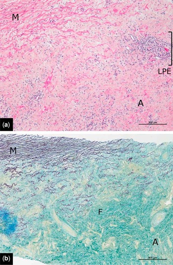
Steroid treatment with prednisone 15 mg twice per day was initiated, and coronary artery bypass surgery was performed, with the healthy left mammary artery supplying the left anterior descending artery. The postoperative course was uneventful. Histological analysis of biopsy specimens revealed a vasculitis pattern of the aorta characterised by intimal hyperplasia and mononuclear cell infiltration (Fig 4). The mammary artery and the pericardium were histologically normal. There was no family history of tuberculosis, and QuantiFERON-TB was negative.
Biopsy of the aorta showing the outer part of the media (M) and the markedly thickened and fibrous adventitia (A). Fibrosis (F) contained lymphoplasmacytic inflammatory elements (LPE). There was no necrosis, no giant cells, and no tuberculoid granuloma. ( a ) Haematein and eosin stain. ( b ) Fuschin and resorcin stain for elastic fibres.

Takayasu’s arteritis, also called women’s pulseless disease, is a panarteritic inflammation of large elastic arteries. It causes marked thickening of the affected artery walls, resulting in narrowing or even occlusion of these vessels in the late stages of the disease. Any large artery may be affected, resulting in high clinical variability. Clinical presentations range from simple absent radial pulse (subclavian arteries) to manifestations of severe ischaemia such as acute limb ischaemia (iliac arteries), stroke (carotid arteries), renal infarction (renal arteries), coronary syndrome (ostial coronary stenosis), pulmonary artery stenosis, etc.Reference Weyand and Goronzy 1 The incidence of Takayasu’s arteritis ranges from 1/1,000,000 to 2.6/1,000,000 in adults, but is unknown in children. In children, the heart and brain are affected in, respectively, 19 and 31% of the cases.Reference Brunner, Feldman and Tyrrell 2 The coronary disease found in young women with Takayasu’s arteritis contrasts with the usual epidemiology of coronary disease affecting mainly men with cardiovascular disease risk factors.
The sensitivity and specificity of clinical and angiographic diagnostic criteria of Takayasu’s arteritis are good, although only in the late forms of the disease.Reference Roghi, Pedrotti and Milazzo 3 , Reference Arend, Michel and Bloch 4 Non-invasive imaging techniques such as MRI or CT scans thus play a crucial role in the early diagnosis of Takayasu’s disease.Reference Hartlage, Palios and Barron 5 Indeed, in our case, there was neither hypertension nor absence of radial pulse, because there was no stenosis of the aorta or of the subclavian arteries. The most severe stenosis affected the carotid arteries. CT imaging specifies the vascular characteristics necessary for severity classification: arterial wall thickening, aneurysm, and stenosis of the aorta or branch vessels. Coronary CT scan, indicated in a young adult with acute coronary syndrome, reveals the typical aortitis-associated ostial coronary stenosis, and rules out other possible mechanisms such as congenital coronary anomaly or plaque rupture.Reference Roghi, Pedrotti and Milazzo 3 In our case, the aorta was thickened on its entire ascending portion. Takayasu’s disease classification was 2AC+, P+– thickening of the entire ascending aorta+coronary disease+pulmonary arteries thickening – according to the 1994 Takayasu conference and 2B – severe single complication – according to the Ishikawa clinical classification.Reference Arend, Michel and Bloch 4 Fluorine-18-fluorodeoxyglucose positron-emission tomography as well as MRI imagery are helpful to diagnose and to evaluate the level of activity of the disease, particularly during the inflammatory phase. The fluorine-18-fluorodeoxyglucose uptake in vessel walls and the presence of oedema on “oedema-weighted” T2 sequences in MRI imagery result from inflammation. Gadolinium contrast-enhanced MRI can identify mural fibrosis.Reference Hartlage, Palios and Barron 5 In these usually young patients, non-irradiating MRI is thus the method of choice to monitor the activity of the disease and to detect a relapse. In our case, MRI and positron-emission tomography (PET) were essential to determine the level of activity and the extension of the disease before considering surgical strategy.
Acknowledgement
None.
Conflicts of Interest
None.
Supplementary material
To view supplementary material for this article, please visit http://dx.doi.org/10.1017/S1047951116001505