Introduction
Children with CHD represent a uniquely complex cardiovascular population. Their care depends on multimodal imaging, serial electrocardiographies, invasive and noninvasive monitoring, and rapidly evolving perioperative physiology. In the cardiac ICU, clinicians must synthesise continuous data from electrocardiography, arterial pressure waveforms, pulse oximetry, near-infrared spectroscopy, laboratory values, medications, and ventilator settings. That combination of physiologic complexity and data density makes paediatric cardiology and cardiac intensive care a natural setting for artificial intelligence applications. Recent reviews specifically focused on CHD, paediatric echocardiography, paediatric electrocardiography, and congenital heart surgery all conclude that artificial intelligence has substantial potential but is still in an early translational stage. Reference Holt, Lee and Punn1–Reference Leone, Miller and Silvani3
Broadly, current paediatric cardiac artificial intelligence work falls into five overlapping domains: automated interpretation of echocardiography and cardiac magnetic resonance; artificial intelligence-enhanced electrocardiography analysis; prediction of deterioration in the cardiac ICU; postoperative risk prediction after congenital heart surgery, especially low cardiac output syndrome and acute kidney injury; and advanced waveform analytics using electrocardiography and arterial pressure morphology. Across these domains, the central clinical question is the same: can computational models detect clinically meaningful patterns earlier, more consistently, or more precisely than current workflows allow? Existing evidence suggests the answer may be yes, but only under specific conditions and not yet with sufficient external validation for routine broad deployment. Reference Holt, Lee and Punn1,4
Methods
This manuscript is a narrative review. The literature base was drawn from peer-reviewed reviews and primary studies identified through PubMed/MEDLINE and Scopus searches using combinations of the terms artificial intelligence, machine learning, analytic algorithm, deep learning, paediatric cardiology, CHD, echocardiography, electrocardiography, cardiac intensive care, predictive analytics, high-frequency physiologic data, waveform, low cardiac output syndrome, acute kidney injury, extubation, lactate, and congenital heart surgery. Priority was given to paediatric- or CHD-specific studies, multicentre investigations, systematic reviews, and primary studies with direct clinical relevance. Candidate studies were retained when they addressed diagnosis, monitoring, risk prediction, physiologic interpretation, or implementation in paediatric/CHD populations; purely technical studies without a clinically interpretable paediatric cardiac use case, non-peer-reviewed reports, and studies lacking sufficient methodological detail were not used as core evidence. Adult studies were included selectively when they established a foundational method with direct relevance to paediatric translation.
Because this is a narrative rather than a systematic review, a formal PRISMA-style duplicate screening and adjudication process was not performed. Study selection was therefore vulnerable to author-selection bias. To reduce this risk, inclusion was organised around predefined clinical domains, and emphasis was placed on studies with transparent methods, clinically relevant endpoints, paediatric or CHD populations, multicentre validation when available, and explicit reporting of limitations. This selection process and the absence of formal systematic-review screening should be considered limitations of the review. All authors participated in study selection and jointly reviewed the included literature for relevance to the predefined clinical domains.
Because the literature is heterogeneous in population, input features, outcomes, and performance reporting, formal meta-analysis is generally not appropriate at present. Recent systematic reviews in congenital heart surgery and paediatric post-cardiac surgery acute kidney injury reach the same conclusion: performance is often promising, but study heterogeneity and frequent lack of external validation limit pooled inference and bedside generalisability. Reference Mohammadi, Rajai Firouzabadi and Hosseinpour5,Reference Cheong, So, Lal and Coveliers-Munzi6
Results
Artificial intelligence in paediatric and fetal echocardiography
Echocardiography is the main imaging modality in fetal and paediatric cardiology and is one of the technically most developed areas of cardiovascular artificial intelligence. However, technical maturity in adult echocardiography should not be interpreted as clinical maturity in children with CHD. Paediatric-focused reviews describe applications in acquisition guidance, view classification, segmentation, measurement automation, and congenital lesion detection, but paediatric datasets remain smaller and anatomically more heterogeneous. Accordingly, these tools should currently be viewed as investigational or augmentative in complex paediatric CHD rather than ready for routine autonomous acquisition or measurement. Reference Wang, Doan, Reddy and Jone2
At the methodological level, Ouyang and colleagues provided a highly influential demonstration that video-based deep learning can segment the left ventricle and estimate ejection fraction directly from echocardiographic clips. Because EchoNet-Dynamic was developed predominantly in adults, its performance should not be extrapolated directly to paediatric CHD, where ventricular morphology, surgical reconstruction, loading conditions, and imaging windows differ substantially. Paediatric translation therefore requires dedicated paediatric/CHD training sets and external validation rather than simple transfer of adult algorithms. Reference Ouyang, He and Ghorbani7
Paediatric- and CHD-specific evidence is emerging. Arnaout and colleagues showed that an ensemble of neural networks could identify standard fetal cardiac views and support prenatal CHD detection from human-acquired ultrasound images, demonstrating lesion-detection feasibility in a directly relevant population. More recent paediatric and prenatal studies have evaluated external assessment of fetal CHD screening models and paediatric echocardiographic view-classification/diagnostic-support pipelines. These studies strengthen paediatric relevance, but they remain constrained by disease prevalence, lesion heterogeneity, image-quality variation, and limited prospective multicentre testing. Reference Arnaout, Curran and Zhao8,Reference Athalye, van Nisselrooij, Rizvi, Haak, Moon-Grady and Arnaout17,Reference Jabarulla, Uden, Beerbaum and Oeltze-Jafra18
Recent paediatric/fetal echocardiography reviews emphasise that the current evidence base remains developmental. Many studies are single-centre, disease-specific, or based on relatively small annotated datasets; external validation and prospective workflow evaluation remain uncommon. Thus, although imaging artificial intelligence is technically advanced relative to other paediatric cardiac domains, routine use for complex CHD acquisition, automated measurement, or independent diagnosis is not yet supported. Reference Wang, Doan, Reddy and Jone2,Reference Cao, Zhong, Ma, Li and Huang19
Artificial intelligence in cardiac magnetic resonance for CHD
cardiac magnetic resonance is central to longitudinal assessment of complex CHD, but manual segmentation and reconstruction are time intensive. Recent CHD-focused cardiac magnetic resonance reviews show that artificial intelligence is being used across acquisition, reconstruction, segmentation, and reporting. The most clinically relevant gains are in reduced acquisition burden, improved image reconstruction from undersampled data, and more reproducible ventricular quantification. Reference Gearhart, Anjewierden, Buddhe and Tandon9
Bai and colleagues provided an early landmark demonstration that deep learning could automate cardiovascular MRI analysis with strong agreement to expert methods. More recent CHD-specific reviews suggest that these methods are especially valuable in congenital populations because serial volumetric assessment is common, manual contouring is laborious, and reproducibility matters greatly for operative timing and longitudinal follow-up. Still, like echocardiography, cardiac magnetic resonance artificial intelligence in CHD is limited by variable anatomy, small paediatric datasets, and the need for robust multicentre generalisation. Reference Mohammadi, Rajai Firouzabadi and Hosseinpour5,Reference Bai, Sinclair and Tarroni10
Artificial intelligence in paediatric electrocardiography
Paediatric electrocardiography is an especially attractive artificial intelligence target because it is inexpensive, ubiquitous, and already used in screening, perioperative care, and longitudinal surveillance. The most up-to-date paediatric-specific review notes that artificial intelligence-enhanced electrocardiography interpretation in children is promising but faces unique challenges related to age-dependent waveform evolution, small datasets, and the lower prevalence of many target diseases compared with adult populations. Reference Leone, Miller and Silvani3
Hannun and colleagues demonstrated cardiologist-level arrhythmia classification with deep neural networks in large electrocardiography datasets, establishing the feasibility of high-performance artificial intelligence-electrocardiography classification. Although that foundational study was not paediatric-specific, it laid the groundwork for paediatric artificial intelligence-electrocardiography development. More recently, Mayourian and colleagues reported expert-level automated diagnosis of paediatric electrocardiographies, marking a major step toward clinically relevant paediatric electrocardiography interpretation. Reference Leone, Miller and Silvani3,Reference Hannun, Rajpurkar and Haghpanahi11,Reference Mayourian, La Cava and de Ferranti12
The paediatric ECG literature is expanding beyond rhythm interpretation, but age-dependent physiology creates a major generalisability problem. Normal intervals, repolarisation, conduction patterns, and waveform morphology change across infancy, childhood, and adolescence; consequently, adult algorithms cannot be assumed to perform equivalently in children. This concern is particularly relevant for tasks such as corrected QT interval assessment and recognition of pre-excitation/Wolff-Parkinson-White patterns, where age, heart rate, lead morphology, and threshold definitions influence interpretation. Paediatric-specific training and age-stratified validation are therefore essential. The likely near-term role of paediatric artificial intelligence-electrocardiography is triage, screening, and decision support rather than replacement of expert interpretation. Reference Leone, Miller and Silvani3
Analytic algorithms for cardiac ICU deterioration and postoperative risk
Given the emphasis of this review on cardiac critical care, the remaining results focus on analytic algorithms that use static and longitudinal data to characterise overlapping cardiac ICU outcomes: deterioration/cardiac arrest, postoperative low cardiac output physiology, hyperlactatemia, extubation failure, acute kidney injury, and de-escalation of vasoactive or respiratory support. These outcomes are presented in related subsections because their clinical labels differ, but they share a common translational problem: abundant physiologic data are available, yet most studies still evaluate retrospective or observational model performance rather than real-time model-guided care. Importantly, multicentre evidence now exists for several high-frequency risk-analytics applications, including hyperlactatemia, extubation failure, and vasoactive weaning. Reference Yu, Skinner and Esangbedo13,Reference Asfari, Wolovits and Gazit25–Reference Goldsmith, Nadkarni and Futterman27
A particularly relevant paediatric cardiac study is the cardiac arrest prediction work by Yu and colleagues. In a single-centre cardiac ICU cohort, an analytic algorithm using 11 electronic health record-derived variables achieved an area under the receiver operating characteristic curve of 0.85 one hour before cardiac arrest. The study demonstrates a retrospective predictive signal, but the algorithm was not deployed in real time and therefore did not test whether an alert could prevent arrest or change care. Model discrimination alone is consequently insufficient evidence of clinical benefit; the next step is prospective silent validation followed by carefully monitored real-time implementation to determine whether the signal provides actionable lead time without unacceptable false-alert burden. Reference Yu, Skinner and Esangbedo13
The broader cardiovascular artificial intelligence literature, including a recent AHA review on artificial intelligence for improving outcomes in heart failure and critical care, frames paediatric cardiac ICU predictive analytics as highly promising but not yet implementation-ready at scale. The promise lies in earlier recognition; the challenge lies in translating statistical prediction into better bedside decisions without adding noise, alarm fatigue, or opaque risk scores that clinicians do not trust. Reference Armoundas, Narayan and Arnett14
Low cardiac output physiology and low cardiac output syndrome prediction after congenital heart surgery
low cardiac output syndrome is an important postoperative syndrome and a meaningful target for analytic algorithms, but its label is unstable across studies because definitions combine different thresholds for lactate, urine output, vasoactive support, haemodynamics, or clinical judgement. Baloglu and colleagues developed single-centre supervised models in 181 neonates, 14.9% of whom met the study low cardiac output syndrome definition; reported area under the receiver operating characteristic curve values ranged from 0.91 to 0.98 across 2- to 12-hour forecasting horizons, with vasoactive-inotrope score, urine output, and lactate among the most influential features. Reference Baloglu, Wang and Marino15 These findings show that the model can forecast the study-defined low cardiac output syndrome label, but they do not prove that it detects a distinct latent physiologic state before clinicians can recognise it. The distinction between earlier physiology and earlier labelling is therefore a physiologic hypothesis and translational goal, not a proven property of the Baloglu algorithm. Prospective validation using independently defined physiologic endpoints is needed to establish whether the algorithm identifies deterioration before conventional bedside recognition.
low cardiac output syndrome prediction should be interpreted within the broader postoperative-risk literature rather than as an isolated artificial intelligence task. Adverse outcomes of interest include low cardiac output syndrome, prolonged ventilation or extubation failure, extracorporeal membrane oxygenation requirement, acute kidney injury, cardiac arrest, and mortality. Large retrospective cohorts demonstrate that analytic models can stratify these risks, but performance varies by endpoint and dataset; for example, Tong and colleagues evaluated five algorithms in approximately 23,000 paediatric congenital heart operations. Static perioperative models remain clinically useful for resource allocation, family counselling, ICU staffing, and identifying patients who warrant enhanced monitoring, even when they do not identify a directly modifiable physiologic target. Continuously updating models may add value by incorporating changing bedside variables, but they also introduce challenges from missing or noisy data, alarm fatigue, model drift, interpretability, and workflow integration. Reference Mohammadi, Rajai Firouzabadi and Hosseinpour5,Reference Tong, Du and Chen16
Other postoperative outcomes: acute kidney injury and major adverse outcomes
Beyond low cardiac output syndrome, analytic algorithms have been applied to AKI, hyperlactatemia, extubation failure, and de-escalation of support. Cheong and colleagues found promising discrimination for post-cardiac-surgery acute kidney injury but substantial risk of bias and limited external validation. In contrast, some high-frequency physiologic analytics have already been evaluated across multiple centres: Asfari and colleagues validated a near real-time hyperlactatemia index in 3,496 children from 10 cardiac ICUs (58,168 lactate measurements; AUC 0.95); Hames and colleagues evaluated IDO2 and IVCO2 in a multicentre cohort of neonates after congenital cardiac surgery, where elevated indices before extubation were associated with extubation failure; and Goldsmith and colleagues studied IDO2 during vasoactive weaning across three paediatric cardiac ICUs, finding higher values in failed than successful weans. Reference Cheong, So, Lal and Coveliers-Munzi6,Reference Asfari, Wolovits and Gazit25–Reference Goldsmith, Nadkarni and Futterman27 These studies demonstrate that the field has progressed beyond exclusively single-centre model development, but most remain observational and do not yet establish that algorithm-guided action improves outcomes.
High-frequency physiologic data and waveform intelligence in the cardiac ICU
High-frequency physiologic data are particularly relevant to the cardiac ICU because conventional monitors compress rich beat-to-beat signals into intermittent or summary values. Published paediatric work has used continuously streamed monitor data and near real-time analytic indices to characterise oxygen-delivery risk, hyperlactatemia, extubation readiness, vasoactive weaning, and deterioration. For example, multicentre studies of the Etiometry platform have validated associations between continuously updated risk indices and elevated lactate, extubation failure, and failed vasoactive weaning, while prospective observational work has shown that adding IDO2 to clinician-identified high-risk status can reduce the number of alerts without losing detected cardiac-arrest events. Reference Asfari, Wolovits and Gazit25–Reference Dewan, Cooper and Tegtmeyer28 These studies are important because they move beyond static prediction toward longitudinal physiologic interpretation; however, many algorithms were calculated retrospectively or evaluated observationally, so prospective interventional evidence remains limited.
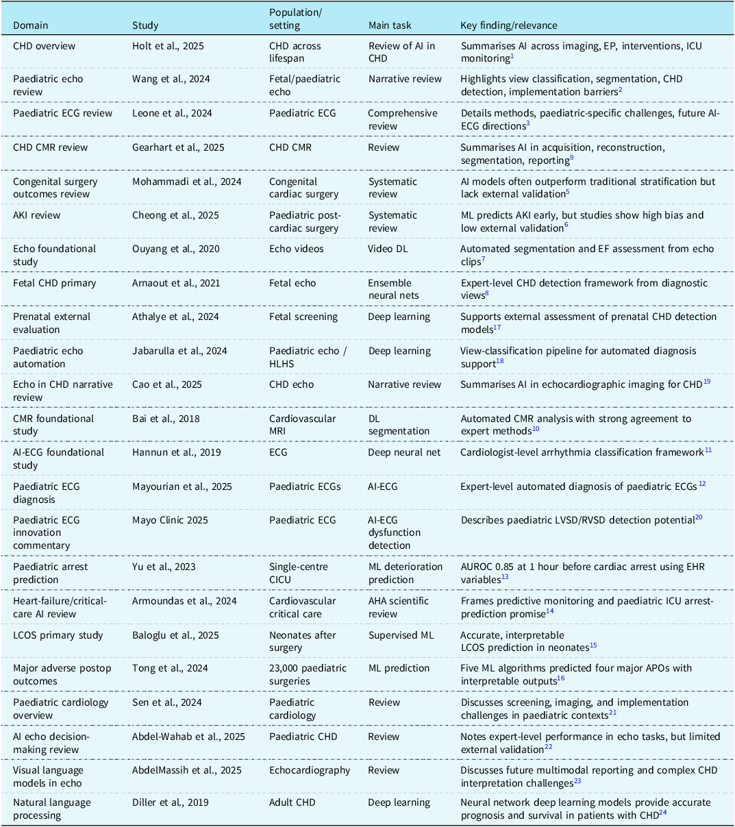
Comprehensive representative table of paediatric/CHD-focused artificial intelligence studies (Table 1).
Discussion
The strongest current evidence supports analytic algorithms as augmentative rather than autonomous technologies in paediatric cardiology. Imaging and electrocardiography demonstrate technical feasibility, but the most distinctive translational opportunity in this review is the cardiac ICU, where continuously changing physiology creates a need for longitudinal interpretation. The current literature includes multicentre validation for several risk-analytics applications, yet much of the field remains concentrated in model development, retrospective testing, or observational validation. The central challenge is therefore not simply to generate another accurate model but to demonstrate that a validated signal can be understood, safely integrated into care, and used by clinicians to improve outcomes.
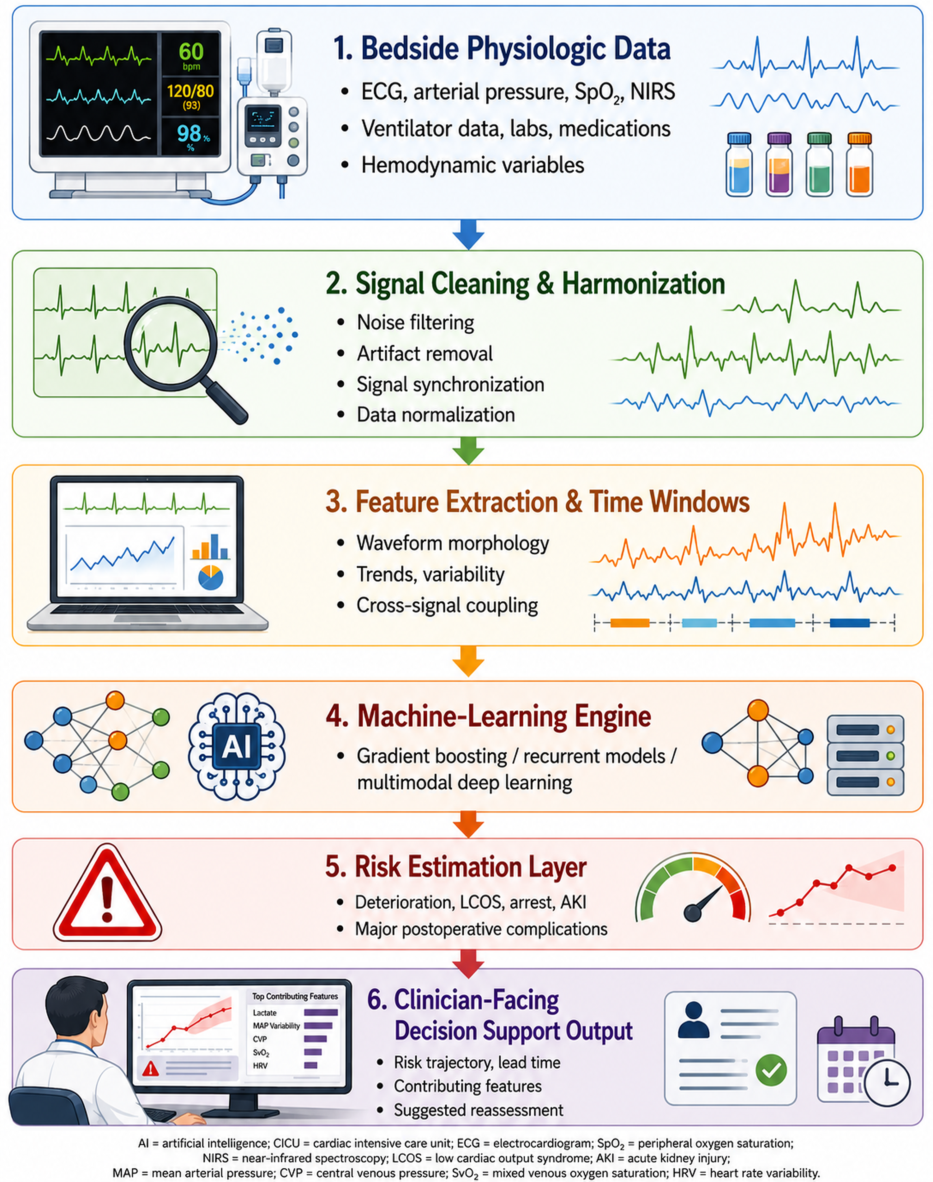
A useful translational framework for cardiac ICU analytic algorithms is prediction -> physiologic interpretation -> prospective validation -> clinician-guided clinical action -> outcome improvement. This translational pathway and the integration of multimodal cardiac ICU data into clinically interpretable risk trajectories are summarised in Figure 1. Prediction establishes that a signal exists; physiologic interpretation determines whether the signal is clinically coherent; prospective silent validation tests calibration and transportability before the model influences care; only then should real-time clinical action be evaluated. The final standard is whether model-informed care improves meaningful processes or patient outcomes. This sequence is consistent with broader implementation principles in clinical decision support, but it is presented here as a pragmatic cardiac ICU-specific framework rather than as a previously validated formal framework. Institutional culture, data infrastructure, staffing, workflow, and clinician trust can create barriers at every stage, and these factors should be evaluated explicitly during implementation.
AI architecture for predictive monitoring in the CICU. Proposed workflow for real-time paediatric cardiac critical care AI. The key translational step is not model generation alone but the conversion of continuously acquired multimodal signals into interpretable risk trajectories with clinically actionable lead time. Supported by recent paediatric CICU prediction studies and CHD-focused reviews.

The same translational logic applies to low cardiac output syndrome and other postoperative-risk models. High discrimination does not by itself establish clinical usefulness, especially when outcome labels vary or influential features overlap with variables clinicians already monitor. The potential added value of an analytic algorithm is its ability to integrate trajectories and interactions consistently over time and to present them with sufficient lead time for clinicians to reassess physiology. Whether that earlier signal changes management safely must be established prospectively rather than inferred from retrospective prediction. Reference Baloglu, Wang and Marino15
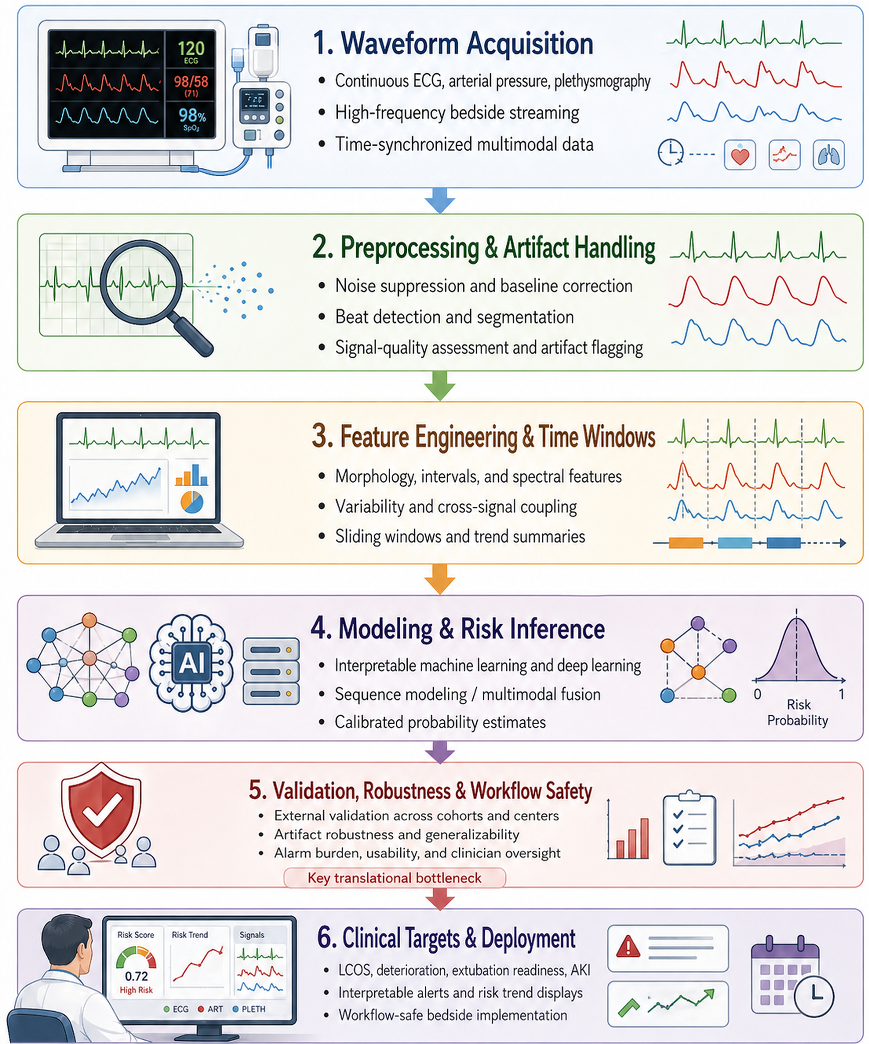
Waveform and high-frequency physiologic analytics may be among the most distinctive contributions of computational methods to paediatric cardiac critical care. Traditional monitoring reduces rich signals to summary values, whereas continuous analytics can preserve temporal structure and integrate oxygen delivery, ventilation, pressure, and laboratory trajectories. Multicentre studies of hyperlactatemia, extubation failure, and vasoactive weaning demonstrate that such approaches can generalise beyond a single centre, although performance can still vary with patient mix, monitoring practice, data completeness, and institutional workflows. Reference Asfari, Wolovits and Gazit25–Reference Goldsmith, Nadkarni and Futterman27 A conceptual pipeline from high-frequency waveform acquisition through preprocessing, feature extraction, model analysis, and bedside implementation is shown in Figure 2. Technical and implementation vulnerabilities include artefact, missingness, hardware variability, alarm fatigue, model drift, and differences in clinician trust or local practice.
Waveform analytics pipeline for paediatric cardiac critical care. Conceptual waveform-AI pipeline for paediatric cardiac intensive care. The translational bottlenecks are artefact robustness, external validation, and workflow-safe implementation rather than model architecture alone.

Across all domains, several limitations recur. Paediatric datasets remain smaller and more heterogeneous than adult datasets; external validation is improving but remains inconsistent; outcome definitions vary, particularly for low cardiac output syndrome and AKI; and implementation science lags behind model development. Multicentre data sharing and harmonisation require substantial technical, governance, and financial resources. Even a prospectively validated model may perform differently across institutions because case mix, monitoring strategies, thresholds for intervention, staffing, and workflows differ. For this reason, multicentre validation should include calibration and centre-level performance, and deployment should include local monitoring for drift, subgroup performance, false-alert burden, and unintended workflow effects. Reference Mohammadi, Rajai Firouzabadi and Hosseinpour5,Reference Cheong, So, Lal and Coveliers-Munzi6,Reference Asfari, Wolovits and Gazit25–Reference Goldsmith, Nadkarni and Futterman27
A practical way forward is to align future studies with bedside use cases rather than data availability alone. For imaging and electrocardiography, this means paediatric/CHD-specific multicentre validation and clinically meaningful deployment targets. For cardiac ICU analytics, the priority should be prospective silent validation before bedside action, followed by staged real-time implementation with prespecified safety, calibration, alert burden, and workflow metrics. Comparative studies should test whether model-informed care adds value beyond clinician assessment and existing risk tools. The goal is precision support of bedside decision-making, not automated replacement of clinical judgement.
Conclusion
Analytic algorithms in paediatric cardiology and cardiac intensive care have progressed from proof-of-concept studies to paediatric-specific models and several multicentre validation studies. The evidence is strongest for demonstrating that clinically relevant signals can be extracted from imaging, electrocardiography, electronic health record variables, and high-frequency physiologic data; evidence that real-time use improves outcomes is much more limited. For cardiac ICU applications, the appropriate translational sequence is prediction, physiologic interpretation, prospective validation, clinician-guided action, and outcome assessment. Even when this pathway is completed, analytic algorithms should remain adjunct tools that enhance bedside critical-care reasoning and monitoring rather than autonomous decision-makers. The next decisive advances will require multicentre collaboration, data harmonisation, prospective validation, implementation science, and demonstration of benefit across diverse clinical environments.
Representative paediatric and congenital-heart-disease AI studies and reviews

Table 1. Long description
The table has 14 rows and 4 columns. The columns are labeled Domain, Study, Population/setting, Main task, and Key finding/relevance. Each row provides details about a specific study or review. Row 1: Domain, CHD overview; Study, Holt et al., 2025; Population/setting, CHD across lifespan; Main task, Review of AI in CHD; Key finding/relevance, Summarises AI across imaging, EP, interventions, ICU monitoring. Row 2: Domain, Paediatric echo review; Study, Wang et al., 2024; Population/setting, Fetal/paediatric echo; Main task, Narrative review; Key finding/relevance, Highlights view classification, segmentation, CHD detection, implementation barriers. Row 3: Domain, Paediatric ECG review; Study, Leone et al., 2024; Population/setting, Paediatric ECG; Main task, Comprehensive review; Key finding/relevance, Details methods, paediatric-specific challenges, future AI-ECG directions. Row 4: Domain, CHD CMR review; Study, Gearhart et al., 2025; Population/setting, CHD CMR; Main task, Review; Key finding/relevance, Summarises AI in acquisition, reconstruction, segmentation, reporting. Row 5: Domain, Congenital surgery outcomes review; Study, Mohammadi et al., 2024; Population/setting, Congenital cardiac surgery; Main task, Systematic review; Key finding/relevance, AI models often outperform traditional stratification but lack external validation. Row 6: Domain, AKI review; Study, Cheong et al., 2025; Population/setting, Paediatric post-cardiac surgery; Main task, Systematic review; Key finding/relevance, ML predicts AKI early, but studies show high bias and low external validation. Row 7: Domain, Echo foundational study; Study, Ouyang et al., 2020; Population/setting, Echo videos; Main task, Video DL; Key finding/relevance, Automated segmentation and EF assessment from echo clips. Row 8: Domain, Fetal CHD primary; Study, Arnaout et al., 2021; Population/setting, Fetal echo; Main task, Ensemble neural nets; Key finding/relevance, Expert-level CHD detection framework from diagnostic views. Row 9: Domain, Prenatal external evaluation; Study, Athalye et al., 2024; Population/setting, Fetal screening; Main task, Deep learning; Key finding/relevance, Supports external assessment of prenatal CHD detection models. Row 10: Domain, Paediatric echo automation; Study, Jabarulla et al., 2024; Population/setting, Paediatric echo / HLHS; Main task, Deep learning; Key finding/relevance, View-classification pipeline for automated diagnosis support. Row 11: Domain, Echo in CHD narrative review; Study, Cao et al., 2025; Population/setting, CHD echo; Main task, Narrative review; Key finding/relevance, Summarises AI in echocardiographic imaging for CHD. Row 12: Domain, CMR foundational study; Study, Bai et al., 2018; Population/setting, Cardiovascular MRI; Main task, DL segmentation; Key finding/relevance, Automated CMR analysis with strong agreement to expert methods. Row 13: Domain, AI-ECG foundational study; Study, Hannun et al., 2019; Population/setting, ECG; Main task, Deep neural net; Key finding/relevance, Cardiologist-level arrhythmia classification framework. Row 14: Domain, Paediatric ECG diagnosis; Study, Mayourian et al., 2025; Population/setting, Paediatric ECGs; Main task, AI-ECG; Key finding/relevance, Expert-level automated diagnosis of paediatric ECGs.
AKI = acute kidney injury; AUROC = area under the receiver operating characteristic curve; CMR = cardiac magnetic resonance; ECG = electrocardiography; EHR = electronic health record; LCOS = low cardiac output syndrome.