INTRODUCTION
During its life-cycle, Trypanosoma cruzi, the aetiological agent of Chagas disease, alternates between a triatomine insect vector and a mammalian host. This protozoan possesses 3 main stages: the epimastigote, non-infective replicative form found in the gut of the insect vector, the trypomastigote, infective form able to invade nucleated mammalian cells and the amastigote, infective and intracellular replicating form found in the mammalian host (de Souza, 2000). Along this life-cycle, T. cruzi has to adapt to different environments through morphological and functional changes that involve complex networks of enzymatic pathways (de Souza, 2000). In this regard, we previously found a fatty acid exchange between phospholipids and neutral lipids, when T. cruzi epimastigotes grown at 28 °C were transferred to 37 °C, suggesting the action of phospholipases, lipases and acyl transferases as part of a deacylation-reacylation adaptive cycle (Florin Christensen et al. 1997). In further studies, we demonstrated that Plase A1 was the only phosphatidylcholine-degrading activity detected in T. cruzi, being remarkably higher for the infective stages than for the non-infective epimastigotes (Wainszelbaum et al. 2001).
In African trypanosomes, Plase A1 was also intensively studied. In pathogenic T. brucei high levels of enzyme activity were found while low levels were detected in T. lewisi, the non-pathogenic trypanosome of rats, suggesting the involvement of this enzyme in the pathology of African Trypanosomiasis (Hambrey et al. 1981). In addition, in T. brucei it has been proposed that Plase A1 could induce cell damage either directly, or through free fatty acids and lysophosphatidylcholine generated from parasite or host phospholipids (Tizard et al. 1978). Accordingly, in T. cruzi, we reported upon parasite autolysis, a rapid accumulation of free fatty acids and lysophospholipids, suggesting the action of Plase A1, among other enzymes (Wainszelbaum et al. 2001). Moreover, it has been shown that these compounds released from lysed T. cruzi trypomastigotes caused toxic effects on culture cells (Asahi et al. 1986).
Considering the possible roles of Plase A1 in T. cruzi biology and its potential pathogenic effects, we here study Plase A1 localization in T. cruzi infective stages and whether this enzyme is involved in host cell lipid modifications leading to protein kinase C activation.
MATERIALS AND METHODS
Materials
[1-14C] oleate (56 mCi/mmol), [32P] sodium orthophosphate (10 μCi/μl), 1-palmitoyl-2-[1-14C] palmitoyl-phosphatidylcholine, 1-palmitoyl-2-[1-14C] araquidonyl-phosphatidylethanolamine (40–60 mCi/mmol) and Sephadex G-50 (medium) were obtained from Amersham Biosciences (Arlington Heights, IL, USA). Soybean phosphatidylcholine type II-S, bovine serum albumin (fraction V), lipid standards, EGTA, Grace medium, carboxyfluorescein diacetate, Triton X-100 and dimethyl sulfoxide were from Sigma Chemical Co. (St Louis, MO, USA). Phosphate-free RPMI 1640 medium and fetal bovine serum, were from GIBCO BRL (Rockville, MD, USA). Silica Gel 60 thin-layer chromatography plates and solvents were from Merck (Darmstadt, Germany). Sulfosuccinimidobiotin was from Pierce (Rockford, Il, USA). Streptavidin MicroBeads and MACS Separation Columns were from Miltenyi Biotech (Bergisch Gladbach, Germany). Centriplus Centrifugal Filter Devices were purchased from Millipore Corporation. The peroxidase-conjugate antibody was from Santa Cruz biotechnology, the anti-phospho-serine antibody was from Calbiochem (Darmstadt, Germany) and the anti-phospho protein kinase C antibody was purchased from Cell Signaling Technology (Danvers, MA, USA). ECL Western Blotting Detection System was purchased from Amersham Biosciencies (USA).
Cells and cultures
Trypanosoma cruzi RA lethal strain was used (González Cappa et al. 1981). Epimastigotes were axenically grown at 28 °C in a biphasic medium for 48 h, as previously described (Isola et al. 1986) and harvested by centrifugation at 1000 g for 15 min. Amastigotes and trypomastigotes were purified from Vero cells (African green monkey kidney cells, non-phagocytic cell line), grown in 199 medium+10% fetal bovine serum, at 37 °C with 5% CO2 (Moreno et al. 1994).
Preparation of parasite homogenate supernatants
For gel filtration analyses, parasite homogenate supernatants were prepared as follows: amastigotes or trypomastigotes (1×10⁸/ml) were resuspended in 10 mM Tris/HCl, pH 7·4, in the presence of protease inhibitors (0·01% leupeptin; 2 mM phenylmethanesulfonyl fluoride; 25 U/ml aprotinin; 1 mM benzamidine; 0·5 mM Nα-p-tosyl-L-lysine chloromethyl ketone hydrochloride) and subjected to 4 cycles of freezing and thawing. Disrupted parasites were then centrifuged at 27000 g for 30 min and the resultant supernatants (homogenate supernatants) stored at −80 °C until used for Sephadex chromatography.
Preparation of parasite culture supernatants
To evaluate if Plase A1 is secreted into the culture medium, the different parasite stages (3×10⁸/ml per assay) were incubated in 199 medium +5% fetal bovine serum for 1 h, at 37 °C for amastigotes and trypomastigotes or at 28 °C for epimastigotes. After this, parasite suspensions were centrifuged at 12100 g (amastigotes and trypomastigotes) and 3200 g (epimastigotes), for 30min. Aliquots of the supernatants (culture supernatants) were filtered through a Centrifugal filter device (100000 kDa) to ensure that Plase A1 activity was not associated to fragments of lysed cells, and then stored at −80 °C, in the presence of the protease inhibitors described above, for further Plase A1 activity determination. To rule out the possibility that the secreted Plase A1 activity detected was due to cell breakage, parasite integrity was tested using 2 different approaches. (1) For immunoblot analysis, culture supernatants (60–100 μg of protein/lane) were loaded on SDS-PAGE gels (Laemmli, 1970), transferred to nitrocellulose membranes and incubated with specific anti-glutamate dehydrogenase (cytosolic marker) (Barderi et al. 1998). Immunoblots were visualized using ECL as substrate with a peroxidase-conjugate secondary antibody. Chemiluminescence was detected using the Storm® Gel and Blot Imaging System. (2) For carboxyfluorescein diacetate staining, aliquots of parasite suspensions after 1 h incubation were stained with 5% (v/v) of carboxyfluorescein diacetate (Goodall and Johnson, 1982). A UV light microscope (Eclipse E600, Nikon) was used to visualize the cells and images were recorded using Spot RT software, version 3.4 (Spot Diagnostic Instruments Inc).
Plase A1 activity determination
Plase activity was assayed by analysing the products of hydrolysis of radio-isotope labelled phosphatidylcholine (PC) or phosphatidylethanolamine (PE), as previously described by Wainszelabum et al. (2001). Briefly, the incubation mixture consisted of 140 μl of enzyme sample or corresponding buffer/culture media (controls), 40 μl of substrate sonicated mixture (0·01 μCi/assay of 1-palmitoyl-2-[1-14C]-oleoyl PC and 0·6 mg/assay of soybean PC or 0·01 μCi/assay of 1-palmitoyl-2-[1-14C] araquidonyl-PE and 0·6 mg/assay PE) and 20 μl of 1 M sodium acetate, pH 4·7. Incubations were carried out for 60 min at 37 °C, lipids were then extracted according to the method of Bligh and Dyer (1959) and separated by thin-layer chromatography (TLC) on Silica Gel plates using chloroform/methanol/water (65[ratio ]35[ratio ]2·5, v/v). Lipids were identified by comparison with non-radioactive standards run simultaneously and revealed by iodine vapours. Radioactivity was detected using a Storm® Gel and Blot Imaging System (Amersham Biosciences, Piscataway, NJ, USA), and the lipid spots scrapped off from the plates were measured by liquid scintillation (Rackbeta, Liquid Scintillation Counter, Amersham-Pharmacia). The amount of radioactivity present in the lysophosphatidylcholine (LPC) (or lysophosphatidylethanolamine) spot, together with the specific radioactivity of PC used in the assays, were employed to calculate the amount of LPC produced, which is a direct measurement of Plase A1 activity.
Gel filtration analysis of homogenate supernatants from T. cruzi infective stages
Homogenate supernatants (2–2·5 mg protein) were loaded into a Sephadex G-100 column, previously equilibrated with Tris-buffered saline (pH 7·4). Elution was carried out in the same buffer, with collecting fractions of 1·5 ml, monitored by protein spectrophotometry and Plase A1 activity was assayed every 2 fractions. The column was previously calibrated with the following molecular weight markers: bovine serum albumin (67 kDa), ovalbumin (43 kDa), and myoglobin (18 kDa).
Plase A1 characterization
Each peak of Plase A1 activity eluted from the Shepadex chromatograms was analysed to determine the following parameters. (i) Optimum pH: Plase activity was assayed with a universal buffer 0·1 M Tris-HCl/0·1 M sodium citrate (pKa 3·1, 4·8, 6·4, 8·1), adjusted to different pH values (3–8). (ii) Requirement of bivalent cations: to analyse their effect on the enzyme activity, 2 mM Na2EDTA or CaCl2 or MnCl2 or MgCl2 was added to the Plase assay described above. (iii) Effect of detergents: 0·1%, 0·3%, 1% and 10% Triton X-100 was added to the samples and Plase A1 activity was determined.
Preparation of T. cruzi membrane-enriched, organelle-enriched and soluble fractions
Trypomastigotes, amastigotes or epimastigotes (1−3×10⁸/ml) were resuspended in PBS, containing the protease inhibitors described above and disrupted by 4 freezing and thawing cycles. The samples were centrifuged at 600 g for 10 min and the resultant supernatants were then centrifuged at 15 000 g for 5 min at 4 °C. The 15000 g pellets obtained (organelle-enriched fraction) were stored and the corresponding supernatants were further centrifuged at 105000 g for 60 min at 4 °C (Wilkowsky et al. 2001). The 105000 g pellets obtained (membrane-enriched fraction) and their corresponding supernatants (soluble fraction), as well as the 15000 g pellets (organelle-enriched fraction), were aliquoted and stored at −80 °C for further Plase A1 determinations.
Parasite membrane preparation by protein biotinylation and affinity chromatography
To corroborate Plase A1 membrane localization, another experimental method was performed to obtain the membrane fraction. Parasites from the different stages (5×10⁷/ml) were incubated with 0·5 mg N- Sulpho Biotin for 30 min on ice for biotinylation of parasite surface proteins (Altin and Pagler, 1995). The reaction was stopped with 10 mM Tris/HCl, and cells were washed thrice with cold Tris-buffered saline (pH 7·4) to block any remaining reagent, and then disrupted by 4 freezing/thawing cycles. The samples were incubated with magnetically labelled Streptavidin MicroBeads and loaded on MACS Separation Columns (Miltenyi Biotech, Germany) that were placed in a strong permanent magnet. The magnetizable column matrix retains the magnetically labelled membranes on the column. Bound membranes were washed thrice with ice-cold Tris-buffered saline (pH 7·4) and, after removal of the column from the magnetic field, the retained membrane fraction was eluted separated from the unlabelled cell fractions, which passed through the column. The parasite membranes (attached to the column) were then magnetically separated, aliquoted and stored at −80 °C for further Plase A1 determinations.
Metacyclogenesis assay and preparation of metacyclic supernatants
To analyse if Plase A1 was secreted along the differentiation process from the epimastigote stage into metacyclic trypomastigotes, metacyclogenesis assays were performed as described by Isola et al. (1986). Briefly, epimastigotes (5×10⁶/ml) resuspended in Grace medium (Junker et al. 1967) were stimulated to differentiate with Triatoma infestans intestinal extract (protein concentration 2 mg/ml) for 15 min at 28 °C and then transferred to modified Grace medium at 28 °C. Control parasites (non-stimulated) were incubated at 28 °C in modified Grace medium alone. Differentiation curves were obtained by counting the parasites in a Neubauer chamber and metacyclogenesis was evaluated by parasite motility and shape in live samples and shape and relative kinetoplast-nucleus position in wet fixed-stained (May Grünwald-Giemsa) preparations, every 24 h. At least 200 cells were examined in each case. From days 4 to 10 samples of 5 ml each were taken off from stimulated and control cultures, centrifuged at 12000 g for 30 min and the resultant supernatants (metacyclic supernatants) were filtered through a Centrifugal filter device (100000 kDa) and stored at −80 °C, in the presence of the protease inhibitors described above, for further determination of secreted Plase A1 activity. Parasite integrity was also evaluated by Immunoblot analysis and carboxyfluorescein diacetate staining, as described above.
Purification of Plase A1 from T. cruzi amastigotes
To analyse the different effects of T. cruzi Plase A1 (Tc-Plase A1) on host cells, the enzyme purification was performed from amastigotes following the method of Wainszelbaum et al. (2001). Briefly, parasite homogenates (1010 cells) were prepared by freezing and thawing, in the presence of the protease inhibitors described above, and centrifuged for 45 min at 15000 g, at 4 °C. The 15000 g supernatants were subjected to sequential DEAE-Trisacryl, Con A-Sepharose 4B and CM-Sepharose chromatography. After each step, samples containing Plase A1 activity were pooled and dialysed against the buffer used in the following chromatographic procedure. The active fractions from the last chromatographic step (CM-Sepharose) were pooled and lyophilized (purified Tc-Plase A1) and stored at −80 °C until used in host cell stimulation assays.
Vero cell lipid profile determination upon stimulation with intact parasite stages, or purified Tc-Plase A1
Vero cells (4×10⁶/plate) were labelled with 1 μCi [14C] oleic acid for 4 h at 37 °C under 5% CO2. Labelled oleic acid was then removed and cells were incubated with 4×10⁷intact parasites from the different T. cruzi stages (ratio 10[ratio ]1 parasites/cell) or 100 μg protein of purified Tc-Plase A1 (protein amount that possesses a similar specific activity that is present in 4×10⁷ infective T. cruzi stages), for 10–30 min. The different stimuli were then removed and lipids from Vero cells were extracted (Bligh and Dyer, 1959), separated by TLC on Silica Gel plates using chloroform/methanol/water (65[ratio ]35[ratio ]2·5, v/v). The lipids were allowed to run up to the middle of the plate, to separate polar lipids and hexane/diethylether/acetic acid (70[ratio ]30[ratio ]1, v/v), then allowed to run up to the top to separate neutral lipids in the upper half of the plate (Wainszelbaum et al. 2001). Radioactivity was detected with the Storm® Gel and Blot Imaging System (Amersham Biosciences, Piscataway, NJ, USA). Lipids were identified by comparison with non-radioactive standards as described above. Lipid spots were scraped off from the plates and radioactivity was measured by liquid scintillation (Rackbeta, Liquid Scintillation Counter, Amersham-Pharmacia). For mass determination, plates were sprayed with 10% CuSO4 in 8% H3PO4, exposed to 150 °C for 13 min for charring (Baron et al. 1984) and then scanned in an Umax-Astra 610S apparatus.
Analysis of free fatty acids from Vero cells upon stimulation with purified Tc-Plase A1
To determine the identity of the free fatty acids (FFA) released by the action of purified Tc-Plase A1 on Vero cells, gas chromatographic analyses were performed. Cells (4×10⁶/plate) were incubated with 100 μg of purified Tc-Plase A1 or 0·5% dimethyl sulfoxide (vehicle) for 30 min. Lipid extraction and TLC separation was performed as described above. The portion of silica gel corresponding to FFA was scraped off the plate and the lipids were eluted with chloroform/methanol (2[ratio ]1 v/v). Methylation was performed with 20% v/v BF3-CH3OH for 2 min at 100 °C. After adding 2 vols of water and 2 vols of chloroform, the fatty acid methyl esters extracted from the upper phase were analysed by gas chromatography in a Hewlett Packard 5890 equipment. The identity of each peak was confirmed by gas chromatography – mass spectrometry analysis using a GCMS-QP 5050a Shimadzu spectrometer.
Determination of Vero cell protein phosphorylation patterns and host cell protein kinase C activation upon stimulation with purified Tc-Plase A1
In order to study the effect of purified Tc-Plase A1 on host cell protein kinase C (PKC), in vivo activation of Vero cells protein kinases was analysed with 3 different experimental approaches. (1) Autoradiographic analysis of host cell protein phosphorylation patterns after Tc-Plase A1 stimulation. Briefly, Vero cells (5×10⁶/plate) were incubated with 150 μCi/ml [32P] sodium orthophosphate for 4 h, at 37 °C in RPMI 1640 free-phosphate medium (Brattsand et al. 1989). After this, cells were washed and incubated for 15, 30 and 60 min, at 37 °C with (a) 0·5% dimethyl sulfoxide (vehicle), (b) 32 nM PMA (phorbol 12-myristate 13-acetate) and c) 100 μg purified Tc-Plase A1. Cells were then scraped off and resuspended in 100 μl of Laemmli sample buffer (Laemmli, 1970), analysed by SDS-PAGE electrophoresis and transferred to nitrocellulose membranes. Radioactivity was detected as described above. (2) Immunoblot analysis of host cell protein phosphorylation patterns after PKC down regulation. Vero cells were previously incubated with 100 nM PMA for 24 h, to induce PKC down-regulation (Hansra et al. 1999) and then treated with the same stimuli described in (1) for 30 min, at 37 °C. Cells were then scraped off and resuspended in 100 μl of Laemmli sample buffer (Laemmli, 1970), run in SDS-PAGE gels and analysed by immunoblot using an anti phospho-serine antibody and revealed by chemiluminescence using the Storm® Gel and Blot Imaging System. (3) Host cell PKC activation determined by immunoblot using an anti phospho-PKC antibody. Vero cells were treated, processed and analysed by immunoblot, as described in (2). PKC activation was detected using an anti phospho-PKC (pan) antibody. Images were revealed by chemiluminescence as described above.
Protein determinations
Protein concentrations were determined by the method of Lowry et al. (1951) using BSA as standard.
RESULTS
Plase A1 activity in T. cruzi infective forms is predominantly membrane-associated
Previous studies in our laboratory demonstrated that Plase A1 activity was higher in the infective stages, amastigote and trypomastigote, than in epimastigote; this non infective stage showed a single peak of enzyme activity on Shephadex G-75 chromatograms with an apparent molecular mass of 40 kDa (Wainszelbaum et al. 2001). To elucidate if T. cruzi infective stages possessed any Plase A1 isoforms, preliminary studies were performed by gel filtration analysis on Sephadex G-100 columns. The results showed that homogenate supernatants from amastigotes and trypomastigotes had a main peak of Plase A1 activity in the void volume, suggesting that Plase A1 could be present as a particulated-associated form. Only in amastigotes, a second peak of activity of approximately 40 kDa was detected (data not shown), similar to that described for epimastigotes Plase A1 (Wainszelbaum et al. 2001).
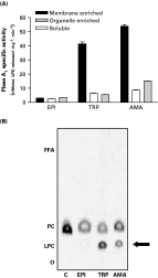
The distribution of Plase A1 in the different subcellular fractions obtained by differential centrifugation was analysed in the 3 parasite stages. As shown in Fig. 1A, Plase A1 was mainly detected in membrane-enriched fractions of the infective stages and the specific activities (nmoles of lysophosphatidylcholineċmin−1ċmg−1 protein±S.D., n=3) were approximately 20-fold higher in trypomastigotes (45·7±1·3) and amastigotes (55·4±0·9) than in epimastigotes (3·18±0·03). Furthermore, in parasite membranes obtained by biotinylation of surface proteins with subsequent affinity chromatography, Plase A1 activity was also detected, being remarkably higher in amastigotes and trypomastigotes (Fig. 1B). These results confirmed that Plase A1 activity was membrane associated in T. cruzi infective stages. This membrane associated activity hydrolysed 1-palmitoyl-2-[1-14C] palmitoyl-phosphatidylcholine and generated only lysophosphatidylcholine (LPC) with no release of labelled free fatty acid (FFA) (Fig 1B). Similar results were obtained when 1-palmitoyl-2-[1-14C] araquidonyl-phosphatidylethanolamine was assayed as substrate (data not shown), confirming the presence of a Plase A1 activity acting on zwitterionic phospholipids.

Fig. 1. Plase A1 activity is predominantly membrane-associated in Trypanosoma cruzi infective forms. (A) Plase A1 specific activity. Plase A1 activity was assayed in the following fractions: soluble (105000 g supernatant, □), organelle-enriched (15000 g pellet), and membrane-enriched (105000 g pellet, [squf ]) obtained by differential centrifugation of the 3 parasitic stages. Lipids from the samples were extracted and separated by TLC. Radioactivity was measured by liquid scintillation. Results are expressed as nmoles of LPCċmin−1ċmg−1 protein. Values represent means±S.D. of 3 independent experiments. (B) PC is efficiently hydrolysed by membrane associated Plase A1. This enzyme activity was assayed in parasite membrane fractions (obtained by biotinylation of surface proteins followed by affinity chromatography) from the 3 T. cruzi stages, using as substrate: 1-palmitoyl-2-[1-14C]oleoyl-phosphatidylcholine. Lipids from the samples were extracted and separated by TLC. Autoradiograms were scanned using the Storm® Gel and Blot Imaging System. The experiment was performed thrice with similar results. C: control, EPI: epimastigotes, TRP: trypomastigotes, AMA: amastigotes, O: origin, LPC: Lysophosphatidylcholine, PC: Phosphatidylcholine, FFA: Free fatty acids.
In the organelle-enriched fraction, Plase A1 activity was also higher in the infective stages compared to epimastigotes but significantly lower with respect to those found in membrane enriched fractions (Fig. 1A). In the soluble fractions, even when the lowest levels of Plase A1 activities for the 3 parasite stages were detected, they were also higher in the infective forms (Fig. 1A).
As reported for epimastigotes (Wainszelbaum et al. 2001), Plase A1 activity from T. cruzi infective stages proved to be independent of bivalent cations such as Ca2+, Mn2+ and Mg2+, with an optimum acidic pH (4–5) and to be activated in the presence of Triton X-100 (up to 1%). In amastigotes, the Plase A1 activity corresponding to the void volume fraction from Sephadex G-100 chromatograms described above, had a wider optimum pH, ranging from 4 to 7 (data not shown).
Plase A1 activity is secreted by T. cruzi infective stages
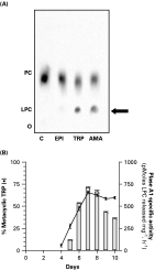
To determine if T. cruzi Plase A1 was also secreted to the extracellular media as it has been reported for other membrane-localized enzymes like cruzipain and trans-sialidase (Parussini et al. 1998; Gruppi et al. 1997), this activity was assayed in culture supernatants from the different parasite stages. The specific Plase A1 activities for amastigotes and trypomastigotes expressed as pmol of LPCċmin−1ċmg−1 protein±S.D. (n=3), were 450±3·5 and 850±4·7 respectively (Fig. 2A). By contrast, under the same conditions no significant Plase A1 activity was detected in epimastigote supernatants (Fig. 2A). To eliminate the existence of parasite lysis, other experimental approaches to determine parasite integrity were performed. Culture supernatants were tested by immunoblot using glutamate dehydrogenase as cytosolic marker. No reactivity was observed, indicating that the Plase A1 activity detected in these samples was due to parasite secretion (data not shown). Carboxyfluorescein diacetate staining of parasite suspensions clearly demonstrated the integrity of parasite cell membrane; viability was also indicated by parasite motility (data not shown).

Fig. 2. Plase A1 activity is secreted to the medium by Trypanosoma cruzi infective stages. (A) The different parasite stages (3×10⁸/ml per assay) were incubated in 199 medium+5% fetal bovine serum for 1 h, at 37 °C for amastigotes and trypomastigotes or at 28 °C for epimastigotes. Cells were then centrifuged and the different parasite culture supernatants obtained were used to determine Plase A1 activity. TLC autoradiograms are representative of 3 independent experiments, and the image was scanned using Storm® Gel and Blot Imaging System. C: control, EPI: epimastigotes, TRP: trypomastigotes, AMA: amastigotes, O: origin, LPC: Lysophosphatidylcholine, PC: Phospatidylcholine. (B) Secreted Plase A1 activity increases simultaneously with metacyclogenesis. Epimastigotes (5×10⁶/ml) were stimulated to differentiate by incubation with Triatoma infestans intestinal extract, for 15 min at 28 °C (day 0). The parasites were then transferred to modified Grace medium at 28 °C. Growth and differentiation curves were determined every day and Plase A1 activity was assayed in the metacyclic supernatants. The points represent the mean from three parallel experiments, with differences between samples being less than 15%. -[bull ]- percentage of metacyclic trypomatigotes, □ specific Plase A1 activity.
Considering that Plase A1 activity was secreted by the infective stages, we studied the release of the enzyme along with the differentiation process from the non-infective epimastigote into the infective metacyclic trypomastigote, with the purpose of applying this property as a stage marker. Plase A1 activity was detected from day 5 as morphogenesis progressed, the enzyme activity remarkably increased in metacyclic supernatants, reaching a peak on day 7 when maximal differentiation was taking place (Fig. 2B). In control supernatants (epimastigotes without stimulation to differentiate) no metacyclic forms were observed and no Plase A1 activity was detected (data not shown), in agreement with the above results demonstrating that epimastigotes do not secrete significant amounts of Plase A1 (Fig. 2A). Parasite integrity was confirmed as described above, remaining functionally and morphologically viable through the whole experiment (data not shown) and supporting that the detected activity was due to secreted Plase A1.
Vero cell lipid profile is modified by the interaction with T.cruzi infective stages, or purified T. cruzi Plase A1
Since Plase A1 was a membrane-associated activity and also secreted by the T. cruzi infective stages, we analysed the host cell lipid composition after interaction of the Vero cells with the different intact parasite stages. Previous infection assays on these cells determined that at least 1 h was required for a successful parasite invasion (RA strain) (Isola et al. unpublished observations). Therefore, in order to avoid this event the assays here described were performed between 10 and 30 min.
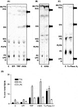
Vero cell lipid modifications were already detected after the first 10 min of parasite-host cell interaction (data not shown). The lipid profiles were remarkably modified when cells were exposed to intact amastigotes or trypomastigotes for 30 min, with significant generation of FFA, diacylglycerol (DG) and LPC (Fig. 3A). By contrast, epimastigotes failed to induce these changes (Fig. 3A). Charring of the plate revealed that the mass of LPC, DG and FFA increased concomitantly with labelling; simultaneously a diminishing in phosphatidylcholine (PC) mass was observed (Fig. 3B). When Vero cells were exposed to purified Trypanosoma cruzi Plase A1 (Tc-Plase A1) (Fig. 3C), the modifications induced on lipid profiles were similar to those obtained with the intact infective parasites (generation of FFA, DG and LPC) (Fig. 3A). The gas chromatography – mass spectrometry analysis of the fraction corresponding to the FFA released by the action of Tc-Plase A1, indicated that stearic and palmitic acids were the most abundant (47·6% and 42·4%, respectively), followed by myristic acid (10%), with respect to the total FFA present in these samples (data not shown).

Fig. 3. Vero cell lipid profile changes upon stimulation with intact Trypanosoma cruzi infective stages or purified parasite Plase A1. Vero cells (4×10⁶/plate) labelled with [14C] oleate were incubated for 30 min with intact parasites (A and B) or Tc-Plase A1 (C). The stimuli were removed and lipids separated by TLC. For mass determination (B), plates were then sprayed with 10% CuSO4 in 8% H3PO4, exposed to 150 °C for 13 min for charring. TLC plates are representative of 3 independent experiments. Images were scanned using Storm® Gel and Blot Imaging System. C: control, EPI: epimastigotes, TRP: trypomastigotes AMA: amastigotes, Tc-Plase A1: purified T. cruzi Plase A1, O: origin, LPC: lysophosphatidylcholine, PC: phosphatidylcholine, PE: phosphatidylethanolamine, PI/PS: phosphatidylinositol+phosphoserine, MG: monoacylglycerol, ST/DG: free sterols+diacylglycerol, FFA: free fatty acids, TG: triacylglycerol, SE: steryl esters. (D) Lipids spots from (A) and (C) plates were scraped off and radioactivity was measured by liquid scintillation. The bars represent the percentage of: (□) LPC, ( ) DG and ([squf ]) FFA, with respect to the total radioactivity detected for each stimulus (EPI, TRP, AMA and Tc-Plase A1). Values represent the mean±S.D. of 3 experiments independently.
It was noteworthy that no PC-Plase A1 activity was detected in Vero cells, supporting the concept that the modifications observed on these cells were mainly due to Tc-Plase A1 (data not shown). Quantitative analysis of the lipids expressed as a percentage over the total radioactivity showed a substantial increase in DG, FFA and LPC when Vero cells were exposed to intact T. cruzi infective stages as well as Tc-Plase A1, with respect to control and to epimastigotes (Fig. 3D).
Purified T. cruzi Plase A1 stimulates host cell PKC
The rise in cellular FFA, DG and LPC generated by Tc-Plase A1 on Vero cells described above, suggested that these lipids can act as second messengers and stimulate host cell protein kinase C (PKC) (Nishizuka, 1995; Prokazova et al. 1998). To ascertain this hypothesis, different experimental approaches were used i.e. detection of protein phosphorylation patterns in Vero cells by autoradiography and immunoblot analysis to determine serine phosphorylated proteins and PKC phosphorylation.
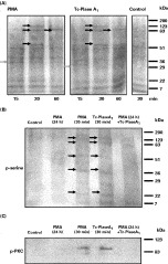
As indicated in Fig. 4A, phosphorylation patterns of [32P] Vero cells stimulated with Tc-Plase A1 (15–30 min) showed an enhancement in band labelling corresponding to 120, 85 and 55 kDa, similar to those observed with PMA (phorbol 12-myristate 13-acetate) (black arrows), a known PKC activator (Liu and Heckman, 1998). After 60 min of stimulation, a unique band of approximately 85 kDa remained phosphorylated in both Tc-Plase A1 and PMA stimulated cells. The only differences observed were 2 bands of approximately 30 kDa in the Tc-Plase A1 stimulated cells and of 35 kDa in the PMA treated cells, detected at 15 min (grey arrows). The control represents the basal phosphorylation pattern induced by the vehicle and no changes were observed at the different times assayed.

Fig. 4. Purified Trypanosoma cruzi Plase A1 activates host cell PKC(A) Protein phosphorylation patterns indicate that Vero cell PKC is activated upon stimulation with Tc-Plase A1. [32P] Vero cells were stimulated with: 100 nM PMA or 100 μg purified Tc-Plase A1 for 15, 30 or 60 min. Protein-phosphorylation patterns were analysed by SDS-PAGE and autoradiography. Control shows the basal phosphorylation profile. Black arrows indicate PKC phosphorylated proteins and grey arrows show the differences. SDS-PAGE autoradiograms are representative of two independent experiments. Images were scanned using Storm® Gel and Blot Imaging System. (B) Host cell PKC down regulation abrogates the phosphorylation profiles induced by Tc-Plase A1. Vero cell PKC was down regulated by exposure of Vero cells to 100 nM PMA (24 h) and then treated with 32 nM PMA or 100 μg Tc-Plase A1 for 30 min. Samples were analysed by immunoblot and revealed by chemiluminescence using an anti phospho-serine antibody with the Storm® Gel and Blot Imaging System. Images are representative of 3 independent experiments. Black arrows indicate PKC phosphorylated proteins. (C) Host cell PKC activation was determined by immunoblot using an anti phospho-PKC antibody. Vero cells were treated, processed and analysed by Immunoblot, as described in B. Activated PKC was detected using an anti phospho-PKC (pan) antibody. Images are representative of 2 independent experiments and were revealed by chemiluminescence as described above. PMA: phorbol 12-myristate 13-acetate, Tc-Plase A1: purified T. cruzi Plase A1, kDa: molecular weight in kiloDalton.
To confirm that the phosphorylation profiles were due to host cell PKC activation, this enzyme was down regulated by prolonged treatment of Vero cells with PMA (Fig. 4B, lane PMA 24 h). The blockade of host cell PKC abrogated the characteristic protein phosphorylation patterns. When these PKC down-regulated cells were stimulated with Tc-Plase A1 (Fig. 4B, lane PMA 24 h+Tc-Plase A1), the enzyme failed to induce the enhanced phosphorylation profiles observed in non-down-regulated cells (Fig. 4B, lanes PMA 30 min and Tc-Plase A1 30 min). These treated cells were also analyzed by immunoblot using an anti-phospho PKC (pan) antibody that recognizes exclusively phosphorylated PKC (Fig. 4C). Stimulation of Vero cells with Tc-Plase A1 (30 min) induced PKC phosphorylation, similar to that obtained with PMA (30 min), confirming that Tc-Plase A1 is involved in host cell PKC activation (Fig. 4C, lanes PMA 30 min and Tc-Plase A1 30 min). In contrast, in down-regulated cells (PMA 24 h) PKC phosphorylation was almost undetectable (Fig. 4C, lanes PMA 24 h and PMA 24 h+Tc-Plase A1).
DISCUSSION
Our previous studies on T. cruzi homogenates demonstrated a PC-Plase A1 activity that was up to 20-fold higher in amastigotes and trypomastigotes than in the non-infective epimastigotes (Wainszelbaum et al. 2001). In agreement, the higher levels of Plase A1 activity in the different subcellular fractions here analysed were always associated to the infective stages. It is noteworthy that in these stages, membrane-bound Plase A1 activity was remarkably higher, than the activities detected in organelle-bound or soluble fractions. In contrast, in T. brucei the major proportion of Plase A1 activity (more than 90% of the total) corresponds to a soluble cytosolic fraction, thus this localization does not appear to have a similar counterpart in T. cruzi (Opperdoes and van Roy, 1982). In epimastigotes, almost undetectable levels of membrane PC-Plase A1 activity were found in accordance with our previous results, where the main enzyme localization was lysosomal (Wainszelbaum et al. 2001). However, it must be mentioned that a membrane Plase A1 activity has been previously reported in epimastigotes, but acting only on anionic phospholipids such as inositolphospholipids and inositolphosphoceramides (Bertello et al. 2000). Phospholipases that have anionic phospholipids as substrates are different from those cleaving zwitterionic substrates (Van den Bosch, 1974). In this respect, T. cruzi PC-Plase A1 was able to hydrolyse zwitterionic phospholipids such as PC and PE and it appears to display a preference for physiological phospholipids containing sn-1-saturated acyl chains, mainly stearic and palmitic acid for the cell type here assayed.
Gel filtration analyses of parasite homogenate supernatants suggest that Plase A1 could be present as a particulated form associated with cell membranes. The detection in amastigotes in a second peak of activity suggests either the existence of at least 2 isoforms in this stage, or that Plase A1 could be eluted partially associated with membrane particles/substrate and as a free monomer in solution. Considering the low Plase A1 activity and the subcellular distribution in epimastigotes with respect to the infective stages, it is possible that the presence of Plase A1 isoforms are being differentially expressed in T. cruzi stages. In addition, its isoforms could process the substrate with different efficiency. The possible existence of Plase A1 isoforms is supported by the presence of at least 16 different genes encoding putative lipases in T. cruzi data base (www.tcruzidb.org); however, some of these genes could be haplotype variants. Most of these sequences present a high degree of similarity (70–80%) and their comparison with other eukaryotic Plases A1 demonstrates 30–50% homology. At present, we are performing further studies to elucidate the identity of the genes encoding for T. cruzi Plase A1.
The biochemical characteristics (optimal pH, ion dependence and effect of detergents) of Plase A1 activity from the infective stages were similar to those previously reported for epimastigote lysosomal Plase A1 (Wainszelbaum et al. 2001) and for other lysosomal phospholipases A1 already described (Hostetler et al. 1982; Pete et al. 1994). Only in amastigotes a wider range of optimal pH (4 to 7) was detected for the void volume fraction (Sephadex G-100 chromatograms), possibly related to its intracellular environmental requirements.
It is well known that parasite host-cell interaction could be modulated by the influence of secreted parasite enzymes (Burleigh and Woolsey, 2002). In T. cruzi, it has been reported that lysosomal enzymes like cruzipain, participate in the endocytic pathway, although they can also be localized in plasma membranes and be secreted to the extracellular media (Aparicio et al. 2004; Gruppi et al. 1997; Souto-Padron et al. 1990). In this regard, we also here demonstrate that Plase A1 is secreted by T. cruzi infective stages. Moreover, biochemical and electron microscopical data strongly suggest that most of T. cruzi surface antigens are released as plasma membrane vesicles (Gonçalves et al. 1991). Thus, as Plase A1 is mainly membrane localized in the infective stages, this mechanism could partially explain the release of the enzyme to the extracellular medium.
During the metacyclogenic process, in which epimastigotes differentiate into metacylic trypomastigotes, an increase in secreted Plase A1 activity was detected together with the appearance of metacyclic forms. In this respect, it has been reported in T. cruzi that membrane phospholipases A1, A2 and C, may act in remodelling reactions needed for plasma membrane transformation during cell differentiation (Salto et al. 2003). Therefore, detection of secreted Plase A1 could be used as a biochemical marker for T. cruzi infective stages.
In T. cruzi, as in many other successful intracellular parasites, the ability to modify or alter complex biochemical signalling pathways on the host cell plays a critical role (Wilkowsky et al. 2001; Burleigh and Woolsey, 2002). In this sense, we have previously shown that only membranes of the infective stages can activate the host cell phosphatidylinositol 3-kinase pathway and downstream protein kinase B (Akt) (Wilkowsky et al. 2001). In addition, they can also induce a Ca2+ signal (Tardieux et al. 1994; Wilkowsky et al. 1996) required for typical PKC isoform activation (Nishizuka, 1995; Liu and Heckman, 1998). Moreover, it has been reported that T. cruzi induces signals on macrophages that activate PKC with the consequent upregulation of parasite invasion (Villalta et al. 1999). Here, we determined that either intact infective parasites or purified Trypanosoma cruzi Plase A1 (Tc-Plase A1), triggered off, on Vero cells, the generation of DG, FFA and LPC. It is well known that these lipid second messengers activate host cell PKC (Nishizuka, 1995; Liu and Heckman, 1998) and in the case of LPC, this lysophospholipid can also prolong the effect of direct PKC activators (Prokazova et al. 1998). The rise in lipid second messengers observed in Vero cells stimulated with Tc-Plase A1 indicated host cell PKC activation since the phosphorylation patterns were similar to those observed with PMA, a known PKC activator (Hansra et al. 1999). In addition, Vero cell PKC down regulation abrogates phosphorylation induced by Tc-Plase A1. The comparison of Vero cell phosphorylation patterns induced by short stimulation with PMA or Tc-Plase A1 showed striking similarities, especially at the level of the bands corresponding to 120, 85 and 55 kDa. Some differences observed in the bands of the region of about 30–35 kDa may reflect the activation of different Vero cell PKC isoforms induced by the different stimuli used (Nishizuka, 1995; Liu and Heckman, 1998). Taking in consideration these findings, we propose that host cell PKC activation induced by Tc-Plase A1 could participate in the signalling events that modulate parasite invasion, a role for PKC that it has been already reported (Villalta et al. 1999).
As regards DG, the increase observed in stimulated Vero cells could be due to de novo DG biosynthesis induced by FFA, a fact that has been demonstrated in eukaryotic cells with consequent PKC activation (Yu et al. 2001).
On the other hand, the usefulness of lipid biosynthesis inhibitors (lysophospholipid analogues) as therapeutic agents has been demonstrated in T. cruzi (Lira et al. 2001). With respect to T. cruzi Plase A1, up to now there are no effective specific inhibitors. In previous work we found that only chlorpromazine has an inhibitory effect (Wainszelbaum et al. 2001), but at concentrations that induce cell toxicity (Tsao et al. 1982). Plase A1 appears as the only enzyme present in T. cruzi that hydrolyses PC. Meanwhile, in Vero cells, different PC degradation pathways have been described with the participation of a cytosolic and a lysosomal Plase A1 but with different substrate preference, particularly phosphatidylinositol (Suzuki and Matsumoto, 1978). This distinctive biochemical characteristic could provide novel targets for the development of chemotherapeutic agents.
Collectively, these results describe a novel role for Plase A1 from T. cruzi infective stages, since this enzyme contributes to lipid modifications on host cells and concomitantly activates PKC signalling pathway. The understanding of these early complex mechanisms triggered off during T. cruzi-host cell interaction could provide original targets for the development of chemotherapeutic agents.
We thank Dr J. J. Cazzulo for the generous gift of the anti-glutamate dehydrogenase antibody, Dr V. Duschak and Dr C. Alba Soto for their technical support and Dr M. Jacobsen for her helpful criticisms of the manuscript. This work was supported by grants from the Universidad de Buenos Aires (UBA), Agencia Nacional de Promoción Científica y Tecnológica (FONCYT), Consejo Nacional de Investigaciones Científicas y Técnicas (CONICET) and UNDP/World Bank/WHO Special Programme for Research and Training in Tropical Diseases (TDR).