Introduction
Nematodes show a very broad spectrum of feeding habits and/or relationships with other living beings. Among them, a good number of species are regarded as active entomopathogenic forms, even being used and commercialized for biological control of insect species causing plant diseases. Spiders have not received much attention as hosts of parasitic/parasitoid nematodes. Nevertheless, several rhabditid taxa have already been recorded in association with this kind of chelicerate arthropod, for instance steinermatid (Poinar, Reference Poinar and Nentwig1987), panagrolaimid (Pizzi & Carta, Reference Pizzi and Carta2003, Pizzi, Reference Pizzi2009, Wyrobist et al., Reference Wyrobist, Kowal, Kornaś, Basiaga, Nowik, Pałasz and Nosal2015) and heterorhabditid (Poinar, Reference Poinar and Nentwig1987) species, as well other unidentified forms (Platnick & Sedgwick, Reference Platnick and Sedgwick1984).
The family Brevibuccidae Paramonov, 1956 is a rare (very infrequent) nematode taxon of the superfamily Panagrolaimoidea Thorne, 1937. Its representatives are characterized and distinguishable from other close families by, among other features, their post-equatorial vulva. Until now, this family included three genera that differ in the lip-region morphology and vulva position. Actually, the vulva–anus distance is shorter than tail length in Brevibucca Goodey, 1935 and Cuticonema Sanwal, 1959 but longer in Plectonchus Fuchs, 1930. Besides, Brevibucca and Plectonchus have reduced lips whereas Cuticonema bears flap-like lips. The three genera lack a post-vulval sac. Members of Brevibuccidae occur mainly in tunnels of bark beetles or compost (Andrássy, Reference Andrássy1984, Reference Andrássy, Csuzdi and Mahunka2005), but they have never been reported in association with spiders.
Numerous nematode specimens collected from the oral opening of a tarantula spider were sent to the authors for their examination and identification. The subsequent study revealed that they belonged to a non-described genus and species of Brevibuccidae. This contribution aims to characterize these specimens.
Materials and methods
Nematode extraction and processing
Nematodes were submitted by Mr Maciej Szudarski, who recovered them directly from the oral opening of a subadult male of the tarantula Chromatopelma cyaneopubescens (Theraphosidae), originating from Venezuela, and bred in captivity in Poland. Specimens were first fixed in 70% ethanol, sent to the authors, then fixed in 4% formalin, transferred to pure glycerine following Siddiqi's technique (Siddiqi, Reference Siddiqi1964), and mounted on permanent glass slides.
Light microscopy (LM)
Measurements were taken on specimens mounted in permanent slides. Drawings were made using a drawing tube (camera lucida) attached to a Leitz Laborlux S microscope (Leitz, Wetzlar, Germany). Pictures were taken with a Nikon Eclipse 80i light microscope provided with differential interference contrast optics (DIC) and a Nikon Digital Sight DS-U1 camera (Nikon, Tokyo, Japan). Micrographs were edited using Adobe® Photoshop® CS.
Scanning electron microscopy (SEM)
Specimens preserved in glycerine were selected for observation under SEM according to Abolafia (Reference Abolafia2015). The nematodes were hydrated in distilled water, dehydrated in a graded ethanol–acetone series, critical-point dried, coated with gold, and observed with a Zeiss Merlin microscope (5 kV) (Zeiss, Oberkochen, Germany).
The terminology used for the morphology of stoma and spicules follows the proposals by De Ley et al. (Reference De Ley, van de Velde, Mounport, Baujard and Coomans1995) and Abolafia & Peña-Santiago (Reference Abolafia and Peña-Santiago2006), respectively.
DNA extraction, PCR and sequencing
DNA was extracted from single individuals preserved in ethanol using the proteinase K protocol. Nematode material was cut in small pieces using a sterilized needle on a clean slide with a small drop of AE buffer (10 mm Tris-Cl + 0.5 mm EDTA; pH 9.0), transferred to a microtube containing 18 μl AE buffer and 2 μl proteinase K (600 μg ml−1) (Roche, Basel, Switzerland), and stored at −80°C within 15 min (for several days). The microtubes were incubated at 65°C (1 h), then at 95°C (15 min) and finally at 80°C (15 min) (Castillo et al., Reference Castillo, Vovlas, Subbotin and Troccoli2003). The microtube was centrifuged at 13,000 rpm for 3 min and 2 μl of extracted DNA was transferred to a microtube containing: 2.5 μl 10 × polymerase chain reaction (PCR) reaction buffer, 5 μl 5 × Q-solution, 0.5 μl deoxyribonucleoside triphosphates (dNTPs) (10 mm each), 1 μl of each primer (10 mm), 0.2 μl Taq DNA polymerase (Qiagen, Venlo, The Netherlands) and double-distilled water (ddH2O) to a final volume of 25 μl. The primers used for amplification of the D2–D3 region of the 28S rRNA gene were D2A (5′-ACAAGTACCGTGAGGGAAAGTTG-3′) and D3B (5′-TCGGAAGGAACCAGCTACTA-3′) (De Ley et al., Reference De Ley, Felix, Frisse, Nadler, Sternberg and Thomas1999). PCR cycle conditions were as follows: one cycle of 94°C for 3 min; followed by 35 cycles of 94°C for 1 min + annealing temperature of 55°C for 45 s + 72°C for 2 min; and finally one cycle of 72°C for 10 min. After DNA amplification, 5 μl of product was loaded on a 1% agarose gel in 0.5% Tris–acetate–EDTA (40 mm Tris, 20 mm glacial acetic acid and 2 mm EDTA; pH = 8) to verify the amplification using an electrophoresis system (Labnet Gel XL Ultra V-2. Progen Scientific, London, UK). The bands were stained with RedSafe (20,000×) previously added to the agarose gel solution, and visualized and photographed under a UV transilluminator (Vilbert Lourmat E-Box-VX2/20MX, Thermo Fisher Scientific, Waltham, Massachusetts, USA). The product was stored at −20°C prior to sequencing. PCR products were purified using the QIAquick PCR purification kit (Qiagen), quantified using a spectrophotometer (Synergy HT, BioTek, Winooski, Vermont, USA) and used for direct sequencing in both directions using the primers referred to above. The sequencing reactions were performed at ‘Centro de Instrumentación Científico-Técnica (CICT)’ of the University of Jaén (Spain) using an Applied Biosystems Hitachi 3500 Genetic Analyzer (Applied Biosystems, Waltham, Massachusetts, USA). The sequence obtained (535 bp) was submitted to the GenBank database under accession number MF177710.
Phylogenetic analyses
The newly obtained sequence was aligned with other D2–D3 expansion segments of the 28S rRNA gene sequences available in GenBank using ClustalX alignment in the program MEGA7 (Kumar et al., Reference Kumar, Stecher and Tamura2016). Outgroup taxa were chosen according to the results of previously published data (Lewis et al., Reference Lewis, Dyal, Hilburn, Weitz, Liau, LaMunyon and Denver2009). Sequence alignments were edited manually using BioEdit 7.2. (Hall, Reference Hall1999). The sequence dataset was analysed with Bayesian inference (BI) using MrBayes 3.2.6 (Huelsenbeck & Ronquist, Reference Huelsenbeck and Ronquist2001; Ronquist & Huelsenbeck, Reference Ronquist and Huelsenbeck2003), respectively. The analysis under GTR model was initiated with a random starting tree and run with the Markov chain Monte Carlo (MCMC) for 106 generations. For phylogenetic relationships, analyses were based on 28S rDNA. Zeldia punctata (DQ145664) was used as the outgroup. The tree was visualized and saved with the program FigTree v1.4.3 (http://tree.bio.ed.ac.uk/software/figtree/).
Descriptions
Family Brevibuccidae Paramonov, 1956
Diagnosis (emended)
Panagrolaimoidea. Small body, up to 2.5 mm long, cuticle poorly annulated, lateral field inconspicuous, lip region with six separated lips, panagrolaimoid stoma with well-developed gymnostom having refringent rhabdia, panagrolaimoid pharynx with subcylindrical pharyngeal corpus and isthmus variable in length, female reproductive system monodelphic–prodelphic, ovary extending anterior or posterior to vulva level, post-vulval sac present or absent, female tail conical or conical–elongate, male tail conical with a long mucro, spicules curved or bent ventrad with rounded manubrium, and thick gubernaculum.
Tarantobelus gen. n.
Diagnosis
Brevibuccidae. Small body, up to 1.0 mm long, cuticle nearly smooth, lateral field inconspicuous, lip region with six separated lips, each bearing a small cuticular flap topping it, panagrolaimoid stoma with well-developed gymnostom having broad and refringent rhabdia, panagrolaimoid pharynx with isthmus slightly longer than basal bulb, excretory pore situated at level of isthmus, female reproductive system monodelphic–prodelphic, length of post-vulval sac less than the corresponding body diameter, vulva distinctly protruding, female tail conical with acute tip, male tail conical with a long and thin mucro, spicules curved ventrad with rounded manubrium, and thick gubernaculum.
Taxonomic summary
Type and only species
Tarantobelus arachnicida gen. n., sp. n.
Etymology
The generic name is composed by ‘taranto’ (from tarantula, this from the Medieval Latin ‘tarantola’, the wolf spider Lycosa tarantula, described originally from the Italian city of Taranto, which gave name to the true tarantulas) and the genus (Panagro)belus.
Relationships
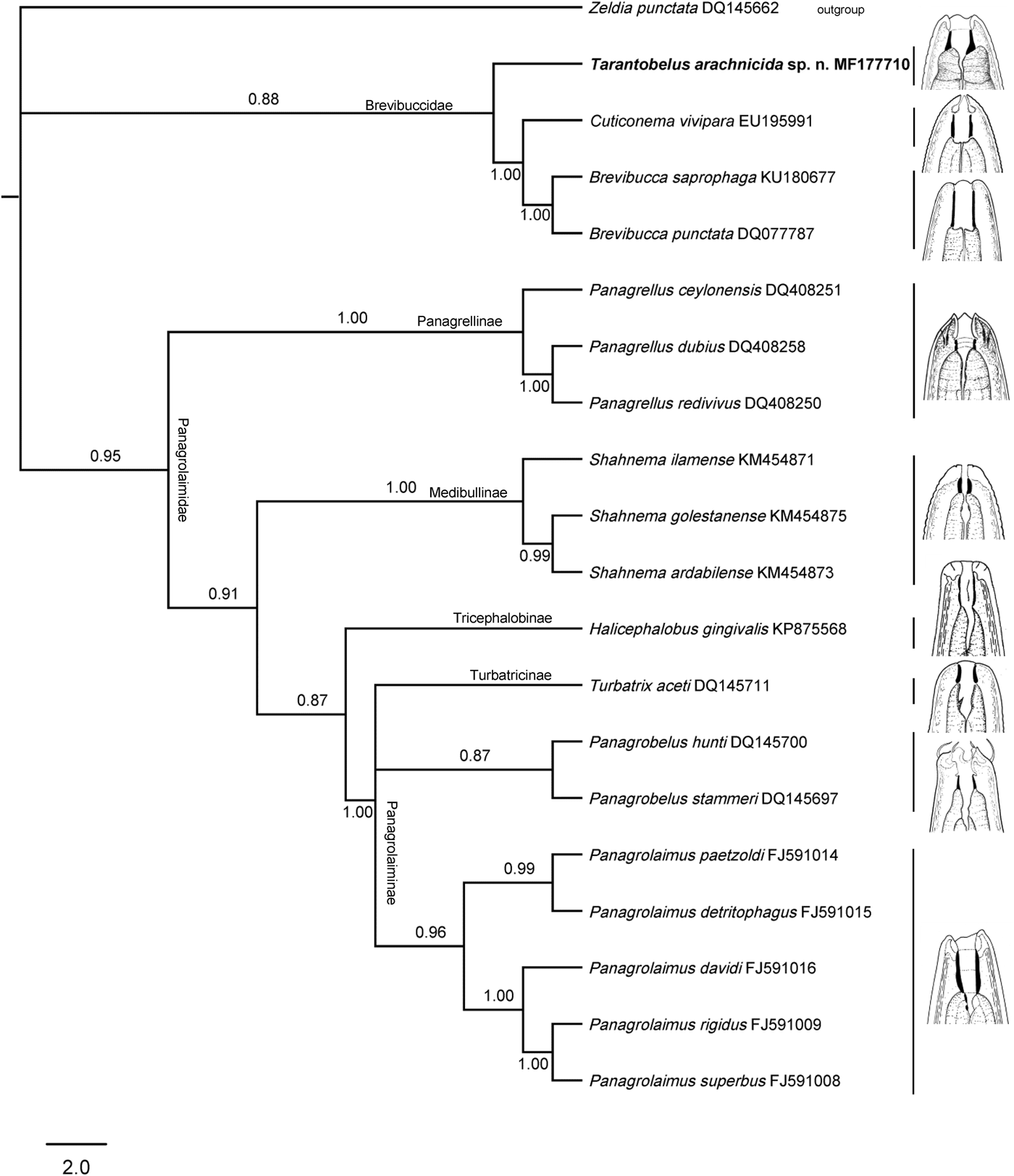
The new genus much resembles the other members of the family, namely the genera Brevibucca, Cuticonema and Plectonchus, all of them having a post-equatorial vulva position. Nonetheless, it differs morphologically from them in its shorter isthmus (as long as vs. longer than basal bulb), more posterior location of the excretory pore (at isthmus level vs. at corpus level) and presence (vs. absence) of post-vulval sac. (These morphological differences are supported by molecular analyses, as shown in fig. 6.) Besides, Tarantobelus gen. n. can be distinguished from Brevibucca in its smaller gymnorhabdia (c. one-third vs. one-half of the total length of stoma) and larger vulva–anus distance (more vs. less than tail length); from Cuticonema in the morphology of lips (low conoid vs. flap-like) and larger vulva–anus distance (more vs. less than tail length); and from Plectonchus by having a more posterior location of the excretory pore (at isthmus level vs. at pharyngeal corpus), post-vulval sac present (vs. absent) and ovary extending beyond the vulva (vs. not extending beyond the vulva).
Remarks
With a cladistic evolutionary approach, the new genus might be considered the most primitive member of the family Brevibuccidae. Thus, the position of the excretory pore (posterior, at level of the pharyngeal isthmus) and the presence of a post-vulval sac should be interpreted as plesiomorphic conditions of their respective characters. The anterior position of the excretory pore (at the level of the pharyngeal metacorpus) as well the absence of a post-vulval sac, are the corresponding apomorphic conditions of these characters, which are shared by the remaining three genera of the family, namely Brevibucca, Cuticonema and Plectonchus. On the other hand, Brevibucca and Cuticonema might share a second synapomorphy regarding the vulva position at a short distance (less than tail length) from the anus, while in Plectonchus and Tarantobelus gen. n. the vulva is at a long distance (more than tail length) from the anus (plesiomorphic condition).
The results of the molecular analysis (see fig. 6) provisionally support the morphological observations. Nevertheless, more sequences of other representatives of the family should be studied to confirm these findings.
Tarantobelus arachnicida gen. n., sp. n.
Material examined
Thirty-one females and 26 males; in general, in acceptable condition.
Measurements
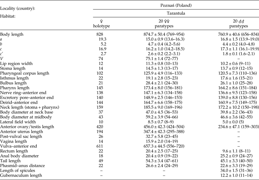
See table 1.
Morphometrics of Tarantobelus arachnicida gen. n., sp. n. Measurements in μm and in the form mean ± standard deviation (range) where appropriate.

Demanian indices (de Man, Reference de Man1880): a = body length/body diameter; b = body length/pharynx length; c = body length/tail length; c' = tail length/anal body diameter; V = (distance from anterior region to vulva/body length) × 100.
Description
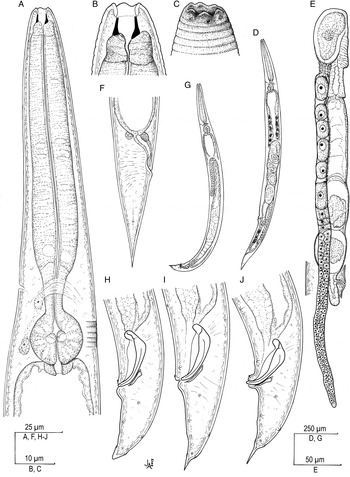
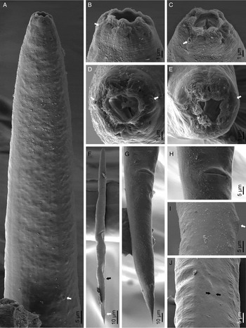
Adult (figs 1–5). Stout nematodes (a = 14–19) of small size, body 0.66–0.95 mm long. Upon fixation, habitus nearly straight or slightly curved ventrad in females, and visibly more curved ventrad in the posterior region (J-shaped) in males. Cuticle bearing very weak transverse incisures, only perceptible with SEM at anterior region (fig. 3B, C); annuli 2 μm wide. Lateral field inconspicuous, hardly appearing as a shallow longitudinal band. Lip region continuous with the adjacent body, consisting of six moderately separated lips, each bearing a small flap topping it; labial and cephalic papillae buttom-like, not especially protruding; oral aperture triangular when observed in frontal view; amphidial aperture oval, slightly oblique. Stoma typical panagrolaimoid: cheilostom short, lacking refringent rhabdia; gymnostom well developed, hardly longer than broad, slightly widened at its posterior part, and with distinct and refringent rhabdia; stegostom short, funnel-like and with poorly refringent rhabdia. Pharynx also typical panagrolaimoid: corpus 5.6–8.3 times longer than isthmus, robust, cylindrical, slightly widening at its posterior part, but pro- and metacorpus are not easily distinguishable; isthmus nearly as long as the basal bulb, and comparatively broad; basal bulb spheroid. Cardia short, conoid, surrounded by intestinal tissue. Nerve ring located at 74–81% of neck length from the anterior end, surrounding the isthmus. Excretory pore at isthmus level too, at 73–86% of neck length. Deirid somewhat inconspicuous, situated at level of basal bulb, at 82–93% of neck length. Intestine with cardiac (anterior) and rectal (posterior) regions made of thin walls.
Tarantobelus arachnicida gen. n., sp. n.: (A) neck; (B) stoma; (C) lip region; (D) entire female; (E) female reproductive system; (F) female posterior end; (G) entire male; (H–J) male posterior end.

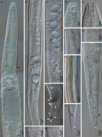
Tarantobelus arachnicida gen. n., sp. n. (light microscope): (A) neck (arrow pointing at the excretory pore); (B, C) female reproductive system; (D, E) anterior end at lip and stoma levels, respectively; (F, G) needle crystal packs and lipid droplets; (H) testis; (I, K) female posterior end in lateral and ventral views, respectively (white arrow pointing at the phasmid, black arrow pointing at the anus); (J) male posterior end (white arrows pointing at the genital papillae, black arrow pointing at the phasmid); (L) vulva, ventral view; (M) vaginal musculature.

Tarantobelus arachnicida gen. n., sp. n. (scanning electron microscope): (A) anterior end (arrow pointing at deirid); (B–D) female lip region in ventral, right subventral and frontal views, respectively (arrows pointing at the amphids); (E) male lip region in frontal view (arrow pointing at the phasmid); (F) entire female (black arrow pointing at the vulva, white arrow pointing at the anus); (G) female posterior half in ventral view; (H) vulva; (I) vulval region, lateral view (arrow); (J) lateral field (between arrows).

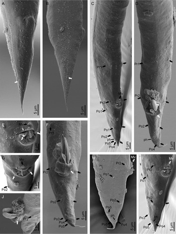
Tarantobelus arachnicida gen. n., sp. n. (scanning electron microscope): (A, B) female posterior end in subventral and right lateral views, respectively (arrows pointing at the phasmid); (C, D, F, H–J) male posterior end in subventral (C, I), ventral (D, F) and lateral (H, J) views (black and grey arrows pointing at the genital papillae, white arrow pointing at the gubernaculum in D, F, J); (E, G) cloacal opening (white arrows pointing at the circum-cloacal papillae-like processes, black arrows pointing at the genital papillae); Pr, pre-cloacal papilla; Po, post-cloacal papilla; Mv, mid-ventral papilla; ph, phasmid.

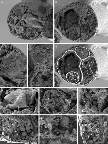
Tarantobelus arachnicida gen. n., sp. n. (scanning electron microscope, female): (A, B, E) transverse section at mid-body length in anterior (A) and posterior (B, E), views (int, intestine; lar, larva; lu, intestine lumen; ov, ovary; ut, uterus); (C) detail of an oocyte in the ovary; (D) ovary–uterus–intestine junction; (F) uterine egg; (G) larval intestine (arrow); (H–K) intestinal cells with lipid droplets (black arrows) and needle crystals (white arrows).

Female. Intestinal cells including special bodies appearing as small packs of needle-like crystals (fig. 2 F, G). Reproductive system monodelphic–prodelphic, dextral position in respect of the intestine; ovary straight; oviduct somewhat longer than the corresponding body diameter, made of prismatic cells; uterus c. six times the length of the corresponding body diameter, tubular, containing uterine eggs 25–62 × 26–67 μm in different developmental stages, occasionally with emerged larvae inside the female body (endotokia matricida); post-vulval sac 0.4–0.8 times the length of the corresponding body diameter, with very thick walls; vagina extending inwards to 21–33% of the body, its walls weakly refractive; vulva a transverse slit occupying more than one-half of the body diameter, distinctly protruding. Rectum 0.8–1.3 times the length of the anal body width, visibly sigmoid when observed in lateral view, and with three rectal glands. Posterior lip of anus often more prominent than the anterior one (fig. 4A, B). Tail conical with acute tip; phasmids situated at 58–62% of tail length.
Male. Reproductive system monorchic, testis ventrad, reflexed anteriorly. Cloacal opening with circum-cloacal papillae-like processes (fig. 4E, G). Genital papillae arranged as follows: two reduced pre-cloacal pairs (Pr), one mid-ventral (Mv) situated in front of the cloacal aperture, and five post-cloacal pairs (Po) of which one is lateral anterior (Po1), two subventral (one about the middle of the tail, Po2, and another almost terminal, Po4) and two subdorsal (one in the posterior half of tail, Po3, and another nearly terminal, Po5). Spicules curved ventrad, having rounded manubrium, short calamus, ventrad curved lamina with small dorsal hump, well-developed ventral velum, and finely rounded tip. Gubernaculum thick, almost straight, with small conical manubrium and fine acute tip. Tail conical, slightly curved ventral, especially at its posterior part, ending in a variably (usually well-) developed thin mucro, which exceptionally may be absent; phasmids very posterior, close the most posterior genital papillae.
Diagnosis
The new species is characterized by its 0.66–0.95 mm body length, cuticle with very fine transverse striation, inconspicuous lateral field, lip region 9–13 μm broad and consisting of six lips separated at each end with a small cuticular flap, stoma 12–17 μm long with gymnostom made of strong and refringent rhabdia, neck 150–198 μm long and isthmus slightly longer than basal bulb, intestine with differentiated cardiac and rectal regions, mature females bearing needle-like crystalline structures inside the intestinal cells, female reproductive system monodelphic–prodelphic, post-vulval sac 0.4–0.8 times the length of the corresponding body diameter, V = 72–77, vulva very prominent, female tail conical with acute tip (47–61 μm, c = 14–19, c′ = 2.2–3.1), male tail also conical (40–50 μm, c = 16–20, c′ = 1.6–2.1) but bearing a terminal mucro, and spicules 31–36 μm long.
Taxonomic summary
Etymology
The specific name, composed by the ancient Greek ‘arakhne’ (= spider) and the Latin suffix ’-cida’ (= killer), refers to the fact that this species kills spiders.
Type locality and habitat
Specimens collected from the oral opening of captive C. cyaneopubescens (Arachnida, Theraphosidae) imported from Venezuela and bred in Poznań (Poland). Tarantula specimens infected with this nematode stop feeding, they go into a state of lethargy and finally they die. It is unknown whether these aetiological symptoms are due to direct causes of this nematode (impeding their feeding or damaging some organ) or to indirect factors (inoculating bacteria or some toxic substance).
Type material
Thirty females (holotype and paratypes) and 25 males (paratypes) deposited in the Departamento de Biología Animal, Biología Vegetal y Ecología, Universidad de Jaén, Spain. One female and one male (paratypes) deposited in the nematode collection of the Swedish Museum of Natural History, Stockholm (Sweden).
On the taxonomy and systematics of the superfamily Panagrolaimoidea
Figure 6 shows the evolutionary relationships between the members of the superfamily Panagrolaimoidea, including representatives of its two families, Brevibuccidae and Panagrolaimidae, nine genera that have hitherto been sequenced and Z. punctata (Cephaloboidea, Cephalobidae) as the outgroup. The two panagrolaimoid families are well defined and present good molecular support. Their main morphological difference is the location of the vulva, equatorial in Panagrolaimidae and post-equatorial in Brevibuccidae, the latter hence probably representing the apomorphic condition of the character. The relationships between the four brevibuccid genera have already been discussed (see the remarks on the new genus, above).
The Bayesian tree inferred from known and the newly sequenced Tarantobelus arachnicida gen. n., sp. n. based on sequences of the 28S rDNA region.

Within Panagrolaimidae, the most primitive genus might be Panagrellus Thorne, 1938, a member of the subfamily Panagrellinae Andrássy, 1976, which also includes Baujardia Bert, Tandingan De Ley, van Driessche, Segers & De Ley, 2003, both very similar taxa (Bert et al., Reference Bert, Tandingan De Ley, Van Driessche, Segers and De Ley2003; Abolafia et al., Reference Abolafia, Alizadeh and Khakvar2016) that are characterized by their very short gymnorhabdia (possible apomorphic condition) and long post-vulval sac (probably plesiomorphic). Meiostoma Andrássy, 2007, currently classified under Panagrellinae, is a poorly characterized taxon, needing further studies to confirm its relationships.
Shahnema Siddiqi, 2014, the only member of the subfamily Shahnematinae Siddiqi, 2014 (see Abolafia & Peña-Santiago, Reference Abolafia and Peña-Santiago2016), constitutes a solidly supported clade. From a morphological perspective, this genus is characterized by its specially differentiated metacorpus (rounded and valvate) and non-valvate basal bulb, probably two apomorphic conditions within the superfamily. Besides, Shahnema shows reduced gymnorhabdia and short post-vulval sac. Medibulla Siddiqi, 1993 (Cephaloboidea, Osstellidae Heyns, 1962, Medibullinae Siddiqi, 1993) is very similar to Shahnema, both having in common the mentioned apomorphies as well their large amphids with aperture slightly smaller than the lip region width, and short gymnorhabdia. Besides, Medibulla lacks a sac-like spermatheca, as it occurs in panagrolaimoid taxa. All this evidence suggests that, provisionally, Medibulla should be classified together with Shahnema under the same subfamily, Medibullinae, Shahnematinae becoming its junior synonym, until additional data are available.
The remaining subfamilies and genera of Panagrolaimidae form a definite clade, although its support is not very high and the relationships between its components are more difficult to elucidate. This group is characterized morphologically by having well-developed gymnorhabdia and a short post-vulval sac, both probably apomorphic conditions. The subfamily Tricephalobinae, represented in the tree by the genus Halicephalobus Timm, 1956, but also including Tricephalobus Steiner, 1936, is characterized by having a swollen metacorpus (apomorphic). A reasonable doubt persists concerning the true identity of the genus Indocephalobus Mondal & Manna, Reference Mondal and Manna2014. It was classified originally under Tricephalobinae (Mondal & Manna, Reference Mondal and Manna2014), probably due to its swollen metacorpus, but several relevant features suggest that it might be identical to Diplogastrellus (Diplogasteromorpha). Actually, its stoma bears a dorsal tooth (not illustrated in the line drawing but distinct in LM pictures), the pharyngeal basal bulb lacks valves according to LM pictures (their presence, however, is mentioned in the description), and the vulva is situated very close to the anus. Besides, and taking into account that the spicules are thin and long, and the gubernaculum sigmoid, the type species of the genus, I. zebrae Mondal & Manna, Reference Mondal and Manna2014, does not differ from D. gracilis (Bütschli, Reference Khan, Bajaj, Sultana and Tahseen1876) Paramonov, 1952 (see Timm, Reference Timm1961; Khan et al., Reference Khan, Bajaj, Sultana and Tahseen2008) in any relevant character. According to this, Indocephalobus is considered a junior synonym of Diplogastrellus and I. zebrae as a junior synonym of D. gracilis.
The relationships between Turbatrix Peters, 1927, Panagrobelus Thorne, 1939 and Panagrolaimus Fuchs, 1930 are not well resolved in the molecular tree. Turbatrix, the only member of the subfamily Turbatricinae, is distinguished by its reduced cheilostom, well-developed and strongly sclerotized gymnostom, and stegostom with a metastomatal setose dorsal tooth (possibly apomorphic characters), meanwhile Panagrobelus and Panagrolaimus, both members of Panagrolaiminae, are distinguished by their well-developed cheilostom (plesiomorphic), very conspicuous gymnorhabdia (probably apomorphic), and stegostom with small metastomatal tooth-like projections (probably plesiomorphic). The relationships of the remaining genera classified under Panagrolaiminae and not yet sequenced, namely Panagrobelium Andrássy, 1984, Procephalobus Steiner, 1934 and Propanagrolaimus Andrássy, 2005, cannot be discussed in depth.
The superfamily Panagrolaimoidea hence includes 17 genera, grouped in two families (Brevibuccidae and Panagrolaimidae), listed below and summarized in the key:
Brevibuccidae Paramonov, 1956
Brevibucca Goodey, 1935
Cuticonema Sanwal, 1959
Plectonchus Fuchs, 1930
= Anguilluloides Rühm, 1956
Tarantobelus gen. n.
Panagrolaimidae Thorne, 1937
= Shahnematidae Siddiqi, 2014
Baujardia Bert, Tandingan De Ley, van Driessche, Segers & De Ley, 2003
Halicephalobus Timm, 1956
= Micronema Körner, 1954, nec Bleeker (Reference Bleeker1858)
= Phytorhabditis Lordello & De Oliveira, 1963
Medibulla Siddiqi, 1993
Meiostoma Andrássy, 2007
= Brevistoma Mukhina, 1981, nec Tjeder (Reference Tjeder1967)
= Mukhina Ozdikmen, 2010
Panagrellus Thorne, 1938
= Turbator Goodey, 1943
= Tylorhabdus Sukul, 1971
Panagrobelium Andrássy, 1984
Panagrobelus Thorne, 1939
Panagrolaimus Fuchs, 1930
= Cephalobus (Neocephalobus Steiner, 1929)
= Neocephalobus Steiner, 1929
= Pseudorhabditis Kreiss, 1929, nec Perroncito (Reference Perroncito1880)
= Asymmetricus Kreis, 1930
= Panagrodontus Thorne, 1937
= Panagrolaimoides Ivanova, 1958
= Panagromacra Massey, 1964
= Eminensia Mahajan, 1979
Procephalobus Steiner, 1934
Propanagrolaimus Andrássy, 2005
Shahnema Siddiqi, 2014
= Baldwinema Atighi, Pourjam, Bert, Pedram, Ghaemi & Panahandeh, 2015
Tricephalobus Steiner, 1936
= Trilabiatus Goodey, 1963
Turbatrix Peters, 1927
Key to genera identification

Acknowledgements
The authors thank the Polish spider breeder Maciej Szudarski for providing the material examined, and Alba N. Ruiz-Cuenca for assisting with the DNA extraction. SEM pictures and DNA sequencing were obtained with the assistance of technical staff and equipment of ‘Centro de Instrumentación Científico-Técnica (CICT)’, University of Jaén.
Financial support
The present work was supported financially by the ‘University of Jaén/Fundación Caja Rural Jaén’ for the project entitled ‘Filogeografía de nematodos rabdítidos (Nematoda, Rhabditida) en ambientes xerofíticos del sur de la península ibérica’ (UJA2014/03/01).
Conflict of interest
None.