Introduction
Recent evidence has indicated that the functional activity in white matter (WM) reflected in blood oxygenation level-dependent (BOLD) signals is related to physiological activity in WM fiber tracts (Gawryluk, Mazerolle, & D'Arcy, Reference Gawryluk, Mazerolle and D'Arcy2014; Peer, Nitzan, Bick, Levin, & Arzyt, Reference Peer, Nitzan, Bick, Levin and Arzyt2017; Wu et al., Reference Wu, Wang, Li, Schilling, Gao, Anderson and Gore2019), and thus may provide a useful biomarker for WM function. This provides a new opportunity to investigate how microstructural alterations in fiber tracts are related to their function, and how alteration in the function of fiber tracts is related to gray matter (GM) function and WM fiber tract integrity.
Whether alterations of the functional activity of WM contribute to the pathophysiology of schizophrenia is yet unclear, as is its relation to GM function and WM fiber integrity (Moskowitz & Heim, Reference Moskowitz and Heim2011). Multiple prior studies document structural alterations in WM integrity in schizophrenia utilizing diffusion tensor imaging (DTI) (Cetin-Karayumak et al., Reference Cetin-Karayumak, Di Biase, Chunga, Reid, Somes, Lyall and Kubicki2019; Meng et al., Reference Meng, Li, Li, Xiao, Lui, Sweeney and Gong2019; Xiao et al., Reference Xiao, Sun, Shi, Jiang, Tao, Zhao and Lui2018). Most previous studies included chronically ill patients previously treated with antipsychotic medication for years, a factor which has been suggested to exert effects on brain function and both GM and WM structure (Bartzokis et al., Reference Bartzokis, Lu, Nuechterlein, Gitlin, Doi, Edwards and Mintz2008; Meng et al., Reference Meng, Li, Li, Xiao, Lui, Sweeney and Gong2019; Xiao et al., Reference Xiao, Sun, Shi, Jiang, Tao, Zhao and Lui2018).
Mazerolle et al. (Reference Mazerolle, Beyea, Gawryluk, Brewer, Bowen and D'Arcy2010) and Ding et al. (Reference Ding, Huang, Bailey, Gao, Cutting, Rogers and Gore2018) reported that GM and WM functional networks were activated simultaneously in healthy participants. Altered GM–WM functional connectivity has been reported recently in schizophrenia patients who were taking antipsychotic medication (Jiang et al., Reference Jiang, Luo, Li, Li, Yang, Li and Yao2019) and suggested its foundational value of disruptions in brain connections.
The present cross-sectional study acquired resting-state functional magnetic resonance imaging, DTI, and high-resolution T1-weighted images in a large sample of antipsychotic-naïve schizophrenia patients and healthy comparison subjects. Our aim was to test for alterations in BOLD signals in WM tracts, and their temporal synchrony with BOLD signals in GM regions and their relation to fiber tract integrity assessed by using DTI. We also investigated relations between these brain measures and current illness symptom severity and duration of untreated psychosis (DUP).
Methods and materials
Participants
This investigation included 153 antipsychotic-naïve schizophrenia patients recruited from the Psychiatric Outpatient Clinic or the Mental Health Screening Program of West China Hospital. The Nottingham Onset Schedule was used to evaluate DUP based on information provided by patients, family members, and medical records (Norman & Malla, Reference Norman and Malla2001). Clinical diagnoses were established using the Structured Interview for DSM-IV Axis I Disorders (SCID), and the Positive and Negative Syndrome Scale (PANSS) was used to assess current symptom severity. Healthy comparison subjects (n = 153) were recruited through advertisements and assessed using the non-patient version of the SCID to confirm lifetime absence of psychotic, mood, and substance use disorders. Healthy comparison subjects with a known history of a psychiatric disorder in their first- or second-degree relatives were excluded. Additional exclusion criteria in both groups included a history of traumatic brain injury, or significant systemic or neurological disorder. All participants were right-handed and of Han ancestry (Table 1). This investigation was approved by the research ethics committee of West China Hospital of Sichuan University. Written informed consent was obtained from all study participants.
Demographic and clinical characteristics of antipsychotic-naïve schizophrenia patients and healthy comparison subjects

MRI data acquisition
All MRI data were collected on the same 3-T scanner (EXCITE, General Electric, Milwaukee). Padding within the head coil limited subject's head motion and earplugs were provided. During scanning, subjects were instructed to relax with eyes closed, without falling asleep or systematic thinking (confirmed by a self-report of subjects immediately after scans). All scans were reviewed by an experienced neuroradiologist to rule out gross brain abnormalities.
Functional imaging studies used a gradient-echo echo-planar imaging sequence with TR = 2s, TE = 30 ms, flip angle = 90°, 64 × 64 matrix size, and a field of view of 240 × 240 mm2 resulting in a voxel size of 3.75 × 3.75 × 5 mm3. Each functional run comprised 200 image volumes and each brain volume contained 30 axial slices with no interslice gap.
DTI data were acquired using a bipolar diffusion-weighted spin-echo echo planar imaging sequence with TR = 1s, TE = 70 ms, and a 128 × 128 matrix over a field of view of 240 × 240 mm resulting in 50 axial slices of 3 mm thickness covering the whole brain without gap. Each DTI data set included 15 images of unique diffusion directions (b = 1000 s/mm2) and a non-diffusion image (b = 0).
For registration purposes, high-resolution anatomical T1-weighted images were acquired with a three-dimensional spoiled gradient sequence: TR = 8.5 ms, TE = 3.5 ms, TI = 400 ms, flip angle = 12°, with a 240 × 240 matrix over a field of view of 240 × 240 mm resulting in 156 axial slices of 1 mm thickness.
It was confirmed that all study participants did not have head translation movements >2 mm or rotations >2°.
Functional data preprocessing
The BOLD image preprocessing was carried out using Statistical Parametric Mapping software (SPM12). The analysis pipeline was in accordance with one reported previously (Ding et al., Reference Ding, Huang, Bailey, Gao, Cutting, Rogers and Gore2018). The main procedures were as follows:
First, the first five volumes of each scan were discarded to allow for T1 equilibration. Second, the remaining functional images were slice-time corrected and realigned using a six-parameter (rigid-body 6) linear transformation. In order to reduce the effects of head motion and non-neuronal BOLD fluctuations, the Friston's 24-parameter model (Friston, Williams, Howard, Frackowiak, & Turner, Reference Friston, Williams, Howard, Frackowiak and Turner1996) was included as a covariate in image analysis. Third, T1-weighted images were segmented into GM, WM, and cerebrospinal fluid and registered to the mean BOLD image. After that, images were normalized into Montreal Neurological Institute (MNI) space, each voxel was resampled to 3 × 3 × 3 mm3. Slow signal drift and physiological noise in BOLD images were removed by temporal band-pass filtering within 0.01–0.1 Hz.
The amplitude of low-frequency fluctuations (ALFF) of each WM bundle and GM region was obtained from the averaged time series within each region of interest (ROI).
Functional synchrony analyses
The GM and WM masks were separated into 84 regions (GM) and 48 tracts (WM), respectively, by applying the PickAtlas tool (Maldjian, Laurienti, Kraft, & Burdette, Reference Maldjian, Laurienti, Kraft and Burdette2003) based on Brodmann's areas and the JHU ICBM-DTI-81 WM atlas (Mori et al., Reference Mori, Oishi, Jiang, Jiang, Li, Akhter and Mazziotta2008). To avoid confounds of signal corruption by neighboring GM and vascular structures, the WM mask threshold was set at a high value of 0.80. Mean time series were constructed by averaging BOLD signals in voxels of every GM region and WM tract before conducting pairwise correlation analyses between WM tracts and GM regions. The correlation coefficients of BOLD signals in pairwise WM bundle and GM region were assessed by Pearson correlation analysis. The functional correlation or synchrony was adopted as a brain measure and reflected by the correlation coefficients. In this study, the functional correlation or synchrony was used to reveal the synchronous degree of BOLD signals in WM tracts and neural activity within GM regions.
Diffusion data preprocessing
Routine DTI preprocessing was performed with the Full Pipeline of the Pipeline for Analyzing Brain Diffusion Images toolkit (PANDA), including head motion and eddy current correction. Then, non-linear registration was used to register the individual fractional anisotropy (FA) map into MNI space. Finally, the atlas-based analysis was performed by calculating the regional averages of voxels in the transformed FA map according to the JHU ICBM-DTI-81 WM atlas (Mori et al., Reference Mori, Oishi, Jiang, Jiang, Li, Akhter and Mazziotta2008).
Statistical analyses of brain measures
The group differences of ALFF in WM ROIs, and correlation coefficients of BOLD signals in pairwise WM bundle and GM region, and FA were identified using analysis of covariance (ANCOVA) in SPSS (Statistical Product and Service Solutions for Windows version 24.0) with age and gender as covariates. Bonferroni correction was used to correct for multiple comparisons in all analyses.
For exploratory purposes, comparison of schizophrenia patients and healthy comparison subjects in voxel-based ALFF maps was performed using ANCOVA in SPM12 software with age and gender as covariates. A minimum cluster size of 20 contiguous voxels preserved cluster significance at p < 0.05, FDR-corrected.
Partial correlation analyses between FA and ALFF, and GM–WM correlation coefficients
For patient group, the associations of FA to ALFF, and GM–WM correlation coefficients were assessed in WM regions with significant group differences by partial correlation analyses with age and gender as covariates. Bonferroni correction was used to correct for multiple comparisons.
Partial correlation analyses between brain measures and age, DUP, symptom severity
Partial correlation analyses were performed between brain measures that showed significant group differences and age, and DUP, and symptom severity scores. Age and gender were covariates in the analyses of DUP and symptom correlations, and gender was a covariate in the analyses of age effects. Bonferroni correction was used to correct for multiple comparisons.
Results
Group differences of WM function
In exploratory analyses, compared to healthy comparison subjects, schizophrenia patients showed lower ALFF at voxel-level in the genu and splenium of corpus callosum, right anterior corona radiata, and right cingulum (cingulate gyrus) (p < 0.05, FDR-corrected, Table 2).
Differences in the amplitude of low-frequency fluctuation (ALFF) at voxel-level in white matter (WM) between antipsychotic-naïve schizophrenia patients and healthy comparison subjects

HC, healthy comparison subjects; P, antipsychotic-naïve schizophrenia patients.
Though, we found no significant group differences in ALFF in 48 WM ROIs.
Group differences of functional correlations between GM regions and WM tracts
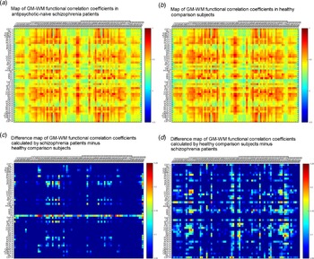
The maps shown in Fig. 1 represent correlations between function in WM tracts with that in GM regions, reflecting GM–WM functional synchrony. The horizontal and vertical stripes shown in Fig. 1a, b reflect the synchronous BOLD responses within distinct pairs of WM tracts and GM regions.
(a–d) Blood oxygenation level-dependent (BOLD) signals reflecting temporal correlations between gray matter (GM) regions and white matter (WM) tracts in antipsychotic-naïve schizophrenia patients (a), in healthy comparison subjects (b), and group differences of GM–WM functional correlation coefficients (c, d). Greater GM–WM functional correlation coefficients in schizophrenia patients compared to healthy comparison subjects (c) and greater GM–WM functional correlation coefficients in healthy comparison subjects compared to schizophrenia patients (d) are indicated by red/warm color, and lower functional correlation coefficients are indicated by blue/cold color. BA indicates Brodmann area. List of abbreviations and full names for WM tracts: CST, corticospinal tract; ML, medial lemniscus; iCBLP, inferior cerebellar peduncle; sCBLP, superior cerebellar peduncle; CBRP, cerebral peduncle; ALIC, anterior limb of internal capsule; PLIC, posterior limb of internal capsule; RLIC, retrolenticular part of internal capsule; ACR, anterior corona radiata; SCR, superior corona radiata; PCR, posterior corona radiata; OR, optic radiation; SS, sagittal stratum; EC, external capsule; CGG, cingulum (cingulate gyrus); CGH, cingulum (hippocampus); FXC, fornix (cres); SLF, superior longitudinal fasciculus; SFO, superior fronto-occipital fasciculus; UF, uncinate fasciculus; TAP, tapetum; mCBLP, middle cerebellar peduncle; PC, pontine crossing tract; GCC, genu of corpus callosum; BCC, body of corpus callosum; SCC, splenium of corpus callosum; FX, fornix (body).

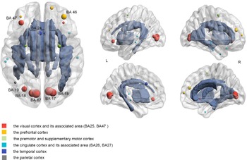
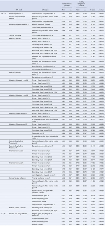
Compared to healthy comparison subjects, schizophrenia patients showed weaker GM–WM functional correlations in widespread regions (p values were thresholded at a Bonferroni-corrected threshold of p < 0.05/82 = 0.0006, Table 3, Fig. 2) including corpus callosum (genu and body), bilateral external capsule, bilateral cingulum, bilateral uncinate fasciculus, bilateral superior longitudinal fasciculus, right sagittal stratum, right cerebral peduncle, right posterior thalamic radiation, left anterior limb of internal capsule with diffuse cortical regions including visual cortex, cingulate cortex, temporal cortex, prefrontal cortex, pars orbitalis, and premotor cortex. Stronger GM–WM functional correlations were observed between fornix (column and body) and temporal cortex, piriform cortex, and angular gyrus in patients compared to healthy comparison subjects.
Differences in gray matter (GM)–white matter (WM) functional correlation coefficients between antipsychotic-naïve schizophrenia patients and healthy comparison subjects. Shown are GM regions and WM tracts for which significant group differences in GM–WM functional correlations were identified. The nodes denote GM regions with their size indicating the frequency of significant correlations with WM tracts. GM regions were labeled if at least three or more correlations were identified. BA indicates Brodmann area. R, right; L, left.

Differences in gray matter (GM)-white matter (WM) correlation coefficients between antipsychotic-naïve schizophrenia patients and healthy comparison subjects

HC, healthy comparison subjects; P, antipsychotic-naïve schizophrenia patients.
Group differences of WM structure
Regarding the integrity of WM structure, schizophrenia patients showed reduced FA in widespread tracts (p values were thresholded at a Bonferroni-corrected threshold of p < 0.05/48 = 0.001, Table 4), including bilateral anterior corona radiata, bilateral cerebral peduncle, corpus callosum, right cingulum (cingulate gyrus and hippocampus), and right fornix(crus)/stria terminalis.
Differences in fractional anisotropy (FA) between antipsychotic-naïve schizophrenia patients and healthy comparison subjects

HC, healthy comparison subjects; P, antipsychotic-naïve schizophrenia patients.
Relations between FA and ALFF, and GM–WM correlation coefficients
In patients, positive associations were found between FA in the genu of corpus callosum and GM–WM correlation coefficients for the association between the genu of corpus callosum and bilateral pars orbitalis (part of the inferior frontal gyrus), and right anterior prefrontal cortex (p < 0.05, uncorrected). Though, we found no significant associations between FA and ALFF neither at voxel-level nor by the ROI analyses.
Relations between brain measures and age, DUP, and symptom severity
In patients, regression analyses revealed a positive linear relationship between age and GM–WM correlation coefficients for the association between the left external capsule and visual cortex (p < 0.05, uncorrected, online Supplementary Fig. S1, Table S1). FA values in the genu of corpus callosum showed a negative association with age in schizophrenia patients (p < 0.05, uncorrected, online Supplementary Fig. S2, Table S2). No significant age-related associations were observed with brain measures in healthy comparison subjects. No significant associations were found between DUP and brain measures in schizophrenia patients.
Regarding the associations between symptom severity and brain measures in the patient group, we observed no significant associations between them.
Discussion
The main purpose of this study was to identify the alterations of functional activity in WM and to examine its relationship to the alterations of functional activity in GM and microstructural WM integrity in schizophrenia patients independently from possible confounds by antipsychotic medication. These aspects have not been reported in depth before. We identified widespread weakening in the synchrony of functional activity of GM and WM in patients, especially between WM tracts connecting fronto-temporal regions, interhemispheric homologous regions, cortico-subcortical circuits, and visual, cingulate, temporal, prefrontal cortex.
Based on the idea that synchronization between GM and WM functional activity in brain systems reflect how well distinct cortical areas are able to communicate with each other through the dense WM fiber network. The widespread weakening of GM–WM functional correlations in schizophrenia patients is consistent with the concept of schizophrenia as a dysconnectivity syndrome. Previous studies reported that BOLD signal changes in WM are related to BOLD signal changes in GM during task-based fMRI, indicating that altered BOLD signals in WM may contribute to behaviorally-relevant alterations of neural activity in brain GM (Gawryluk et al., Reference Gawryluk, Mazerolle and D'Arcy2014).
Though disruptions of functional correlations in WM tracts were widespread in the present study, their association with alterations in GM regions was mainly distributed in the visual cortex, prefrontal cortex, cingulate cortex, and temporal cortex. Alterations in these GM regions are believed to play important roles in the pathophysiology of schizophrenia. For instance, anterior and posterior cingulate cortex are components of the default mode network that is altered in schizophrenia (Li et al., Reference Li, Hu, Zhang, Tao, Dai, Gong and Lui2019; Lui et al., Reference Lui, Yao, Xiao, Keedy, Reilly, Keefe and Sweeney2015). Deficits in executive function, attention, and working memory have been linked to dysfunction of the dorsolateral prefrontal cortex (Chechko et al., Reference Chechko, Cieslik, Muller, Nickl-Jockschat, Derntl, Kogler and Eickhoff2018; Lencer et al., Reference Lencer, Yao, Reilly, Keedy, McDowell, Keshavan and Sweeney2019); and there are multiple imaging and psychophysical studies showing visual cortex alterations in schizophrenia.
Peer et al. (Reference Peer, Nitzan, Bick, Levin and Arzyt2017) reported the WM functional networks were related to distributed GM networks in healthy individuals in areas including the default-mode, dorsal attention, and frontoparietal control networks. Thus, our findings suggest a role for functional disruptions of WM tracts in schizophrenia with implications for alterations of communication between distant GM regions reflected in the relatively weak spatial coherence GM and WM function.
While there were anatomic dissociations between regional structural and functional alternations as has been reported previously (Liu et al., Reference Liu, Yao, Zhang, Deng, Xiao, Li and Lui2019), there were also anatomic associations of GM and WM function and WM microstructure. The altered connections were found between WM tracts with disrupted structural integrity (e.g. corpus callosum, cingulum) and the GM regions with disrupted functional synchrony (e.g. the prefrontal cortex, visual cortex, temporal gyri, cingulate cortex) in schizophrenia patients. Moreover, both reduced ALFF and FA were observed in the genu and splenium of corpus callosum, right anterior corona radiata, right cingulum (cingulate gyrus) in schizophrenia patients when compared to healthy comparison subjects despite some regional inconsistencies between regional patterns of structural and WM functional correlations.
Right cerebral peduncle, right cingulum, and genu of corpus callosum also demonstrated disruptions in structural integrity and weakening functional synchrony with GM regions to which they project. Based on these findings, our results suggest that disrupted WM integrity might provide the structural basis for the alternations of WM functional measures and the ability of WM to support the functional integration of GM regions to support cognition and behavior. Although our findings showed partially spatial overlapping in altered WM function and fiber tract integrity which was known to be impaired in schizophrenia (Skudlarski et al., Reference Skudlarski, Jagannathan, Anderson, Stevens, Calhoun, Skudlarska and Pearlson2010), we found no significant correlations between ALFF and FA in WM. This might reflect the independence of WM functional activity from WM structural integrity as a novel indicator to reveal the pathophysiology of schizophrenia.
Limitations
There are limitations to this study that should be considered when interpreting the results. First, the majority of patients were young adults with a recent onset of schizophrenia (Flynn et al., Reference Flynn, Lang, Mackay, Goghari, Vavasour, Whittall and Honer2003) while the proportion of chronically ill patients was relatively small. This might have resulted in a bias affecting the age and illness-duration-related alternations in functional correlations in patients. However, the age range in the healthy comparison subject group was similar to that in the patient group and no association with age was observed making a possible sample bias unlikely. Second, subcortical and cerebellar regions were not included in the GM atlas employed; therefore, disruption of GM–WM synchrony involving those areas remains to be determined. Third, with smaller volume, the slight movement of patients might affect the detection of BOLD signals in WM. However, the head movement was integrated with a conservative criterion of 2 mm in this study. Fourth, the neurophysiological basis of the BOLD signal changes in WM is still a matter of debate. More clinical and preclinical investigation is required to fully understand the disease and behavioral significance of alterations in WM function in schizophrenia. Last, the hemodynamic response function in specific WM regions may vary, and impact sensitivity of measured BOLD signal changes to neural activity in WM (Gawryluk et al., Reference Gawryluk, Mazerolle and D'Arcy2014). Other technical factors related to this may impact our findings, such as activations arising from neighboring GM and vascular structure impacting BOLD signals of WM pathways related to partial volume effects (Jiang et al., Reference Jiang, Luo, Li, Li, Yang, Li and Yao2019).
Conclusion
Findings from the present study extend knowledge about functional alterations in brain WM in schizophrenia independently from antipsychotic medication or course of illness effects. Our findings provide new information about the model of schizophrenia as a dysconnectivity syndrome reflected in disruptions of the synchrony between GM and WM function. The specific patterns of altered functional synchrony observed in this study might contribute to the pathophysiology of schizophrenia and highlighted the novelty of GM–WM functional correlation as the indicator to investigate the brain function.
Supplementary material
The supplementary material for this article can be found at https://doi.org/10.1017/S0033291720004420
Acknowledgements
Dr Su Lui acknowledges the support from Humboldt Foundation's Friedrich Wihelm Bessel Research Award. This study was supported by the National Natural Science Foundation of China (Project Nos. 81671664 and 81621003, 81820108018, 81901828), National Program for Support of Top-notch Young Professionals (Project No. W02070140), Fundamental Research Funds for the Central Universities (Project No. 2018SCUH0011), Science and Technology Project of the Health Commission of Sichuan Province (Project No. 18ZD035), Sichuan Science and Technology Program (Project No. 2019YJ0155), and 1.3.5 Project for Disciplines of Excellence, West China Hospital, Sichuan University (Project Nos. ZYYC08001 and ZYJC18020). Drs Su Lui and Qiyong Gong contributed equally as senior authors.
Conflict of interest
Dr Sweeney and Dr Zhang consult to VeraSci. Other authors declare no conflicts.