





Introduction
The rising prevalence of obesity among women of reproductive age increases the risk for gestational diabetes mellitus (GDM), when left untreated can result in negative consequences for both the mother and child. Reference Ogunwole, Zera and Stanford1,Reference Alejandro, Mamerto and Chung2 It is well known that early exposures in the in-utero environment can program fetuses and negatively impact their metabolic health in adulthood resulting in increased susceptibility to obesity, cardiovascular diseases, and type 2 diabetes mellitus (T2DM). Reference Godfrey, Reynolds and Prescott3,Reference Howell and Powell4 GDM, diagnosed by glucose intolerance identified in the second trimester, is associated with dyslipidemia, hyperglycemia, and hyperinsulinemia. 5 Maternal hyperglycemia is the primary contributor for macrosomia and the programmed long-term metabolic dysfunction in offspring by activation of fetal insulin hypersecretion as described by the Pedersen hypothesis. Reference Kc, Shakya and Zhang6,Reference Catalano and Hauguel-De Mouzon7 Independent effects of hyperglycemia have been shown to be harmful not only to the fetus and offspring but the placenta. Reference Shashikadze, Valla and Lombardo8,Reference Aires and Dos Santos9 An adverse outcome in GDM pregnancies is the development of large for gestational age (LGA) infants, defined as those with a birth weight greater than the 90th percentile. Reference Nazeer, Chen and Cornthwaite10 This can increase the risk of childhood obesity and metabolic health complications later in adulthood. Reference Derraik, Maessen, Gibbins, Cutfield, Lundgren and Ahlsson11 Individuals diagnosed with GDM are advised to modify their diet, and if hyperglycemia remains, insulin treatment is recommended to achieve euglycemia. Reference Buchanan, Xiang and Page12,13 Despite achieving optimal maternal glucose concentrations with insulin, adverse outcomes persist in both the placenta and offspring. Reference Thorius, Husemoen and Nordsborg14–Reference Subiabre, Silva and Toledo16 It remains unknown how hyperinsulinemia within maternal obesity and GDM independently contribute to long-term metabolic programming of the offspring. Limited research has examined the long-term effects of exogenous insulin prescribed to women during pregnancy in the context of either GDM or those diagnosed with type 1 diabetes mellitus (T1DM) which can inadvertently lead to maternal hyperinsulinemia and insulin resistance. Offspring of individuals with T1DM and T2DM are also at higher risk for experiencing fetal overgrowth, which like GDM puts them at risk of metabolic diseases later on in life. Reference Thorius, Husemoen and Nordsborg14
The effects of maternal insulin treatment on placental function and fetal development, as well as the long-term outcomes for the offspring, remain incompletely understood. Unlike maternal glucose, maternal insulin does not cross the placenta. Reference Hufnagel, Dearden, Fernandez-Twinn and Ozanne17 Instead, insulin acts on both the insulin receptor (InsR) and the insulin-like growth factor-1 receptor (IGF1R) expressed in the placenta, though it has a higher binding affinity for the InsR. Activation of the InsR signals through the PI3K/Akt/mTOR pathway, which serves to further regulate nutrient transport to the developing fetus. Reference Ruiz-Palacios, Ruiz-Alcaraz, Sanchez-Campillo and Larque18 This raises the critical question of whether maternal insulin treatment, in the context of maternal normoglycemia, may have any long-term consequences on the metabolic health of the offspring. We hypothesized that maternal hyperinsulinemia independently affects offspring glucose homeostasis. Herein, we sought to determine the independent effects of maternal hyperinsulinemia, in the absence of hyperglycemia and obesity on offspring glucose homeostasis into adulthood using two maternal hyperinsulinemic preclinical models. These animal models do not fully recapitulate the pathophysiology of GDM but rather allow us to investigate the direct effect of high circulating insulin during pregnancy.
Materials and methods
Generation of animals
Pancreatic beta-cell specific RIP-βAkt transgenic (RIP-βAkt) male founder mice were generated by Dr Ernesto Bernal-Mizrachi, University of Miami, Florida. Male founders were bred with female C57BL/6J mice purchased from Jackson Laboratory (Stock No: 000664; RRID: IMSR_JAX:000664) to generate WT and RIP-βAkt female mice. WT and RIP-βAkt dams were then bred with WT sires to generate CTRL and βAkt-HIP offspring. To generate RIP-Cre; βTSC2 dams, RIP-Cre containing males were bred to TSC2 flox/flox females. Reference Herrera19,Reference Rachdi, Balcazar and Osorio-Duque20 Line maintenance was performed by breeding RIP-Cre; TSC2 flox/flox sires with TSC2 flox/flox dams to create TSC2 flox/flox and RIP-Cre; βTSC2 flox/flox female mice used for breeding CTRL and βTSC2KO-HIP offspring. All laboratory animals used for the study were housed in the University of Minnesota Twin Cities animal facility on a 14:10 light:dark cycle, with ad libitum access to the normal chow diet (NCD, 2018). All pups, regardless of genotype, were left with the dam for 21 days prior to weaning. CTRL and βAkt-HIP offspring were all fed a NCD for the first 12 weeks and subsequently shifted to a high-fat diet (HFD, D12492) containing 60% kcal from fat. Intraperitoneal glucose tolerance testing (IpGTT) was conducted at 6, 8, and 12 weeks under NCD and 4, 10, and 16 weeks post-HFD. Insulin tolerance testing (ITT) was performed at 10 weeks on NCD and 8 and 12 weeks post-HFD. The first model we established was the βTSC2KO-HIP offspring, where a power analysis was done to determine sample size. For rigor, the RIP-βAkt offspring were studied next to confirm findings in the first model. All animal procedures were approved by the Institutional Animal Care and Use Committee at the University of Minnesota.
Metabolic in vivo mouse studies
Body weight measurements were collected at 1, 3, 6, 8, 10, and 12 weeks while mice were fed a NCD. Body weights were collected weekly up to 12 weeks upon being administered a HFD. Glucose tolerance testing was performed by fasting the mice overnight (16 h) and measuring blood glucose at time 0 min (baseline) and (30, 60, and 120 min) after intraperitoneal (ip) injection with 30% dextrose in the amount of 2mg/kg. Insulin tolerance testing was conducted by fasting mice for 6 h and measuring blood glucose at time 0 and (30, 60, and 120 min) after ip injection of 0.75 U/kg dose of insulin prepared in 0.9% saline. Blood glucose measurements were assessed from tail vein using a handheld glucometer. Insulin concentration was determined from tail vein-collected serum stored at −80° C using the ALPCO Ultrasensitive mouse insulin ELISA, per kit instructions. Analysis was completed by 4-parameter logistic fit utilizing the MyAssays software.
Body composition measurements
Body composition was performed on WT and RIP-βAkt female mice at 85 days of age using magnetic resonance imaging and was completed by the University of Minnesota Metabolic Physiology Core facility.
Statistical analysis
Graphpad Prism (version 10.1.1) software was used to perform statistical analysis of endpoints other than longitudinal body weights. Longitudinal body weight data was analyzed with the SAS 9.4 MIXED procedure. An unpaired, two-tailed Student’s t-test was employed to analyze the difference between groups for one variable between groups. A repeated measures analysis assuming sphericity was conducted with pups as subjects to compare group differences for IpGTT and ITT at each time point, using Sidak’s multiple comparison post hoc test. A mixed model with random mom and pup effects including interactions between mom and the fixed effects of sex, week, and sex*week was utilized for analysis of body weight measurements across time. Statistical significance is denoted as *<0.05, ** <0.01, *** <0.001, ****<0.0001, ns, non-significant.
Results
Maternal hyperinsulinemia with masked hypoglycemia attenuates body weight in male offspring
To investigate the independent effects of maternal hyperinsulinemia on offspring glucose homeostasis, we first used a transgenic hyperinsulinemic mouse model, RIP-Cre; βTSC2 flox/flox mice, where conditional deletion of TSC2 results in overexpression of mTORC1 in a pancreatic beta-cell specific manner. Reference Rachdi, Balcazar and Osorio-Duque20 RIP-Cre; βTSC2 flox/flox mice were previously reported to have increased beta-cell mass. Reference Rachdi, Balcazar and Osorio-Duque20 We performed metabolic phenotyping to fully understand the maternal environment of these dams. RIP-Cre; βTSC2 flox/flox dams exhibit no differences in body weight compared to their control counterparts before pregnancy (Fig. 1a). These dams also present significantly reduced blood glucose concentrations against the βTSC2 flox/flox controls (Fig. 1b). Reduced blood glucose is attributed to elevated circulating insulin concentrations during both non-fasted and fasted (6 h) states (Fig. 1c). The RIP-Cre; βTSC2 flox/flox dams also demonstrate improved glucose tolerance due to high circulating insulin (Fig. 1d), due to increased beta-cell mass. Reference Rachdi, Balcazar and Osorio-Duque20 However, RIP-Cre; βTSC2 flox/flox dams maintain normal insulin sensitivity despite having increased circulating insulin (Fig. 1e). To generate offspring exposed to hyperinsulinemia in pregnancy (βTSC2KO-HIP) and CTRL (βTSC2 flox/flox) offspring, male βTSC2 flox/flox sires were bred with RIP-Cre; βTSC2 flox/flox and βTSC2 flox/flox dams (Fig. 1f). No changes were observed in litter sizes between dams (Fig. 1g) or male βTSC2KO-HIP offspring body weights under a NCD (Fig. 1h). Male βTSC2KO-HIP offspring demonstrated transient glucose intolerance in early adulthood much like the βAkt-HIP offspring under a NCD (Fig. 1i). Their insulin sensitivity remains unchanged (Fig. 1j). We then metabolically challenged the βTSC2KO-HIP offspring with HFD. Under HFD feeding, male βTSC2KO-HIP offspring body weight and glucose homeostasis did not differ from CTRLs (Fig. 1k–1M). We also performed the experiments in βTSC2KO-HIP female offspring which experienced no perturbations in body weight or glucose regulation under both NCD and HFD (Supplemental Fig. 1).
Maternal hyperinsulinemia with masked hypoglycemia attenuates body weight in male offspring. (a) At 65–80 days of age, βTSC2 flox/flox (n = 11) and RIP-Cre; βTSC2 flox/flox (n = 11) dams exhibit comparable body weight. (b) Non-fasting and fasting (6 h during the day) blood glucose concentrations in βTSC2 flox/flox (n = 15) and RIP-Cre; βTSC2 flox/flox (n = 15) dams. (c) Non-fasting and fasting (6 h) insulin concentrations of βTSC2 flox/flox (n = 12–15) and RIP-Cre; βTSC2 flox/flox (n = 12–15) dams. (d) Intraperitoneal glucose tolerance test (IPGTT; 2 g/kg glucose) was performed in βTSC2 flox/flox (n = 15) and RIP-Cre; βTSC2 flox/flox (n = 15) demonstrating no glucose intolerance in RIP-Cre; βTSC2 f/f dams. (e) No changes were observed in insulin sensitivity of βTSC2 flox/flox (n = 5) and βTSC2 flox/flox RIP-Cre (n = 5) dams. (f) Breeding schematic to generate CTRL and βTSC2KO-HIP offspring. (g) No changes in litter size βTSC2 flox/flox (n = 11) and RIP-Cre; βTSC2 flox/flox (n = 12) (h) Body weight trajectory is comparable between CTRL (n = 18) and βTSC2KO-HIP (n = 15) male offspring under NCD. (i) IPGTT (2 g/kg glucose) was performed in CTRL (n = 18) and βTSC2KO-HIP (n = 16). (j) ITT (ip insulin/bw, 0.75 U/kg) of CTRL (n = 17) and βTSC2KO-HIP (n = 16) male offspring under NCD. (k) Male offspring body weight trajectory under HFD, CTRL (n = 18) and βTSC2KO-HIP (n = 15). (l) ITT of CTRL (n = 18) and βTSC2KO-HIP (n = 15) 8 weeks post-HFD. (m) IPGTT of CTRL (n = 18) and βTSC2KO-HIP (n = 15) male offspring 10 weeks post-HFD. Statistical analysis was performed using an unpaired, two-tailed, Student’s t-test or Two-way ANOVA. *p ≤ 0.05, ns, not significant.

Hyperinsulinemic RIP-βAkt dams exhibit insulin resistance and have increased body weight
To validate our findings in the RIP-Cre; βTSC2 flox/flox model, we generated a second model (RIP-βAkt) of maternal hyperinsulinemia containing a constitutively active Akt1, increasing mechanistic target of rapamycin complex I (mTORC1) activity, and repeated the metabolic phenotyping in the dams and offspring. Reference Bernal-Mizrachi, Wen, Stahlhut, Welling and Permutt21 Similar to our RIP-Cre; βTSC2 flox/flox model, the RIP-βAkt mouse is a representative model of hyperinsulinemia as insulin level remains high under both non-fasting and fasting conditions (Fig. 2a). Contrasting to RIP-Cre; βTSC2 flox/flox animals, RIP-βAkt dams are only hypoglycemic when fasted (Fig. 2b). Unlike RIP-Cre; βTSC2 flox/flox mice, RIP-βAkt dams exhibit increased body weight compared to wildtype (WT) dams (Fig. 2c). Body composition analysis revealed that lean rather than fat mass was responsible for the increase in body weight in RIP-βAkt dams (Fig. 2d). RIP-βAkt mice have improved glucose tolerance (Fig. 2e) despite demonstrating mild insulin resistance (Fig. 2f). This suggest that although RIP-βAkt dams may have reduced response in insulin (0.75 Units/Kg) in their peripheral tissues, their significant increase in beta-cell mass (~11 fold increase Reference Alejandro, Bozadjieva and Kumusoglu22 ) permits increased secretion of insulin allowing them to have a higher glucose tolerance. Additionally, RIP-βAkt dams produced smaller litters compared to WT dams (Fig. 2g). We also evaluated maternal liver weight by assessing changes in liver size, as this impacts overall hepatic function and subsequent maternal nutritional balance. However, no changes were observed in the liver (Supplemental Fig. 2).
Hyperinsulinemic dams exhibit normal glucose tolerance with increased body weight and insulin resistance. (a) Fasting insulin (6–16 h) concentrations of RIP-βAkt (n = 7) and WT (n = 10) dams. (b) Non-fasting and fasting glucose (16 h) concentrations of RIP-βAkt (n = 8–11) and WT (n = 10–13) dams. (c) At 50–75 days of age, body weight of RIP-βAkt (n = 20) dams is increased compared to WT (n = 27) controls. (d) Increased body mass in RIP-βAkt (n = 4) dams, compared to WT (n = 4) is attributed to higher lean body mass and not fat mass. (e) Intraperitoneal glucose tolerance test (IPGTT; 2 g/kg glucose) was performed in RIP-βAkt (n = 7) and WT (n = 10) dams at 50–75 days of age. RIP-βAkt dams demonstrate improved glucose tolerance. (f) Assessment of insulin sensitivity was performed via intraperitoneal insulin tolerance test (ITT; ip insulin/bw, 0.75 U/kg) for RIP-βAkt (n = 7) and WT (n = 8). RIP-βAkt dams demonstrate insulin resistance compared to WT dams. (g) WT (n = 11) dams produced larger litter sizes compared to RIP-βAkt (n = 11) dams. Statistical analysis was performed using an unpaired, two-tailed, Student’s t-test or Two-way ANOVA. *p ≤ 0.05, ns, not significant.

Male offspring born to RIP-βakt hyperinsulinemic dams exhibit increased body weight
We then repeated the metabolic phenotyping performed in the βTSC2KO-HIP offspring to confirm the independent effects of maternal hyperinsulinemia in the βAkt-HIP offspring. To generate βAkt-HIP offspring, WT sires were bred with RIP-βAkt dams, which have increased pancreatic beta-cell mass rendering them hyperinsulinemic (Fig. 3a). Controls (CTRL) were generated by breeding WT sires to WT dams. Female βAkt-HIP offspring did not differ in weight gain relative to CTRL offspring under a normal chow diet (NCD) (Fig. 3b). In contrast, male βAkt-HIP offspring demonstrated increased weight starting 8 weeks of age which continues up through 12 weeks of NCD feeding (Fig. 3c). No changes in fasting insulin concentrations seen in 6- or 8-week-old male or female offspring (Supplemental Fig. 2 A-B). Glucose tolerance tests were performed to assess glucose tolerance in offspring. At 6 weeks of age, male βAkt-HIP demonstrated higher glucose level at 30 min post-glucose injection (Fig. 3d). However, the area under the curve is not significant (data not shown). Unlike male offspring, female βAkt-HIP offspring displayed normal glucose tolerance at 6 weeks of age (Fig. 3e). Glucose tolerance was re-assessed at 8 weeks of age under NCD. The glucose intolerance observed in male βAkt-HIP offspring at 6 weeks was not sustained at 8 weeks (Fig. 3f and 3g). We then assessed insulin sensitivity under NCD by performing insulin tolerance tests. Insulin sensitivity in βAkt-HIP offspring were comparable to CTRL offspring in both sexes (Fig. 3h and 3i). Our data demonstrate that male offspring born from a hyperinsulinemic in-utero environment exhibit a mild increase in body weight and experience transient glucose intolerance under NCD, suggesting maternal insulin may be contributing to programming of body weight maintenance in the male offspring.
Maternal hyperinsulinemia induced increase body weight in male offspring with normal glucose homeostasis under a normal chow diet. (a) Breeding schematic for the generation of hyperinsulinemic exposed in pregnancy (βAkt-HIP) offspring using the RIP-βAkt dams. (b) Female βAkt-HIP (n = 5–9) offspring demonstrate no significant body weight changes relative to the controls (CTRL) (n = 16–28) under a normal chow diet (NCD). (c) Male βAkt-HIP (n = 6–11) offspring exhibit body mass not observed in CTRL (n = 15–25). (d) IPGTT at 6 weeks in male offspring (βAkt-HIP n = 4, CTRL n = 9) showed transient glucose intolerance. (e) IPGTT at 6 weeks in female offspring (βAkt-HIP n = 6, CTRL n = 10) showed no differences. (f) IPGTT at 8 weeks in male offspring (βAkt-HIP n = 7, CTRL n = 13), demonstrated the glucose intolerance from 6 weeks diminished. (g) IPGTT at 8 weeks in female offspring (βAkt-HIP n = 5, CTRL n = 11) remain the same between groups. (h) and (i) ITT at 10wks demonstrates both female (βAkt-HIP n = 7, CTRL n = 6) and male βAkt-HIP (βAkt-HIP n = 4, CTRL n = 7) offspring have comparable insulin sensitivity to CTRLs. Statistical analysis was performed using a Two-way ANOVA test. *p ≤ 0.05, ns, not significant.

Offspring from maternal hyperinsulinemic dams maintain normal glucose regulation under HFD
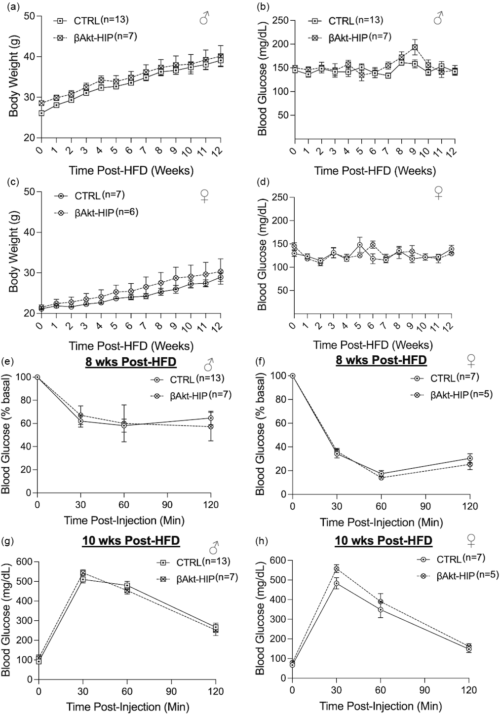
Metabolic phenotyping of offspring born to RIP-βAkt dams did not show sustained glucose intolerance while being fed a NCD despite the body weight difference in males, like offspring born to the RIP-Cre; βTSC2 flox/flox dams. Due to the increased body weight, we sought to determine if glucose regulation would be perturbed under a metabolic challenge such as a high-fat diet (HFD) (D12492) containing 60% kcal of fat 20% kcal of protein, and 20% kcal of carbohydrate. After 12 weeks of NCD feeding, CTRL and βAkt-HIP offspring were transferred to ad libitum HFD feeding for an additional 12 weeks. Body weights were tracked and measured weekly. Interestingly, the body weight difference which was notable under NCD between male CTRL and βAkt-HIP offspring was not seen under a HFD (Fig. 4a). Male βAkt-HIP offspring experience limited weight gain during the duration of HFD feeding. No changes in blood glucose were detected across the 12 weeks of HFD (Fig. 4b). Female βAkt-HIP offspring remain on the same body weight trajectory as CTRLs throughout HFD feeding (Fig. 4c) with no observed changes in blood glucose (Fig. 4d). No changes in fasting insulin concentrations post 8 or 10 weeks of HFD in male or female offspring (Supplemental Fig. 2 C-D). Insulin tolerance testing was repeated at 8 weeks post-HFD demonstrating that βAkt-HIP offspring, male (Figs 4 and 3e) and female (Fig. 4f) do not develop worse insulin sensitivity compared to CTRLs. Glucose tolerance was also reassessed at 10 weeks post-HFD. Neither male (Fig. 4g) nor female (Fig. 4h) βAkt-HIP offspring develop perturbed glucose handling compared to CTRLs. Overall, these data suggest that insulin exposure in-utero drive limited weight gain in βAkt-HIP offspring with negligible impact in female offspring.
Male and female βAkt-HIP offspring have comparable glucose homeostasis and insulin sensitivity to CTRL under a HFD. (a) Body weight trajectory of male βAkt-HIP (n = 7) and CTRL (n = 13) offspring under HFD. (b) Blood glucose trajectory of male βAkt-HIP (n = 7) and CTRL (n = 13) offspring. (c) Body weight trajectory of female βAkt-HIP (n = 6) and CTRL (n = 7) offspring under HFD. (d) Blood glucose trajectory of female βAkt-HIP (n = 6) and CTRL (n = 7) offspring. (e) ITT (ip insulin/bw, 0.75 U/kg) of male offspring 8 weeks post-HFD, CTRL (n = 13) βAkt-HIP (n = 7). (f) ITT (ip insulin/bw, 0.75 U/kg) of female offspring 8 weeks post-HFD, CTRL (n = 7) βAkt-HIP (n = 5). (g) IPGTT (2 g/kg glucose) of male offspring 10 weeks post-HFD, CTRL (n = 13) βAkt-HIP (n = 7). (h) IPGTT (2 g/kg glucose) of female offspring 10 weeks post-HFD, CTRL (n = 7) βAkt-HIP (n = 5). Statistical analysis was performed using a Two-way ANOVA test. *p ≤ 0.05, ns, not significant.

Discussion
Insulin therapy is the gold standard treatment for individuals with GDM or T1DM. However, combating insulin resistance and the demand for insulin by increasing circulating insulin, exogenously, can inadvertently result in a maternal hyperinsulinemic environment. This is especially true since injected insulin does not directly get cleared by the liver as endogenously-produced insulin, but rather by the kidney. Reference Feingold, Ahmed and Anawalt23 Extensive studies have been performed to investigate the consequences hyperglycemia, but there is limited knowledge about the impact of maternal hyperinsulinemia alone on offspring metabolic health. We have shown that male offspring born to multiparous dams, an alternative hyperinsulinemia model, where the InsR is knocked out in the placenta, are protected against diet-induced obesity and glucose intolerance under a HFD challenge. Reference Chung, Mohan, Beetch, Jo and Alejandro24 This demonstrates an important role for insulin in fetal and offspring programming in-utero. Furthermore, a previous study has reported that insulin exhibits toxicity towards first-trimester placental trophoblasts, resulting in increased DNA damage and apoptosis. Reference Vega, Mauro and Williams25 This raises critical questions regarding the effects of insulin as a treatment and the potential long-term metabolic consequences for the offspring. Reference Subiabre, Silva and Toledo16 In the current study, we sought to elucidate the independent effects of maternal hyperinsulinemia on offspring glucose homeostasis in the absence of confounding factors, such as maternal dyslipidemia or hyperglycemia using the RIP-βAkt and RIP-Cre; TSC2 flox/flox transgenic mouse models.
In contrast to maternal glucose, maternal insulin is incapable of crossing the placental barrier and cannot reach the fetus unless bound to an antibody such as IgG. Reference Hufnagel, Dearden, Fernandez-Twinn and Ozanne17,Reference Suffecool, Rosenn and Niederkofler26,Reference Jovanovic and Pettitt27 Indeed, insulin glargine injected in mothers for example cannot be detected in fetal circulation. Reference Pollex, Feig, Lubetsky, Yip and Koren28 However, insulin can act at the maternal-fetal interface via the InsR, in which case it can trigger a cascade of reactions activating the PI3K/Akt/mTOR pathway, a signaling pathway that is highly conserved across species, and regulates nutrient flux and placental function. Reference Ruiz-Palacios, Ruiz-Alcaraz, Sanchez-Campillo and Larque18, Reference Ardestani, Lupse, Kido, Leibowitz and Maedler29 In the current study, the effects of maternal hyperinsulinemia on offspring glucose homeostasis and metabolic trajectory were mild. Additionally, only male offspring were impacted in which case we observed changes in body weight and a transient glucose intolerance, suggesting insulin by itself is not sufficient to induce permanent and major metabolic programming based on the parameters tested. The glucose tolerance test in βAkt-HIP male offspring showed elevated glucose concentrations at 30 min after glucose administration, indicating a potential delay in beta-cell insulin secretion. This time point is critical for assessing the first-phase insulin response. Reference Poon and Tung30 Given that βAkt-HIP males exhibit hyperglycemia compared to controls at this stage, it is likely that they are experiencing a beta-cell functional impairment at this age; however, this remains to be tested. Our findings are not surprising when considering the broader maternal metabolic milieu associated with obesity and GDM. Obesity during pregnancy changes the entire maternal environment shifting towards a low-grade chronic state of inflammation, characterized by inflammatory molecules such as interleukin-6 (IL-6), IL-8, monocyte chemoattractant protein-1 (MCP-1), and tumor necrosis factor alpha (TNFα), all of which can regulate placental function and consequently impact nutrient transport to the fetus. Reference Wallace, Bellissimo and Yeo31,Reference Louwen, Kreis, Ritter and Yuan32 GDM has also been associated with low-grade chronic inflammation and elevated oxidative stress. Reference Sha, Zeng, Zhao and Jin33,Reference Barke, Goldstein and Sundermann34 Additionally, both maternal obesity and GDM are often associated with other metabolic alterations, including dyslipidemia, Reference Zimmerman, Briffa and Kong35 which may collectively contribute to the adverse maternal intrauterine environment.
We observed a mild but significant change in body weight in male offspring born to RIP-βAkt dams. The divergence in body weight of offspring between the experimental groups manifests at 8 weeks of age and is sustained throughout the duration of the mice being fed a normal chow diet. However, it is currently unclear if the weight gain is from increased adiposity, as is seen in humans when babies are born to mothers with GDM. In the future, we anticipate performing body composition of the male βAkt-HIP offspring to determine the driver of this weight difference. Interestingly, when these βAkt-HIP male offspring are placed on a HFD, their weight becomes comparable to the CTRL offspring. Thus, CTRL mice gain more weight on a HFD than Akt-HIP offspring themselves, resulting in similar weight trajectory across the time in which HFD was administered. We do not have a clear explanation for why the body weight phenotype observed under the normal chow diet disappeared during the 60% kcal HFD challenge. One possibility is that the dietary challenge overrides or masks the baseline metabolic phenotype. This could indicate that the HFD produces a ceiling effect, in which both groups gain sufficient weight to minimize detectable differences. Alternatively, HIP may exhibit altered susceptibility or adaptation to excess dietary fat. We have published that genetic deletion of TSC2 in the placenta resulting in enhanced insulin/mTORC1 activity in utero, protected adult offspring from diet-induced obesity. Reference Akhaphong, Baumann, Beetch, Lockridge, Jo, Wong, Zemanovic, Mohan, Fondevilla, Sia, Pineda-Cortel and Alejandro36 This supports our data demonstrating normalization of βAkt-HIP male offspring under HFD. However, additional experiments are needed to determine the mechanisms underlying these observations.
We also observed a divergence in our male βAkt-HIP offspring phenotype, in comparison to the βTSC2KO-HIP, which may be caused by the notable differences in the metabolic phenotypes of the dams. We believe the difference in body weights of the HIP offspring between the maternal hyperinsulinemia models could be due to masked hypoglycemia, a condition where asymptomatic hypoglycemia exists, experienced by the RIP-Cre; βTSC2 flox/flox dams and or the difference in insulin sensitivity in both models. Glucose is a critical nutrient in regulating fetal development and supporting fetal growth and mother during gestation. Reference Hay37 Male βTSC2KO-HIP offspring may be subjected to the effects of developing in a low-glucose intrauterine environment with increased hyperinsulinemia. Although their maternal milieu may be characterized by excess insulin, these offspring are also experiencing the effect of reduced glucose availability, potentially lowering their endogenous insulin secretion. Additionally, the disparities in insulin sensitivity between the two models may also account for the divergent male offspring phenotypes, as insulin plays a central role in placental nutrient transport to support the fetus. Reference Ruiz-Palacios, Ruiz-Alcaraz, Sanchez-Campillo and Larque18,Reference Chung, Mohan, Beetch, Jo and Alejandro24,Reference Anam, Cooke and Dratver38 Masked hypoglycemia is a condition seen among individuals with GDM on insulin treatment and has been shown to not have an impact on birthweight. Reference Naik, Hesarghatta Shyamasunder and Doddabelavangala Mruthyunjaya39 Therefore, we suspect it contributed to the unchanged body weight effect in the βTSC2KO-HIP offspring. In contrast, without the effect of masked hypoglycemia in the RIP-βAkt dams, the additive effect of insulin could be responsible for driving the increased body weight observed in the male βAkt-HIP offspring. Moreover, the RIP-βAkt dams are insulin resistant with increased lean mass compared to the RIP-Cre; βTSC2 flox/flox dams which may cause differences in nutrient availability and transfer in the placenta ultimately affecting overall fetal growth and development.
It is important to acknowledge the limitations for our present study. To generate βAkt-HIP and βTSC2KO-HIP offspring, WT or βTSC2 flox/flox sires were bred with maternal hyperinsulinemia dams which could produce RIP-βAkt or βTSC2KO littermates which were excluded. While these hyperinsulinemic offspring were excluded from the current study, the localization in the uterus and the ratio of different genotypes in a litter could alter the maternal intrauterine environment and may be impacted by fetal insulin originating from the hyperinsulinemic fetuses, a case that would not be anticipated in human singleton pregnancies. Reference Hufnagel, Dearden, Fernandez-Twinn and Ozanne17,Reference Desoye and Hauguel-de Mouzon40 Another caveat of the generated models is that the dams are hyperinsulinemic prior to gestation, which may ultimately change the maternal milieu before fetal development. Thus, to assess the impact of hyperinsulinemia specifically during gestation, an osmotic pump could be used to deliver insulin only within this window. However, this approach may complicate pregnancy maintenance, as achieving proper glucose control is often challenging in this model. To improve the clinical relevance of the study, a model with obesity and hyperinsulinemia can better mimic maternal obesity in humans. Moreover, the insulin concentrations exhibited by our mouse models are not the same as what may been seen in a human pregnancy. Our current data may not fully recapitulate what is seen in humans as insulin treatment of GDM and T1DM and T2DM vary based on the degree of hyperglycemia. Mothers may be treated with long- or short-acting insulin analogues with varying doses. Reference Mukherjee and Dawson41 Data interpretation from our study may also be limited since complete biochemical profiling was not performed. For future studies we plan to assess other important metabolic markers such as, circulating triglycerides, cholesterol, high- and low-density lipoproteins, and ketone bodies to more fully characterize the effects of maternal hyperinsulinemia on the offspring metabolic health. In our study we utilized the HFD – 60 Kcal% from fat – as a metabolic challenge. However, we acknowledge that this diet is not physiologic, nor does it induce the combination of hyperglycemic and lipidemia conditions seen in metabolic syndrome or have the same impact such that chronic obesity does. Statistically, random parental and litter effects were only applied in longitudinal body weight analyses. Additionally, we have not yet explored other potential effects of hyperinsulinemia on the dams in our models, such as alterations in the immune system that could increase susceptibility to infections or cancer. Reference Makhijani, Basso and Chan42 The current study also does not explore how the placenta is altered under maternal hyperinsulinemic conditions.
Conclusion
In conclusion, using two preclinical models, we demonstrated that maternal insulin exposure during normal pregnancy has a mild and transient effect on glucose homeostasis in male offspring. Moreover, an increased body weight under NCD was observed in male offspring born to maternal hyperinsulinemic dams without the effect of masked hypoglycemia. Our data aligns with the current literature, supporting that male offspring are at higher risk of adverse metabolic health outcomes. Reference Li, Zhu and Yeung43–Reference Shook, White and Acharya45 Our current study does not investigate impacts of insulin with other co-founding factors or features present in GDM such as high glucose or hyperlipidemia. In addition, this study did not assess other known effects of maternal obesity and GDM such as offspring cognitive and immunological health. Reference Godfrey, Reynolds and Prescott3,Reference Eleftheriades, Koulouraki, Belegrinos, Eleftheriades and Pervanidou46,Reference Han, Patel, Jones and Dale47 Therefore, further rigorous investigation is warranted to elucidate additional independent effects of maternal hyperinsulinemia on the offspring and mechanisms underlying sexual dimorphism.
Supplementary material
The supplementary material for this article can be found at https://doi.org/10.1017/S2040174426100671.
Acknowledgments
We acknowledgment that all authors have contributed significantly and in keeping with the latest guidelines of the International Committee of Medical Journal Editors. TKH and EUA developed the project. TKH and GC performed experiments and formal analyses. TKH and EUA wrote the manuscript. TKH, GC, and RR were responsible for data visualization. TKH, GC, RR, BG, EBM, SW, and EUA reviewed and edited the manuscript. EUA conceived the study and oversaw the overall direction of this work. All authors have read and agreed to the published version of the manuscript.
Author contribution
This work was supported by National Institutes of Health Grant R01DK136237 to EUA and 1R01HD113553-01A1 to SAW. TKH was supported by R01DK136237-02S1, UMN RAISE, and IMPACT Award to EUA.
Competing interests
All authors have nothing to disclose.




 Open access

