Introduction
Inflammatory disease of the supraglottic larynx is rare. The causes are myriad, and include: reflux,Reference Franco 1 environmental pollutants,Reference Marcotullio, Magliulo and Pezone 2 , Reference Kravos, Zupevc, Cizmarevic and Hocevar-Boltezar 3 granulomatous disorders,Reference Brass and White 5 syphilis,Reference Brass and White 5 , Reference Pace and Csonka 6 fungal infections,Reference Pochini Sobrinho, Della Negra, Queiroz, Ribeiro, Bittencourt and Klautau 7 , Reference Miller, Myer and Gray 8 viral infections,Reference Musharrafieh, Araj and Fuleihan 9 amyloidosis,Reference Eliachar and Lichtig 10 , Reference Hazenberg, Dikkers, Hawkins, Bijzet, Rowczenio and Gilbertson 11 Wegener's granulomatosis,Reference Talerman and Wright 12 chronic relapsing polychondritis,Reference Letko, Zafirakis, Baltatzis, Voudouri, Livir-Rallatos and Foster 13 sarcoidosis,Reference Haar, Chaudhry, Kaplan and Milley 14 systemic lupus erythematosusReference Scarpelli, McCoy and Scott 15 and hereditary angioedema.Reference Markovic, Inwards, Frigas and Phyliky 16 – Reference Wong and Gadsden 18 Chronic non-granulomatous epiglottitis is an extremely rare entity with only a few reports in the literature.Reference Friedman, Toriumi and Strorigl 19 , Reference Schrottenbaum 20 We report two cases of chronic non-granulomatous supraglottitis that repeatedly led to respiratory compromise.
Case reports
Case one
The first patient was a 25-year-old man who presented with the chief complaints of mild dysphonia, globus, dyspnoea and dysphagia. One year prior to his presentation, he had suffered an upper respiratory tract infection, leading to respiratory arrest with emergent tracheostomy. His respiratory status had subsequently stabilised and he had been decannulated. At the time of presentation, the patient reported an unintended 14 kg weight loss over two months, with no other constitutional symptoms.
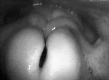
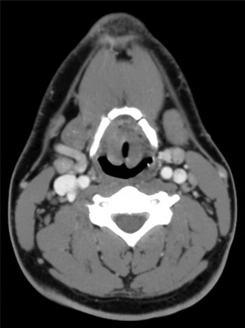
Video-laryngoscopy revealed a severely oedematous, omega-shaped epiglottis (Figure 1). The laryngeal oedema extended down both aryepiglottic folds and included the false vocal folds. The true vocal folds were mobile and without mass or lesion. Computed tomography of the neck demonstrated epiglottic thickening and airway constriction, with no evidence of significant lymphadenopathy (Figure 2).
Video-laryngoscopic view of first patient's epiglottis, which was omega-shaped and severely oedematous.

Axial computed tomography image of first patient, demonstrating the enlarged epiglottis and airway narrowing.

The patient underwent a direct laryngoscopy with biopsies. The biopsies of the epiglottis demonstrated inflammatory infiltrate without evidence of granulomas. Throat culture showed normal respiratory flora. The patient also underwent extensive laboratory investigation, including tests for complement (C) 3 and 4 levels, coccidioides titres, and cytoplasmic antineutrophilic cytoplasmic antibodies (ANCA) and perinuclear ANCA levels, all of which were negative.
Following the results of this investigation, the patient was offered definitive airway management by tracheostomy, but he instead elected for close observation and expectant management. There was a subsequent recurrence of the supraglottic oedema and the patient was admitted to the intensive care unit for antibiotic and steroid treatment, which resolved his symptoms. He endured a prolonged steroid taper and subsequently had an oedematous, but stable, airway. Repeated laryngoscopy continued to reveal evidence of chronic inflammation of the epiglottis and aryepiglottic folds.
Case two
The second patient was a 39-year-old woman who presented with a long history of ‘recurrent throat swelling’. Her symptoms had begun as an upper respiratory tract infection for which she had received treatment with antibiotics. She had improved with antibiotic therapy but the symptoms had subsequently recurred, and she had been seen by an otolaryngologist at another centre. At that time, flexible laryngoscopy had prompted immediate referral to the emergency department, and the patient had been intubated due to extensive epiglottic oedema. She had been extubated five days later following treatment with steroids and intravenous antibiotics.
Over the next 15 years, the patient had suffered seven further episodes of recurrent throat swelling, which had seemed to resolve with antibiotics. However, four months prior to the current presentation, she had suffered another episode of throat swelling; this episode had not responded adequately to antibiotics and the patient had been started on long-term prednisone, which had led to an improvement in symptoms. She had subsequently developed side-effects from the steroids, and the supraglottic oedema had persisted.
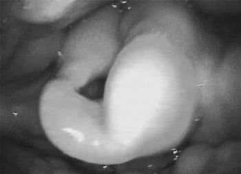
On video-laryngoscopy, the patient was noted to have oropharyngeal and hypopharyngeal oedema (Figure 3). The epiglottis was grossly swollen and the oedema extended to the aryepiglottic folds, and included the false vocal folds and postcricoid region.
Video-laryngoscopic view of second patient's epiglottis, which was grossly swollen and the oedema extended to the aryepiglottic folds.

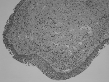
The patient underwent extensive laboratory investigation including cytoplasmic and perinuclear ANCA studies, coccidioides serology, and complement 3 and 4 levels. Her investigation was negative for any abnormalities, and she was taken to the operating theatre for direct laryngoscopy. Biopsies of the epiglottis and aryepiglottic folds revealed chronic inflammation, but no evidence of malignancy, granuloma, immunofluorescence or other abnormality (Figure 4).
Photomicrograph of haematoxylin and eosin staining of the aryepiglottic fold of the second patient, demonstrating chronic inflammation and no evidence of granuloma formation. (×10)

The patient's symptoms improved with prolonged administration of steroids. Despite persistent supraglottic oedema, she had no further respiratory distress.
Discussion
A chronically enlarged epiglottis poses a diagnostic challenge. Patients typically present to their primary care physicians with chronic symptoms of dysphagia, dysphonia, a sore throat, and other non-specific symptoms that provide little assistance in securing a definitive diagnosis. Patients often undergo treatment with antibiotics or therapy for gastroesophageal reflux, which are prescribed by the primary care providers; they do not usually present to an otolaryngologist until symptoms have persisted for some time.
Once neoplasia has been ruled out by biopsy, many disorders must be included in the differential diagnosis.Reference Caldarelli, Friedberg and Harris 21 Laryngeal tuberculosis remains the most common granulomatous disease of the larynx.Reference Caldarelli, Friedberg and Harris 21 It is usually related to active pulmonary tuberculosis and may present with varying degrees of airway obstruction. Initially, there may be generalised supraglottic oedema that may ultimately lead to mucosal ulcerations. Diagnosis is obtained by isolating the acid-fast bacilli on culture and demonstrating the presence of tuberculin granulomas in the tissue. Anti-mycobacterial chemotherapy is the current treatment of choice for this disease.
Syphilitic infection of the larynx is usually noted during the secondary or tertiary phases of the disease, with the presence of diffuse erythematous papules within the larynx.Reference Pace and Csonka 6 , Reference Caldarelli, Friedberg and Harris 21 Diagnosis is based on the demonstration of positive venereal disease research laboratory or fluorescent treponemal antibody-absorption test results, and histology that shows vasculitis and lymphocytic infiltrate, with inconsistent isolation of the causative spirochaete. Treatment with antibiotics is effective in managing syphilis in the secondary phase of disease, but seldom in the tertiary phase of disease.
Infections involving candida species, indolent Haemophilus influenzae type B or actinomycosis can also lead to recurrent episodes of supraglottitis. These are usually successfully treated with antibiotic therapy once identified.Reference Parsons, Smith, Mair and Dlabal 22 – Reference Wilson, Coutras and Tami 24 Viral infections should be ruled out as these may also play a role in recurrent epiglottitis.Reference Musharrafieh, Araj and Fuleihan 9
Patients with hereditary angioedema (C1-esterase deficiency) suffer from recurrent episodes of angioedema that can also involve the larynx and supraglottic structures.Reference Markovic, Inwards, Frigas and Phyliky 16 , Reference Wong and Gadsden 18 These patients will often present with facial swelling and some respiratory complaints, but symptoms rarely include stridor. Diagnosis is made based on a low level of complement 4. Reduced levels of C1-esterase and C1q confirm diagnosis. Treatment is usually directed at the underlying disease process, but if none is discovered, therapy with corticosteroids is usually curative.
Wegener's granulomatosis can also cause epiglottic enlargement with classic ulcerative, necrotic lesions, and oedematous and inflamed mucosa.Reference Talerman and Wright 12 The diagnosis is established by histopathologic findings of vasculitis, and scattered giant cells with poorly formed caseating granulomas.Reference Caldarelli, Friedberg and Harris 21 Wegener's granulomatosis is initially controlled by treatment with steroids, followed by cyclophosphamide or methotrexate for long-term therapy.
Two other cases of non-granulomatous chronic epiglottitis have been reported.Reference Friedman, Toriumi and Strorigl 19 , Reference Schrottenbaum 20 In one case, the patient additionally presented with perioral sores and cystic lesions on the aryepiglottic folds, consistent with a possible diagnosis of benign mucosal pemphigoid.Reference Friedman, Toriumi and Strorigl 19 , Reference Rosenfeld and Biller 25 The second reported case presented with similar symptoms to those of our patients, but there was no mention of the presence or absence of granulomatous changes in the article.Reference Schrottenbaum 20
While antibiotics and steroids have been the mainstay of therapy for oedema of the supraglottic airway, recent literature has examined hydroxychloroquine as a potential steroid-sparing therapy for recurrent airway oedema. Hydroxychloroquine is an antimalarial medication used extensively in the treatment of rheumatologic diseases such as rheumatoid arthritis and systemic lupus erythematosus.Reference Hirshoren, Eliashar and Weinberger 26 , Reference Kalia and Dutz 27 The medication interferes with antigen processing by raising the lysosomal pH and decreasing the function of various immune cells. It thus provides a potent immunomodulatory effect without steroid therapy. A recent report demonstrated the effectiveness of hydroxychloroquine as a steroid-sparing therapy in a case with subglottic stenosis.Reference Hirshoren, Eliashar and Weinberger 26 The patient had been steroid-dependent for over six months and was successfully converted to hydroxychloroquine, which was tapered off over a five-month period. Although this was a single case report, it represents a potential method of treating chronic non-granulomatous supraglottitis.
-
• Chronic non-granulomatous supraglottitis is a diagnosis of exclusion
-
• Patients present with chronic dysphonia, dyspnoea, globus and dysphagia
-
• Diagnosis is made by laryngoscopy, imaging and histopathologic studies
-
• Current management is aimed at airway maintenance with corticosteroids
-
• Possible steroid-sparing therapies for chronic management are being explored
Given the disparate entities that can cause chronic supraglottitis, the investigation of patients with this condition is extensive. After full head and neck examination and flexible laryngoscopy, diagnostic investigation should proceed with basic evaluations, including complete blood count and biochemistry tests. Additional infectious markers should also be evaluated in accordance with the patient history (e.g. coccidioides, histoplasmosis and blastomycosis titres, depending on geographic locale). Ancillary studies such as cytoplasmic and perinuclear antineutrophilic cytoplasmic antibodies, complement 3 and 4 levels, erythrocyte sedimentation rate, C-reactive protein, fluorescent treponemal antibody-absorption, venereal disease research laboratory tests, and purified protein derivative should be carried out as symptoms dictate.
If this preliminary investigation remains inconclusive, evaluation by direct laryngoscopy and biopsy should be performed to rule out neoplasm and establish the presence of granulomas or chondrocyte degeneration. Direct immunofluorescence should be performed to rule out pemphigoid, and positive birefringence under polarised light should be utilised to rule out amyloidosis. If, despite these efforts, the diagnosis is not elucidated, an empirical trial of oral steroids is recommended, with close monitoring to ensure improvement. It is also reasonable to perform ambulatory pH testing to rule out any contribution from laryngopharyngeal reflux. Patients who fail steroid therapy or cannot tolerate the side-effects may also warrant referral to a rheumatologist to discuss hydroxychloroquine therapy as an alternative. Management of the airway is dictated by the severity of the patient's symptoms, wherein a tracheostomy is the gold standard in airway management for recurrent supraglottic oedema.
Conclusion
Chronic non-granulomatous supraglottitis remains a diagnosis of exclusion in patients with chronic inflammation of the supraglottis. Patients may present with chronic dysphonia, dyspnoea, globus and dysphagia. The condition can be exacerbated by infection, leading to respiratory compromise. Diagnosis is made by flexible laryngoscopy, radiological imaging demonstrating epiglottic oedema, and histology suggestive of chronic inflammatory changes in the absence of granulomas, infection, or other diagnosis. Management is primarily focused on airway maintenance with corticosteroids, but possible steroid-sparing therapies for chronic management are being explored.
Acknowledgement
The authors would like to thank Dr Heidi Jess for her assistance in obtaining the photomicrograph for this manuscript.