Introduction
Cisplatin-induced ototoxicity is characterised by a cumulative, dose-related sensorineural hearing loss and tinnitus, and less frequently by vestibular impairment.Reference Schaefer, Wright, Post and Frenkel1 Such hearing loss is progressive, irreversible and bilateral; it usually affects high frequencies first and may gradually spread to lower frequencies.Reference Waters, Ahmad, Katsarkas, Stanimir and McKay2 The major toxic effects of cisplatin include the loss of outer hair cells, degeneration of the stria vascularis and a decrease in the number of spiral ganglion cells.Reference Wright and Schaeffer3, Reference Hoistad, Ondrey, Mutlu, Schachern, Paparella and Adams4
In vitro and in vivo experiments have shown that apoptotic cell death is the primary mechanism of cisplatin toxicity.Reference Boulikas and Vougiouka5 In the cochlea, cisplatin has been shown to induce apoptosis in hair cells, supporting cells, spiral ganglion cells and stria vascularis cells,Reference Alam, Ikeda, Oshima, Suzuki, Kawase and Kikuchi6 by means of activation of caspases, up-regulation of the pro-apoptotic tumour suppressor gene p53, a significant increase in bax-positive cells, a decrease in bcl-2-positive cells and the release of cytochrome C.Reference Devarajan, Savoca, Castaneda, Parks, Esteban-Cruciani and Kalinec7, Reference Zhang, Liu, Ding and Salvi8
Impairment of the K+ recycling process, which is essential to the endocochlear potential, is a known toxic effect of cisplatin induced by apoptosis of type I spiral ligament fibrocytesReference Liang, Schulte, Qu, Hu and Shen9 and by the direct apoptotic effect of cisplatin on the stria vascularis, responsible for decreased cellular Na+/K+ adenosine triphosphatase (ATPase) and Ca2+ ATPase activities.Reference Cheng, Liu, Hsu and Lin-Shiau10
Cisplatin ototoxicity may also result from damage to the structures of the inner ear mediated by free radicals such as reactive oxygen species,Reference Clerici, Di Martino and Prasad11 and from antioxidant depletion, which causes oxidative injury in the rat cochlea.Reference Husain, Scott, Whitworth, Somani and Rybak12, Reference Rybak, Husain, Morris, Whitworth and Somani13
The aim of this descriptive study was to determine the appearance of the cell changes described as blebs in the inner and outer hair cells of the guinea pig following exposure to cisplatin, and to analyse the role of such blebs in the pathogenesis of cisplatin-induced ototoxicity.
Methods
Thirty-one young Hartley guinea pigs weighing 200 to 300 g were used. Twenty-five animals were included in the cisplatin group. Six guinea pigs that had been included in a parallel study of age-related changes of auditory function were used as the control group. This research was performed according to the guidelines of the USA National Institutes of Health Guide for the Care and Use of Laboratory Animals publication, and the Spanish Animal Care and Use Committee. The project was reviewed and approved by our centre's ethics committee. Preyer's test and otoscopy confirmed that the guinea pigs' middle ears were normal.
Experimental design
Animals from the study group were anaesthetised and auditory brainstem responses (ABR) were obtained prior to cisplatin administration. In order to evaluate destructive or degenerative effects in the short, middle and long term, the animals were randomly assigned to three groups. Group A comprised 10 animals that were sacrificed 48 hours after the injection; group B comprised 10 animals that were sacrificed 15 days after the injection; and group C comprised 5 animals that received a second injection of 10 mg/kg of cisplatin 7 days after the first administration and were euthanised 30 days after the initial dose.
Control animals were sacrificed at 48 hours (n = 2), 15 days (n = 2) and 30 days (n = 2).
Procedure
Animals were anaesthetised with an intraperitoneal injection of ketamine 100 mg/kg, diazepam 0.1 mg/kg and atropine 0.02–0.04 mg/kg. Following ABR recording, 5 mg of cisplatin in a 1.0 ml normal solution (0.9 per cent NaCl) was injected intraperitoneally. Five animals were randomly selected to receive a second cisplatin injection of 10 mg/kg one week after the first administration. Animals were observed for poor food and water intake.
Auditory brainstem responses were recorded before drug administration, three days after first administration where applicable (i.e. groups B and C), and before sacrifice.
Auditory brainstem response measurement and interpretation
Animals were tested in an acoustically shielded booth. Subdermal, platinum-alloy needle electrodes were attached, with the active lead at the vertex and referred to a second electrode located at the tip of the nose. The ground electrode was placed on the arm muscles. Ten-microsecond, tone burst stimuli (8 kHz), generated by a Medelec Synergy Generator (Oxford Medical Instruments, Surrey, UK), were delivered monoaurally through a hollow rat ear-bar. Stimuli were repeated five times per second with increasing intensity from 10 to 80 dB (SPL) in 5-dB steps. A range of 375–1221 trials were averaged in order to assure an adequate brainstem response. Hearing threshold was defined as the lowest intensity of stimulation that yielded a recognisable wave.
Pre- and post-treatment ABR thresholds were compared for each ear, and the difference was defined as the threshold change for each time-related group. Auditory brainstem response measurement in control guinea pigs proved to be highly reproducible in the retest schedule, indicating high inter-test reliability.
Sacrifice and specimen preparation
Following anaesthesia and ABR measurement, animals were sacrificed by means of decapitation. Temporal bones were separated from the rest of the cranium and soft tissues, and the cochlear portion was dissected. Under magnification (Zeiss surgical microscope, Carl Zeiss Inc., Jena, Germany), the bony wall of the cochlea was removed with a small pick to expose the spiral ligament. This structure was removed with a pick and forceps to expose the upper aspect of the organ of Corti. Cochlea specimens were then fixed with 2.5 per cent glutaraldehyde in 0.1 M sodium cacodylate buffer. Cochleae were subsequently dehydrated in 10, 30, 70, 90 and 100 per cent concentrations of ethanol (three times each). Critical point was obtained with an Emitec (Ashford, England) 850 system. The samples were splittered with a Bio-Rad (Ashford, England) SC-650 system and observed under a Phillips (Almelo, The Netherlands) XL-30 scanning electron microscope with digital imaging capabilities.
Results
Morphological findings
Scanning electron microscopy of the inner ears of animals from group A (48 hours' survival) showed no blebs.
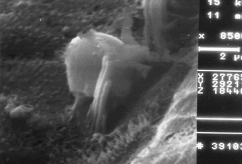
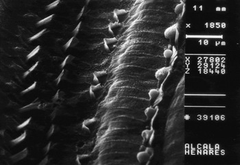
In two animals from group B (15 days' survival), inner hair cells only showed evidence of occasional vesicles, less than 1 µm in diameter, at the junction between the plate and the inner hair cell cuticle (Figure 1). Likewise, apoptotic changes in the outer hair cell cuticular plate were observed, consisting of round microperforations (Figure 2).
Scanning electron photomicrograph of the organ of Corti of a group B animal, showing a bleb emerging from the basal body of the cuticular plate of an inner hair cell (×8500).

Scanning electron photomicrograph of the upper aspect of the organ of Corti of a group B animal, showing microperforations (open arrow) in the outer hair cells (which may correspond to ruptured blebs), the absence of a number of outer hair cells (asterisk), and blebs on inner hair cells (short arrow) (×1850).

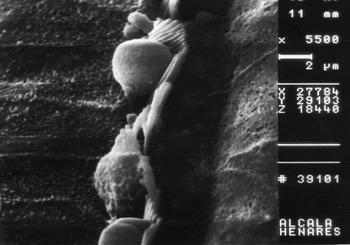
All animals from group C (two cisplatin doses) showed blebs in the region of the primitive kinocilium of most inner hair cells, in different stages of development, as well as the absence of outer hair cells (Figure 3).
Scanning electron photomicrograph of the organ of Corti of an animal from group C, showing inner hair cells blebs at different stages of development (×5500).

Auditory function
One animal from group C died after cisplatin administration. At sacrifice (after the injection of 5 mg/kg of cisplatin), in animals from group B and C which showed inner-ear blebs, average ABR thresholds were 61.6 and 73.3 dB in the right and left ear, respectively (mean of two ears: 67.45 dB). The average ABR thresholds of guinea pigs with no blebs were 36.05 and 37.36 dB in the right and left ear, respectively (mean of two ears: 36.70 dB). Controls did not show any change in auditory function.
Discussion
Blebs are balloon-like protrusions of the plasma membrane which have been associated with cell injury, although they have also been observed in healthy cells during particular stages of the cell cycleReference Porter, Prescott and Frye14 and play a role during embryogenesis.Reference Trinkaus15 Membrane blebs appear when actin attachments between plasma membrane and the underlying cytoskeleton are weakened. Detached blebs are phagocytosed by nearby cells.Reference Zeligs and Wollman16
Blebbing may play a specific role in cell physiology and pathology, such as the regulation of intracellular pressure. During swelling, the geometry of the cell body remains stable, with the extra fluid being diverted to the blebs. On early exposure to lethal insults, cells lose most of their major osmolytes (potassium, chloride and taurine) by a response termed ‘apoptotic volume decrease’. If sodium overload occurs after the cells have exhausted their supply of osmolytes, volume will only be discarded via Na+/K+ ATPase or, in the absence of ATP, by growth and shedding of blebs.
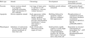
Studies on these types of morphological cell changes have enabled blebs to be identified as either apoptotic, necrotic or physiological (Table I). Blebs were first recognised as a hallmark of apoptosis. In this setting, blebs form within 30 minutes after exposure to diverse stimuli, coinciding with channel-mediated K+ and Cl− loss during apoptotic volume decrease, preceding mitochondrial cytochrome C release and caspase-3 activation.Reference Okada, Maeno, Shimizu, Dezaki, Wang and Morishima17 During apoptosis, caspase-3 cleaves Rho-kinase I, leading to deregulated activity of this kinase and consequently to bleb formation.Reference Coleman, Sahai, Yeo, Bosch, Dewar and Olson18
Characterisation of different bleb types observed
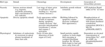
ATP = adenosine triphosphate; min = minute; ROCK = Rho-kinase I; MLCK = myosin light chain kinase; p38MAPK-HSP27 = p38 mitogen activated protein kinase-heat shock protein 27; cAMP = cyclic adenosine monophosphate
Necrotic blebs appear during the last stages of injury prior to cell lysis, and they grow indefinitely without retraction. Necrotic blebbing is associated with a type of cell swelling termed ‘necrotic volume increase’, the execution phase of necrosis, characterised by a rapid influx of Na+. Necrotic blebs are driven by a net gain of sodium and water, and are enlarged by hydrodynamic forces that eject cytosolic material from the cell body, presumably because of cytoskeletal dysfunction.
Blebs have also been observed in physiologically healthy cells at particular stages of the cell cycle, such as when mesoderm cells migrate within the embryoReference Krause and Cutts19 and in neurons differentiating in primary cultures.Reference Olson20
Vesicular structures resembling blebs have been described in the stria vascularis of a patient who had received aminoglycoside antibiotics,Reference Moffat and Kerr21 and in the outer hair cells of a guinea pig treated with neomycin.Reference Friedmann and Kerr22
In a recent study which examined organs of Corti freshly isolated from adult guinea pigs,Reference Shi, Gillespie and Nuttall23 large blebs were observed to form rapidly on the apical membranes of inner hair cells. This bleb development was associated with elevated concentrations of intracellular Na+. Bleb formation was not significantly reduced by disassembling microtubules or decreasing intracellular hydrostatic pressure. Thus, inner hair cell blebs were thought to have arisen as a result of an imbalance of endocytosis and exocytosis in the apical plasma membrane, linked to Na+ loading that occurs in vitro. Phosphatidylserine-rich particles and blebs appeared at the apical membrane of inner hair cells and, more rarely, on outer hair cells, within freshly isolated organs of Corti from adult guinea pigs.Reference Shi, Gillespie and Nuttall23 These particles aggregated at the edge of the cuticular plates of inner hair cells, in the vestigial kinocilium area, and gradually merged into a single, spherical bleb.
Blebbing has been observed to be ATP and cyclic adenosine monophosphate dependent, via a biochemical pathway regulated by phosphatidylinositol 3-kinase activity. Sodium ions can enter hair cells through P2X2 channels located at the apical pole of inner hair cells.Reference Housley, Raybould and Thorne24 Based on the findings of Shi et al.,Reference Shi, Gillespie and Nuttall23 neither the mechanism of blebbing nor the growth in size of inner hair cell blebs is thought to be associated with cellular apoptosis or necrosis. This phenomenon may also occur in vivo under certain pathological conditions, e.g. following loud auditory stimulation.Reference Zeddies and Siegel25
• Blebs are balloon-like protrusions of the plasma membrane, which have been associated with pathological and normal physiological cell processes
• Blebbing is thought to play a specific role in the regulation of intracellular pressure
• The appearance of blebs in the inner ear following cisplatin administration has rarely been described and is difficult to explain
• This animal study found a strong relationship between the number and extension of blebs and the presence of hearing loss, suggesting that these blebs are a manifestation of apoptosis or necrosis secondary to cisplatin administration
The appearance of blebs in the inner ear following cisplatin administration has rarely been described and is difficult to explain. Such blebs have been described as scanning electron microscopy artefacts by some authors.Reference Ibsch, Anken, Vöhringer and Rahmann26 In previously published studies by our group, the presence of blebs in diverse areas of the apical region, such as the kinocilium and other marginal areas, was observed in the inner and outer hair cells of rats following cisplatin administration. These were attributed to early degenerative changes in the inner hair cells, in the region of the primitive kinocilium, which is presumed to be the weakest area of the apical end of inner hair cells.Reference Ramírez-Camacho, García-Berrocal, Buján, Martín-Marero and Trinidad27, Reference Ramírez-Camacho, García-Berrocal, Trinidad, Martín-Marero and Buján28
In the present study, blebs in the region of primitive kinocilium appeared in only a few animals treated with a low dosage of cisplatin (5 mg/kg, single injection). Round microperforations found in the cuticular plate of the outer hair cells could correspond to ruptured blebs. Moderate impairment of hearing, shown by ABR, suggests that this type of bleb could be an early form of adaptive response which develops when damage is still limited. However, blebs in animals treated with higher amounts of cisplatin (group C) were present in a greater number of inner hair cells. Blebs found in group C animals showed different developmental stages; destruction of some blebs could also be seen in the outer hair cells. These morphological findings, together with the severe hearing loss found in these animals, suggests that these blebs are a manifestation of apoptosis or necrosis and not a physiological adaptation, contrary to Shi and colleagues' proposal.Reference Shi, Gillespie and Nuttall23 The absence of such findings in the control group and in most animals from the low dosage groups (A and B) supports this hypothesis. Further studies employing distortion product otoacoustic emissions, which could provide a direct insight into outer hair cell function, are required in order to clarify the role of blebs found in inner-ear structures.
Acknowledgements
This study was supported by grants FIS 05/0673 and CT2005-08593/BQU. The authors would like to thank Dr R Sanz, Head of the ENT Department, University Hospital of Getafe, Dr J A González-García and Dr A Ibáñez of the ENT Department, University Hospital Puerta de Hierro, Professor J Bujan of the Department of Morphology of the University of Alcalá de Henares, and Dr E Jorge-Herrero of the Experimental Surgery Department, University Hospital Puerta de Hierro, for their critical participation in the experimental procedure.