1. Introduction
The first echinoderms appeared during Cambrian time and rapidly diversified, reaching a major diversity peak by Stage 5 – Drumian. However, known echinoderm diversity dramatically decreased by the end of the period (Zamora et al. Reference Zamora, Lefebvre, Álvaro, Clausen, Elicki, Fatka, Jell, Kouchinsky, Lin, Nardin, Parsley, Rozhnov, Sprinkle, Sumrall, Vizcaïno, Smith, Harper and Servais2013), only to rebound during Ordovician time when they reached their highest disparity (Sprinkle & Guensburg, Reference Sprinkle, Guensburg, Albanesi, Beresi and Peralta2003; Lefebvre et al. Reference Lefebvre, Sumrall, Shroat-Lewis, Reich, Webster, Hunter, Nardin, Rozhnov, Guensburg, Touzeau, Noailles, Sprinkle, Harper and Servais2013). Several of the well-defined clades that dominate Ordovician faunas probably have roots in the Furongian Epoch, but the poor record of fossil echinoderms from this stage hampers our ability to identify and interpret plesiomorphic conditions of these important taxa. Sanducystis sinensis n. gen. n. sp. described here is a rare exception.
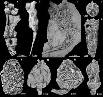
In 1968, Paul revaluated Macrocystella mariae Callaway, Reference Callaway1877 (Fig. 1c), an Early Ordovican ‘cystoid’ of the British Isles, and concluded that it was the earliest known member of Glyptocystitida. This clade was highly successful during Palaeozoic time; it originated at the end of the Cambrian Period and ranged through the Devonian Period. Most Ordovician glyptocystitoids are diagnosed by having a theca composed of four plate circlets including: four basals (one of them, B4, is larger and located in the BC interradius); five infralaterals; five laterals; and four to ten radials. The summit is plated with seven interradial orals with the CD interradius divided into three orals (O1, O6 and O7) associated with the hydropore and gonopore (Sumrall & Sprinkle, Reference Sumrall, Sprinkle, Carnevali and Bonasoro1999). The stem is holomeric and divided into two parts. The highly flexible and expanded proximal part bears thin outer and inner columnals with a large lumen and spiralling pivot points, whereas the stiffer distal portion bears homeomorphic, barrel-shaped columnals with an extremely narrow lumen. Most glyptocystitoids were very likely free living. However, in some callocystitids the presence of cemented holdfasts in adults suggests that the stem was used for permanent attachment in these taxa (Kesling & Mintz, Reference Kesling and Mintz1961; Koch & Strimple, Reference Koch and Strimple1968). The periproct is generally medium to very large and laterally positioned on the side of the theca in the BC interradius; most species have respiratory pectinirhombs positioned along specific plate sutures.
Glyptocystitida and other related stemmed echinoderms. (a) Velieuxicystis ornata Ubaghs, Reference Ubaghs1998, from Furongian deposits of France. Note the four circlets of plates with the feeding appendage facets mounted on radial plates and the periproctal opening lateral. Specimen number FSL 712 765. (b) Barroubiocystis radiata Ubaghs, Reference Ubaghs1998, from Furongian deposits of France, showing a theca composed of four circlets of plates. Specimen number FSL 712 758. (c) Macrocystella mariae Callaway, Reference Callaway1877, a classic Early Ordovician glyptocystitoid. Specimen number NHM UK EE7524. (d, e) Ridersia watsonae Jell, Burrett & Banks, Reference Jell, Burrett and Banks1985 from Furongian deposits of Australia. Note the conical theca organized into three circlets of plates and flat summit with periproct in the CD interray. Specimen numbers NMVP107115 and NMVP107114. (f) Lipsanocystis magnus UMMP 31432 showing the periproctal side of the theca. (g) Pleurocystites squamosus CMC-IP 39537, showing the dorsal surface of the flattened theca. (h) Echinoencrinites lahuseni MCZ 114624 showing the CD interray of the theca and the curved stem. (i) Coronacystis angulatus showing the well-developed pectinirhombs along the radial sutures and low lateral position of the periproct SUI 97605. Photographs taken from latex casts whitened with NH4Cl sublimated.

Since the interpretation of Macrocystella as a plesiomorphic, rhomb-less glyptocystitoid rhombiferan (Paul, Reference Paul1968), several Cambrian stemmed echinoderms that are intermediate in morphology between ‘eocrinoids’ and Glyptocystitida were described. Some of these taxa were assigned to eocrinoids, for example, Ridersia from Australia (Fig. 1d, e) and Cambrocrinus from Poland (Jell, Burrett & Banks, Reference Jell, Burrett and Banks1985; Dzik & Orłowski, Reference Dzik and Orłowski1993). Others were interpreted as plesiomorphic glyptocystitoids, for example, Velieuxicystis (Fig. 1a) and Barroubiocystis (Fig. 1b) both from France (Ubaghs, Reference Ubaghs1998) and the enigmatic middle Cambrian Dibrachicystis (Zamora & Smith, Reference Zamora and Smith2012) from Spain and France. Unfortunately, Dibrachicystis is difficult to interpret because poor preservation prevents a clear understanding of its morphology and consequently its phylogenetic position (Zamora & Smith, Reference Zamora and Smith2012). The other Furongian taxa (Ridersia, Velieuxicystis and Barroubiocystis) have been reviewed, and their morphological information has been included in a phylogenetic analysis (see Section 4.b) to clarify their phylogenetic position.
Unlike other Cambrian glyptocystitoids, Sanducystis sinensis n. gen. n. sp. from the Furongian sediments of South China is abundant, well preserved and provides a combination of characters that elucidates how Glyptocystitida evolved their body plan. The aims of this paper are first to describe the new form and second to provide a phylogenetic analysis of Glyptocystitida to resolve the evolutionary position of Sanducystis.
2. Geological setting and stratigraphy
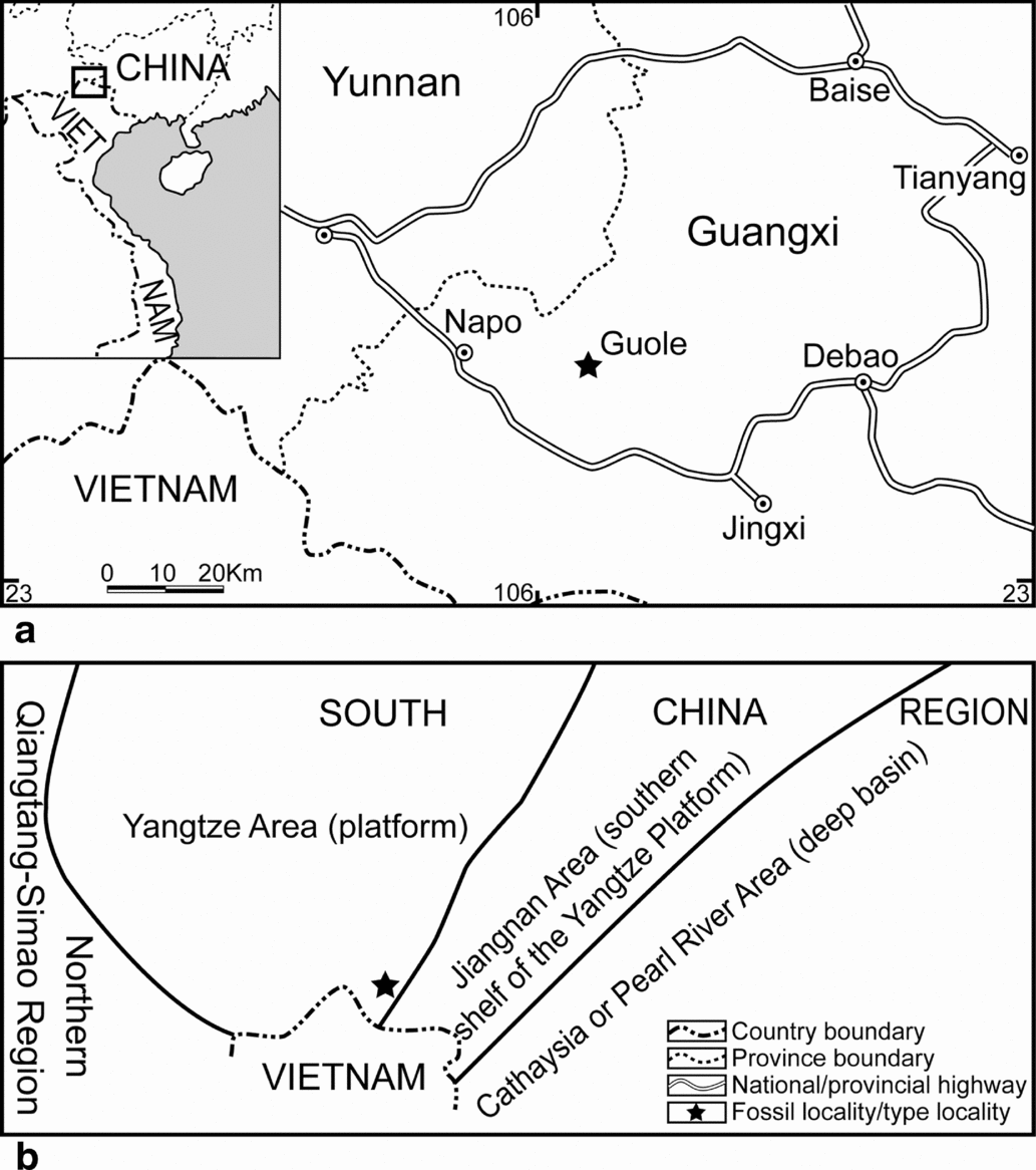
Specimens of Sanducystis sinensis n. gen. n. sp. were collected from mudstones and marls of the Furongian Sandu Formation, Guole Town, Jingxi County, Guangxi Province, SW China (Fig. 2). Guole Town lies in the Yangtze area of the South China Region. During late Cambrian time, the Yangtze area was covered by a shallow marine shelf (Zhou & Zhen, Reference Zhou and Zhen2008). The Sandu Formation has yielded a diverse associated fauna including aglaspididids, brachiopods (e.g. Billingsella, Guoleella, Palaeostrophia, Plectotrophia), echinoderms (e.g. ‘Phyllocystis’ jingxiensis, Cambroblastus guolensis), graptolites (e.g. Callograptus, Dyctionema), hyolithids, trilobites (e.g. Akoldinioidia, Dictyella, Guangxiaspis, Shergoldia, Sinosaukia, Tamdaspis) and undescribed palaeoscolecids (Han & Chen, Reference Han and Chen2008; Zhan et al. Reference Zhan, Jin, Rong, Zhu and Han2010; Lerosey-Aubril, Ortega-Hernández & Zhu, Reference Lerosey-Aubril, Ortega-Hernández and Zhu2013; Zamora, Zhu & Lefebvre, Reference Zamora, Zhu and Lefebvre2013; Zhu, Zamora & Lefebvre, Reference Zhu, Zamora and Lefebvre2014; Zhu et al. Reference Zhu, Peng, Zamora, Lefebvre and Chen2016). Co-occurring trilobites indicate that these deposits are correlative with the Probinacunaspis nasalis – Peichiashania hunanensis Zone of NW Hunan Province, which is part of the Furongian, Stage 9 strata (Zhu, Hughes & Peng, Reference Zhu, Hughes and Peng2007, Reference Zhan, Jin, Rong, Zhu and Han2010). Abundant articulated trilobites strongly suggest that the fauna was buried rapidly, likely as the result of an obrution event (Zhu, Hughes & Peng, Reference Zhu, Hughes and Peng2010). These taphonomic conditions are consistent with the exquisite preservation of the echinoderms.
(a) Geographic location (indicated with the star) and (b) geological setting (star) of the studied site (modified from Zhou et al. Reference Zhou, Zhen, Peng and Zhu2008).
3. Systematic palaeontology
Specimens are deposited in the Nanjing Institute of Geology and Palaeontology, Chinese Academy of Sciences, under repository numbers NIGPAS162387–NIGPAS162401.
Phylum ECHINODERMATA Bruguière, Reference Bruguière1791 (ex Klein, Reference Klein1734)
Subphylum BLASTOZOA Sprinkle, Reference Sprinkle1973
Class ‘RHOMBIFERA’ Zittel, Reference Zittel1879
Order GLYPTOCYSTITIDA Bather, Reference Bather1899
Sanducystis new genus
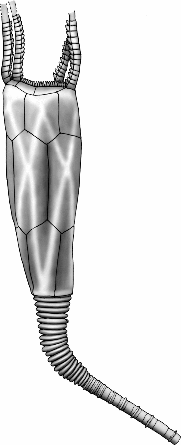
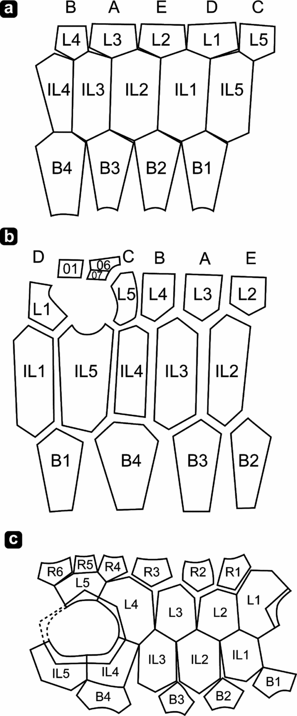
Diagnosis. A genus of Glyptocystitida with a proportionately tall and thin well-organized theca formed from three closed plate circlets (four basals, five infralaterals, and five laterals) (see Fig. 3). Oral surface comprising four ambulacra (B, C, D, E), with the ambulacrum A absent. Anus laterally positioned in CD interray, bordered by oral plates O1 and O7, two laterals (L1, L5) and one infralateral (IL5). Pectinirhombs absent but complex respiratory structures consisting of thin rounded areas bordered by thickened rings visible only on interior of thecal plates.
Reconstruction of Sanducystis sinensis showing its characteristic thecal plating with three circlets of plates, few brachioles arranged in 2-0-2 pattern, proximal stem and part of the distal stem.

Derivation of the name. The name refers to the Sandu Formation from which the specimens were collected.
Discussion. Sanducystis is similar to some Cambrian eocrinoids but bears several derived characters typical of Glyptocystitida (see Section 4.b). Both Ridersia from the Furongian of Australia (Jell, Burrett & Banks, Reference Jell, Burrett and Banks1985) and Sanducystis bear a holomeric stem divided into a proximal portion constructed with outer and inner columnals and a distal portion composed of narrow, homeomorphic, barrel-like columnals. In both taxa the four basal plates form a quadrangular shape proximally around the stem facet, and the exterior surface of the thecal plates is crossed by near-vertical, V-shaped (basals) or X-shaped (infralaterals and laterals) low ridges. Furthermore, they both have tall, narrow thecae constructed with three closed circlets of plates and the same number of plates per circlet. However, while the periproct of Ridersia is within the oral area in the CD interray, it is located laterally on the side of the theca in Sanducystis.
Velieuxicystis and Barroubiocystis from Furongian deposits of France (Ubaghs, Reference Ubaghs1998) are very early glyptocystitoids with four circlets of plates. Velieuxicystis appears to have the periproct positioned laterally, but the oral area is poorly known. The interpreted periproct appears to be positioned above a flat-topped B4 plate, but there are inconsistencies noted between specimens as illustrated in Ubaghs (Reference Ubaghs1998). The oral area of Barroubiocystis is also unknown, but the lack of evidence for the periproct being laterally positioned suggests it was placed within the oral area similarly to Ridersia. Cambrocrinus from Furongian deposits of Poland also bears similarity to Sanducystis in stem morphology, but it has more circlets of plates and the periproct is positioned within the oral area. Other features of Cambrocrinus are poorly documented because of the poor preservation in coarse sandstone.
Several aspects of Sanducystis show similarity to more derived Glyptocystitida such as Macrocystella. Sanducystis shows the same general morphology and theca organized in circlets. However, derived Glyptocystitida have four circlets (basal, infralateral, lateral and radial) while Sanducystis lacks the radial circlet (see ‘Discussion’ above). Derived glyptocystitoids also have the anal opening in a lateral position lying in the BC interray, while it is positioned in the CD interray in Sanducystis. The phylogenetic analysis presented below suggests that the lateral migration of the periproct very likely occurred independently in both Sanducystis and other glyptocystitids (including Velieuxicystis). It is very unlikely (from a constructional point of view) that the periproct in Sanducystis would have migrated first aborally, and then from CD to BC interray. Rather, the periproct in Glyptocystitoids probably had two independent aboral migrations: one within the CD interray as documented only in Sanducystis, and the other within the BC interray as occurred in Macrocystella-like forms.
The plating of the oral area in Sanducystis appears to have resulted from paedomorphic evolution. The typical oral plate-bearing stemmed echinoderm summit bears five ambulacra separated by interradially positioned oral plates arranged in a 2-1-2 pattern (Sprinkle, Reference Sprinkle1973; Sumrall, Reference Sumrall, Harris, Böttger, Walker and Lesser2010; Sumrall & Waters, Reference Sumrall and Waters2012; Kammer et al. Reference Kammer, Sumrall, Zamora, Ausich and Deline2013). The loss of the A ambulacrum in the oral area of Sanducystis is probably the result of paedomorphic ambulacral reduction, as described by Sumrall & Wray (Reference Sumrall and Wray2007). Furthermore, the ambulacra are relatively short, bearing only a few brachioles clustered on the tips of the B–E lateral ambulacra much like the condition in the paedomorphic cheirocrinid Sprinkleocystis (Broadhead & Sumrall, Reference Broadhead and Sumrall2003).
Sanducystis sinensis n. sp
(Figs 3–6)
Diagnosis. Same as genus by monotypy.
Derivation of the name. Named for China.
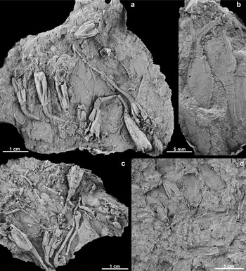
Material and preservation. Large collection of 85 specimens preserved in different taphonomic states, from fully articulated to disarticulated. Thecal flattening seen in many specimens is also variable, suggesting that some specimens preserved with high relief (Fig. 4a, c) were filled with sediment prior to final burial and/or the surrounding sediment underwent rapid lithification. Other specimens are completely flattened (Fig. 4b, d), suggesting no sediment infilling and/or poor lithification of surrounding sediments resulting in thecal collapse during compaction. Morphological characters are more easily interpreted in three-dimensionally preserved specimens, as some features (i.e. suture between plates) become nearly invisible or damaged as specimens become flattened.
Sanducystis sinensis from the Sandu Formation (Furongian Series, upper Cambrian) South China. General views of different slabs showing different states of preservation. (a) Slab NIGPAS163198 with eight partially complete specimens of Sanducystis sinensis preserving original relief. Associated fauna includes trilobite cranidia (centre and right) and the stylophoran ‘Phyllocystis’ jingxiensis (lower left). (b) Small slab with two extremely flattened specimens of Sanducystis sinensis (NIGPAS163199) in which the plate sutures are poorly expressed. (c) Slab preserving six partial specimens (NIGPAS163200). (d) Fragment of a large slab NIGPAS163201preserving six flattened specimens and several isolated plates. Note that in this case, even with specimens preserved flattened, the plate sutures are observable. All photographs were taken from latex casts whitened with NH4Cl sublimated.

Type and figured specimens. The holotype is a complete specimen preserving part and counterpart (Fig. 5d, e, o) which occur in a slab containing eight specimens (Fig. 4a). The specimen museum number is NIGPAS163198. Paratypes include all the other figured specimens, NIGPAS163199–NIGPAS163210. Specimens are deposited in the Nanjing Institute of Geology and Palaeontology, Chinese Academy of Sciences, China (NIGPAS). Other specimens figured are deposited in the following institutions: NHM (Natural History Museum, London); NMVP (Museum of Victoria Palaeontological Collection); FSL (Lyon 1 University Collections); UMMP (University of Michigan Museum of Paleontology); CMC-IP (Cincinnati Museum Center); and MCZ (Museum of Comparative Zoology-Harvard).
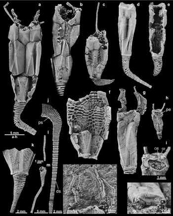
Sanducystis sinensis from the Sandu Formation (Furongian Series, upper Cambrian) South China. Note that all specimens (with the exception of specimens i, l, n and o) are at the same scale to emphasize changes along ontogeny. (a) Large specimen NIGPAS163202 showing its characteristic thecal plating and ornamentation. (b) Large specimen NIGPAS163202 showing the periproctal opening. (c) Medium-sized specimen NIGPAS163203 showing the internal part of a radial plate and a single preserved brachiole. (d, e) Part and counterpart of same unflattened NIGPAS163198 specimen, showing external and internal views of unflattened thecal plates. (f) Preserved specimen NIGPAS163204, showing the internal anatomy of thecal plates with characteristic web of thicker and thinner rods. (g) Medium-sized specimen NIGPAS163205, preserving the brachioles and external growth lines. (h, i) Small specimen NIGPAS163206 showing the anal pyramid, CD orals and C- and D-ray ambulacra with only two small brachioles. (j) Stem from a very large specimen NIGPAS163207, preserving the transition between the curved proximal and straight distal stem. Note how distal columnals increase in height but retain an alternation between flanged and unflanged columnals nearly to the distal tip. (k) Basal plates B3 and B4 (on left) in partial specimen NIGPAS163208 attached to rapidly tapering proximal stem. (l) Distal part of the stem consisting of relatively thin and long holomeric columnals. Specimen number NIGPAS163209. (m) Partially complete stem preserving the transition between the proximal and distal stem. Specimen number NIGPAS163210. (n) Enlargement of specimen in (g) showing summit and details of 5–6 brachioles. (o) Summit view of specimen in (d). All photographs were taken from the latex casts whitened with NH4Cl sublimated. Abbreviations: B, C, D, E – ambulacra; O1–O7 – oral plates; cp – cover plates; ds – distal stem; pe – periproct; ps – proximal stem.

Locality. Sandu Formation, Guole Town, Jingxi County, Guangxi Province, SW China. Co-occurring trilobites indicate an age equivalent to the Probinacunaspis nasalis – Peichiashania hunanensis Zone of NW Hunan of the Furongian, Stage 9 strata (Zhu, Hughes & Peng, Reference Zhu, Hughes and Peng2007, Reference Zhan, Jin, Rong, Zhu and Han2010).
Description.
Stem: Holomeric, long, about three times the length of the theca in the most complete specimen, probably longer. It is very wide proximally but tapers distally, divided in two parts; transition between two parts gradual marked by the increase in height of inner columnals (Fig. 5j, m). The proximal stem is plated with flanged outer columnals and smaller (mostly hidden) inner columnals each bearing a wide circular lumen. The number of proximal columnals is variable, ranging from 19 to 25. The transition from proximal to distal stem is gradational as inner columnals progressively increase their height, outer columnals progressively reduce their flange and stem diameter decreases (Fig. 5j). Distal columnals are nearly homeomorphic, cylindrical, but still show an alternating, low, central flange (Fig. 5j, l). They are slightly wider than tall in the proximal parts and higher than wide in the distal-most portion. Distally, the lumen is very small consisting of a circular perforation occupying one-third of the total columnal diameter. Articulations between distal columnals smooth. No evidence for holdfast structure.
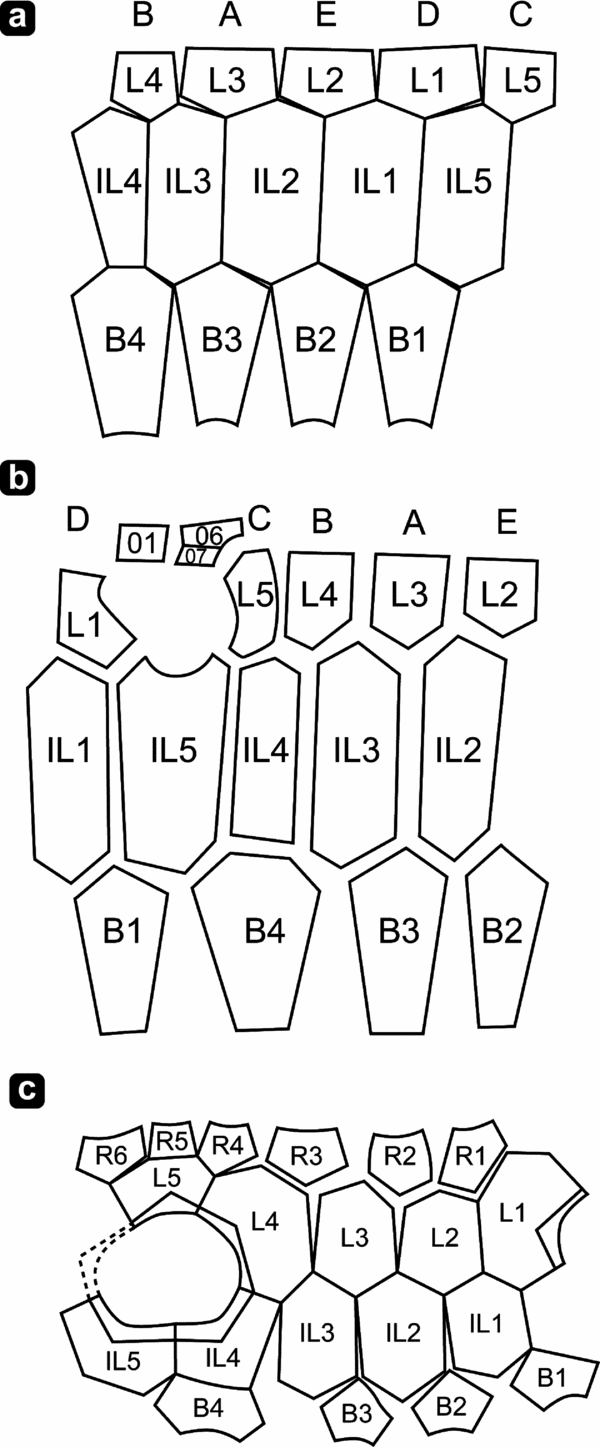
Theca: The theca is elongate and narrow, ranging from 5.5 mm up to 22 mm in height in known material, with an expanded summit and a relatively wide base (Fig. 5a, d, g), two and a half times higher than wide. There are a total of 14 thecal plates organized in three closed (sensu Paul, Reference Paul1968) circlets (four basals, five infralaterals and five laterals), although the lateral circlet is interrupted by the periproct (Fig. 6).
(a) Interpretation of the thecal plating in Ridersia watsonae from Furongian deposits of Australia. (b) Interpretation of the thecal plating in Sanducystis sinensis from Furongian deposits of China. (c) Interpretation of the thecal plating in Early Ordovician Macrocystella mariae from the United Kingdom (after Paul, Reference Paul1968).
The basal circlet (B1–B4) articulates with infralateral plates adorally and accommodates the insertion of the stem aborally. Plates B1, B2 and B3 are very similar in shape, pentagonal, higher than wide, with an expanded adoral portion and tapering distally. These plates typically have a slightly longer basal–infralateral suture either on the left side (B3 and perhaps B4) or on the right side (B1 and B2; Fig. 5b). Plate B4 is hexagonal with a horizontal adoral suture, and is the largest basal. Base of theca marked by well-developed square flange and a lack of a well-developed basal cavity.
The infralateral circlet has five plates (IL1–IL5). They articulate with laterals and basals and also contribute to periproctal frame. Plates IL1, IL2 and IL3 are roughly hexagonal, higher than wide (Fig. 5a, d). Plate IL2 is hexagonal in shape, equally higher than wide. IL4 is smaller than the other infralaterals, pentagonal and with a horizontal base that articulates with B4. Plate IL5 is modified, heptagonal, with an invaginated adoral side to accommodate the periproctal opening, and is the largest infralateral.
The lateral plate circlet has five plates (L1–L5) that articulate adorally with oral plates and aborally with infralateral plates (Fig. 5d). The laterals are approximately half the height of other thecal plates. L1 and L5 have a rounded, invaginated border that accommodates the periproct. L1 articulates adorally with the O1, while L5 articulates adorally with orals O6 and O7. L2, L3 and L4 are pentagonal and slightly wider. All are flat on the adoral side to articulate with oral plates.
The transition between the stem and the theca is abrupt (Fig. 5k). External ornamentation of plates consists of a network of thin, distinct, rounded, near-vertical ridges that extend from a central umbo to the upper and lower sides of the plates, connecting umbos of vertically adjacent plates (Fig. 5a). Between the ridges, the plates are generally flat and smooth with fine external growth lines.
An internal surface of thecal plates consisting of a web of thin rods that generally lie perpendicular to (and cross) sutures along plate margins form a small honeycomb network near plate centers (Fig. 5f). Thin areas between rods may have served as gas exchange areas, whereas the rods act to strengthen the theca.
Subvective system and oral area: Four ambulacra arranged in a 2-0-2 pattern with the A ambulacrum missing (Fig. 5i, o). Summit is plated with interradially positioned oral plates, oval in outline and laterally elongate between the shared ambulacra. Shared ambulacra and peristomial opening covered with small, thin, lathe-shaped ambulacral cover plates (Fig. 5o). Oral plates O1–O7 preserved. O1, O6 and O7 positioned in the CD interambulacrum. O1 and O6 contribute to the peristomial opening while O1 and O7 contribute to the adoral edge of the periproctal opening. O3 and O4 are sutured along the position of the absent A ambulacrum and are twice the size of other oral plates. O2 and O5 are small and positioned in the BC and DE interambulacra, respectively. Ambulacra are extremely short, confined to the summit (Fig. 5o) and bear floor plates although ambulacra are poorly documented in available material. Relatively few brachioles are present on the ambulacra, ranging from two in total in the very small specimens (TH = 5 mm) to as many as five in the largest (TH = 17 mm), probably more in number. The arrangement is typical for short ambulacra as in most cheirocrinids. Brachioles are long (at least two-thirds of thecal height) and relatively stout with large brachiolars and cover plates (Fig. 5c, n). There are approximately two cover plates per brachiolar.
Periproct: Periproctal opening large, circular to elliptical, approximately one-quarter of the thecal height (Fig. 5b, h). It is surrounded by thecal plates L1, L5 and IL5 and oral plates O1 and O7. Anal pyramid occupies half of the periproctal opening bearing small, lathe-shaped plates, at least 14 in number (Fig. 5i). Small periproctal membrane present around periphery of anal pyramid composed of more equant, polygonal plates.
Hydropore and gonopore not observed on studied specimens.
4. Discussion
The origin and basal relationships of major stemmed echinoderm clades is controversial (Sprinkle, Reference Sprinkle1973; Paul, Reference Paul, Paul and Smith1988; Zamora & Smith, Reference Zamora and Smith2012). This is partly because of the difficulty in linking Cambrian groups with more derived and well-defined Ordovician clades (Zamora & Rahman, Reference Zamora and Rahman2014). The Furongian echinoderm record is critical because many Ordovician clades appear to have roots during that time. Unfortunately, the latest Cambrian echinoderm fossil record is very patchy with only few localities worldwide providing articulated specimens (Sprinkle, Reference Sprinkle1973; Jell, Burrett & Banks, Reference Jell, Burrett and Banks1985; Dzik & Orłowski, Reference Dzik and Orłowski1993; Sumrall, Sprinkle & Guensburg, Reference Sumrall, Sprinkle and Guensburg1997; Ubaghs, Reference Ubaghs1998; Zamora, Reference Zamora2012; Zamora et al. Reference Zamora, Lefebvre, Álvaro, Clausen, Elicki, Fatka, Jell, Kouchinsky, Lin, Nardin, Parsley, Rozhnov, Sprinkle, Sumrall, Vizcaïno, Smith, Harper and Servais2013). The Furongian-age Sanducystis displays a morphology that links the stemmed echinoderm Furongian Ridersia and more derived Glyptocystitida, providing a deep root for the glyptocystitoid body plan and helping to define character evolution for the clade.
4.a. Thecal plating of Ridersia, Sanducystis and basal Glyptocystitida
Derived members of Glyptocystitida are diagnosed by having a theca composed of five plate circlets including: four basals (a large one in the BC interradius and three smaller ones); five infralaterals; five laterals; four to ten radials; and seven interradial orals with the CD side bearing three orals (Sumrall & Sprinkle, Reference Sumrall, Sprinkle, Carnevali and Bonasoro1999). The anal opening is consistently positioned on the side of the theca in the BC interray. Macrocystella (Fig. 1c) and Cuniculocystis show this pattern but, in contrast to more derived Glyptocystitida, they lack respiratory structures in the form of pectinirhombs. The thecal plating of this group is very conservative since Ordovician time, but few Cambrian taxa have these defining characters of Glyptocystitida.
Ridersia was originally interpreted as an eocrinoid (Jell, Burrett & Banks, Reference Jell, Burrett and Banks1985). The three plate circlets forming the theca of Ridersia were originally identified as the basal, lateral and radial circlets (Jell, Burrett & Banks, Reference Jell, Burrett and Banks1985, p. 204, fig. 14), by comparison with the situation in other ‘pelmatozoans’ (Fig. 6a). Similarly, the plate pattern of Sanducystis comprises three plate circlets (Fig. 6b) which are here considered homologous to the three plate circlets of Ridersia, interpreted in both taxa as corresponding to the basals, infralaterals and laterals. This interpretation is based on the described ontogenetic series of the derived glyptocystitid Lepadocystis moorei in which a very small specimen lacks the radial circlet and a slightly larger specimen shows the insertion of the radial circlet (Sumrall & Sprinkle, Reference Sumrall, Sprinkle, Carnevali and Bonasoro1999). Both ontogeny and the morphology of Sanducystis suggest that the original plate arrangement for Ridersia was incorrectly identified and that it bears three plate circlets homologous to the thecal plating of Sanducystis. This allows a proper comparison between these genera and the establishment of homologies.
4.b. Phylogenetic position of Sanducystis
To better understand the evolutionary history of early glyptocystitoids, a phylogenetic analysis was conducted on exemplar species of different glyptocystitoid clades. A total of 13 taxa were scored for 34 phylogenetically informative characters that were unordered and equally weighted (see Appendix with character list and codification). Two taxa Hemicosmites and Stephanocrinus were selected as outgroups to polarize the matrix as they are inferred to be closely related to glyptocystitoids (Sumrall & Waters, Reference Sumrall and Waters2012). Trees were searched in Paup* (Swofford, Reference Swofford2002) using maximum parsimony.
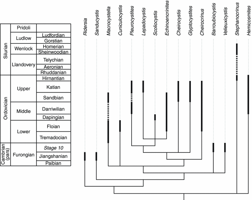
The analysis recovered 44 equally most parsimonious trees of length 62, CI = 0.645, RI = 0.728 and RC = 0.47 (Fig. 7). All of the trees recovered the pectinirhomb-bearing taxa as a clade. The derived taxa lacking pectinirhombs but possessing a radial plate circlet Macrocystella, Cuniculocystis, Velieuxicystis and Barroubiocystis all formed a clade with the pectinirhomb-bearing taxa, but there was no clear relationships inferred among these taxa largely because of the incomplete preservation of both Velieuxicystis and Barroubiocystis. Sanducystis n. gen and sp. and Ridersia are inferred as nested sister taxon to this clade in all most parsimonious trees (MPTs).
Evolutionary tree of Palaeozoic Glyptocystitida and related forms, based on the Adams consensus tree, showing their inferred relationships based on our cladistic analysis and the phylogenetic position of Sanducystis sinensis. Range of fossils indicated in thick black lines (data from Lefebvre et al. Reference Lefebvre, Sumrall, Shroat-Lewis, Reich, Webster, Hunter, Nardin, Rozhnov, Guensburg, Touzeau, Noailles, Sprinkle, Harper and Servais2013 and Zamora et al. Reference Zamora, Lefebvre, Álvaro, Clausen, Elicki, Fatka, Jell, Kouchinsky, Lin, Nardin, Parsley, Rozhnov, Sprinkle, Sumrall, Vizcaïno, Smith, Harper and Servais2013). Discontinuous lines indicate inferred fossil record. Thin black lines indicate ghost lineages base on the phylogenetic hypothesis.
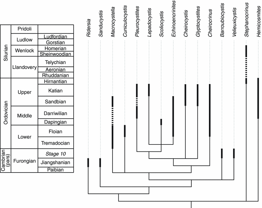
The data matrix relied heavily on characters relating to the placement of pectinirhombs. Although this may seem to heavily weight the matrix, it only had influence over the derived, pectinirhomb-bearing taxa. Furthermore, most of the pectinirhomb-bearing clades such as pleurocystitids, callocystitids and echinoencrinitids show more or less consistent placement of pectinirhombs. Although the plating bordering the periproct is a critical feature of glyptocystitids, the taxa in question only shared a single plate that discriminated taxa informatively (the presence of L1 in the periproctal border). All other periproctal bordering plates were autapomorphies for subclades of glyptocystitoids.
The plate pattern of Ridersia (with only three plate circlets and the periproct in oral position) represents the plesiomorphic condition for glyptocystitoids s.l.; the lateral position of the periproct in more derived taxa with either three or four circlets then results from the aboral migration of the anal opening along plate sutures (in between laterals). In Sanducystis, this migration took place between L1 and L5 (CD interray), whereas in all other glyptocystitids it seems to have occurred in between L5 and L4 (BC interray) as exemplified in (basal) taxa where the periproct is framed by L4-L5/IL4-IL5 (Velieuxicystis and Cuniculocystis). The situation in more derived glyptocystitids, in which the anal opening is framed by five plates (L1-L4-L5/IL4-IL5) results from the secondary expansion of the periproct along the L5-IL5 suture; in these forms, L1 and L5 are also in contact with the anal opening (as in Sanducystis), but this is secondarily acquired according to the phylogenetic results. Two independent, distinct aboral migrations of the periproct occurred in basal glyptocystitids: one in the CD interray and the other in the BC interray.
Acknowledgements
SZ is funded by a Ramón y Cajal Grant (RYC-2012-10576) from the Spanish Ministry of Economy and Competitiveness and project CGL2013-48877. XJZ is funded by National Natural Science Foundation of China (40602002, 41072018, 41330101, 41221001, J0630967). This paper is a contribution by the team ‘Vie Primitive, biosignatures, milieux extrêmes’ of UMR CNRS 5276 and to the ANR project RALI ‘Rise of Animal Life (Cambrian–Ordovician - organization and tempo: evidence from exceptionally preserved biota’. Additional funding was provided by the ‘Assembling the Echinoderm Tree of Life Project’, NSF DEB-1036260. The authors are particularly grateful to Jim Sprinkle (University of Texas) and one anonymous reviewer for their helpful remarks. Daniel Vizcaïno (Carcassone, France) provided access to specimens of Furongian echinoderms from Montagne Noire (France).
Appendix. Character list
-
1. Radial circlet of glyptocystitoid type absent (0) or present (1).
-
2. Basals tall cylindirical (0) or squat and flat bottomed (1).
-
3. Periproct on summit (0) or side of theca (1).
-
4. Stem facet ridge triangular (0), square (1) or absent (2).
-
5. Periproct in CD interray (0) or in BC interray (1).
-
6. Theca with low ridges (0) or heavy network of knife-edge ridges (1).
-
7. Proximal stem lacking inner and outer columnals (0) or present (1).
-
8. Distal stem with discoidal columnals (0) or barrel-shaped columnals (1).
-
9. Pectinirhombs absent (0) or present (1).
-
10. Pectinirhombs lacking vestibule rims (0) or present (1).
-
11. Anus small (0) or greatly enlarged (1).
-
12. Anus lacking periproctal membrane (0) or with periproctal membrane (1).
-
13. Summit large (0) or summit reduced in size (1).
-
14. Erect ambulacra (0) or recumbent ambulacra (1).
-
15. Floor plates forming thecal wall (0) or epithecal (1).
-
16. Pectinirhomb on B2/IL1 present (0) or absent (1).
-
17. Pectinirhomb on L1/L2 present (0) or absent (1).
-
18. Pectinirhomb on L3/L4 present (0) or absent (1).
-
19. Pectinirhomb on L3/R2 present (0) or absent (1).
-
20. Pectinirhomb on L3/R3 present (0) or absent (1).
-
21. Pectinirhomb on L5/R6 present (0) or absent (1).
-
22. Pectinirhomb on R1/R2 present (0) or absent (1).
-
23. Pectinirhomb on R2/R3 present (0) or absent (1).
-
24. Pectinirhomb on R4/R5 present (0) or absent (1).
-
25. Pectinirhomb on B1/B2 present (0) or absent (1).
-
26. Pectinirhomb on B2/B3 present (0) or absent (1).
-
27. Pectinirhomb on L2/R1 present (0) or absent (1).
-
28. Pectinirhomb on L4/R3 present (0) or absent (1).
-
29. Pectinirhomb on L1/R6 present (0) or absent (1).
-
30. Radial circlet bears R5 (0) or absent (1).
-
31. R4 large (0) or distinctly smaller than other radials (1).
-
32. Il2 not sutured to R2 (0) or sutured (1).
-
33. L1 forms portion of periproctal border (0) or absent (1).
-
34. Ambulacra not supported (0) or supported by large radial ridge (1).
Data matrix
-
Ridersia 000100110?0001?00000000000000?????
-
Sanducystis 001100110?1101?00000000000000?????
-
Macrocystella 111110112?110100000000000000000000
-
Cuniculocystis 111111113?1101000000000000000???00
-
Pleurocystites 111??011111111?1001111111111100000
-
Cheirocystis 1111111110110101001110001111100001
-
Lepadocystis 1112101111110111111011111100011010
-
Scoliocystis 11111011111101?1111001111100011000
-
Echinoencrinites 11111011101111?0111011110000111010
-
Glyptocystites 1111101111110110000000001111100110
-
Barroubiocystis 1??1?0110???01?00000000000000?00?0
-
Velieuxicystis 101110110?11???00000000000000???1?
-
Stephanocrinus 000000004?0000????????????????????
-
Hemicosmites 001201005?0000????????????????????
-
Cheirocrinus 1111111110110100000000000011100101