Significant outcomes
-
∙ No group difference was found between controls and obsessive–compulsive disorder (OCD) patients.
-
∙ Correlations between metabolite concentrations and symptom severity, particularly with the Children’s Yale-Brown Obsessive–Compulsive Scale (CY-BOCS) obsession subscore was found, for caudate aspartate (Asp) and choline compounds (tCho) (positively correlated with obsessions) and lipids in thalamus, and occipital glutamine+glutamate (Glx), N-acetylaspartate (NAA)+N-acetylaspartylglutamate (NAAG) (tNAA) and myoinositol (mI) (negatively correlated with obsessions).
Limitations
-
∙ Small sample size.
-
∙ Only four brain regions could be studied due to limited acceptable scan time.
-
∙ Magnetic field strength was 1.5 T. A higher field strength would give increased signal-to-noise ratio (SNR), however, with the technical disadvantages such as larger chemical shift displacements for the volume-of-interest (VOI) excitation and higher sensitivity to susceptibility effects.
Introduction
OCD is a chronic psychiatric disorder often leading to considerable distress and disability. Core features are repetitive, intrusive thoughts with disagreeable or threatening content (obsessions) and ritualistic behaviour, such as checking, counting and/or hand washing (compulsions) or ‘neutralising’ thinking (mental compulsions). The patients are aware that the intrusive thoughts emanate from their own mind and of the exaggerated and meaningless nature of both the obsessions and the compulsions. In about half of the cases OCD is believed to have its onset in childhood, and otherwise usually in early adulthood, and the estimated prevalence in paediatric populations is 0.5–2% (Reference Flament, Whitaker and Rapoport1,Reference Heyman, Fombonne, Simmons, Ford, Meltzer and Goodman2). Although adult and paediatric OCD are very similar phenomenologically (Reference Swedo, Rapoport, Leonard, Lenane and Cheslow3–Reference Geller, Biederman and Faraone5), there are certain differences; particularly aggressive/catastrophic obsessions have been observed to be more prevalent in children than in adults (Reference Geller, Biederman and Faraone5). Moreover, the co-morbidity patterns differ with more tic disorders and less depression observed in paediatric than in adult persons (Reference Geller, Biederman and Faraone5). The phenomenological differences between adult and paediatric OCD, as well as the clinical diversity within these two groups, may, at least in part, reflect pathophysiological heterogeneity (Reference Pittenger, Bloch and Williams6). Glutamatergic dysregulation involving corticostriatothalamocortical (CSTC) circuits has been implicated in OCD [(Reference Carlsson7) and refs therein]. Although ventral (‘affective’) CSTC circuits have attracted most interest in OCD research so far, also the dorsal (‘cognitive’) CSTC as well as the anterior cingulate gyrus – purportedly harbouring an error monitoring system (that may be hyperactive in OCD) – have been studied (Reference Brem, Hauser, Iannaccone, Brandeis, Drechsler and Walitza8).
Although the currently available therapies are effective in a majority of paediatric patients (Reference Watson and Rees9), many get only partial response. Thus, it is important to learn more about the underlying pathophysiology. One way to do that is by means of brain imaging methodology.
Functional brain imaging studies, taken together with the frequently observed reduction of OCD symptomatology following neurosurgery in various stations located along the CSTC pathways are suggestive of excessive glutamatergic signalling in this disorder (Reference Carlsson7).
Many investigations of the pathophysiology of OCD have been performed with 1H magnetic resonance spectroscopy (MRS), however, often with disparate results. This could be due to that most studies were small, average number is 13 subjects/study according to a review of Pauls et al. (Reference Pauls, Abramovitch, Rauch and Geller10), and often together with a heterogenic group composition (comorbidities, medication/treatment, no treatment, etc.). Different spectroscopic methods have also been used, and the results were reported sometimes as metabolic ratios and sometimes as absolute signal intensities. Brennan et al. (Reference Brennan, Rauch, Jensen and Pope11) analysed results from 28 different clinical studies, including both adults and children, different MRS methods and VOI localisations. The most consistent findings in their meta-analysis was that up to 5 of 12 studies showed a difference in specific metabolite concentrations between patients and controls: tCho was increased in thalamus in three of nine studies, Glx was reduced in two of eight studies in the anterior cingulate cortex (ACC), and NAA was reduced in 5 of 12 studies in the ACC and in 4 of 12 studies in the striatum. This indicates that it probably is not only one or two metabolites that are implicated, but rather that OCD manifests itself in alteration of several brain metabolites in several stations of the brain. Therefore, it makes sense to observe as much metabolic and diagnostic detail as possible and search the obtained data for differences in combinations of metabolites and diagnostics (known as latent variables or principal components).
In a previous 1H MRS study in adult persons with OCD, we found significant correlations between MRS-detected metabolites and OCD symptom severity in the right caudate nucleus (CN) and in occipital cortex. The latter was unexpected but showed the strongest correlation with symptom severity. We did not, however, observe any differences with respect to the control group and we hypothesised that this might be caused by the heterogeneity in the patient group (Reference Starck, Ljungberg and Nilsson12). Therefore, we wanted to conduct a new study where we included only newly diagnosed, treatment-naïve adolescent OCD patients and an age- and gender-matched control group.
The aim was to study the underlying pathophysiology of OCD by examining possible group differences between treatment-naïve children/adolescents with OCD and a group of healthy age- and gender-matched subjects using 1H MRS. We also wanted to investigate if results from our previous study of adult OCD patients, regarding correlations between neural metabolites and symptom severity, could be replicated in this group of treatment-naïve children/adolescents.
Methods
Subjects and clinical evaluation
Subjects (n=132) requesting treatment for OCD symptoms at the OCD clinic, Queen Silvia Children’s Hospital, at Sahlgrenska University Hospital (March 2009 to February 2011) were informed about the present study verbally and in writing, if they at their first visit were diagnosed with moderate-to-severe OCD, and if they were 11–17 years old. Exclusion criteria were concomitant attention deficit hyperactivity disorder (ADHD), mental retardation (intelligence quotient below 70) and concomitant disorders with higher treatment priority [e.g. autism, Asperger’s syndrome, primary anorexia nervosa (but anorexia in partial remission where OCD had become the residual and primary disorder was permitted), depression with suicidality that demanded cognitive behavioural therapy (CBT), selective serotonin reuptake inhibitor (SSRI) treatment or inpatient treatment, and psychosis]. However, pervasive developmental disorder not otherwise specified (PDD-NOS) was allowed if the clinical global impression (CGI) score for the PDD-NOS was ≤3 and lower than the CGI for the OCD disorder. Patients already under treatment for OCD with CBT, SSRI or atypical antipsychotics were excluded, as were patients or primary caregivers who could not speak or understand Swedish. For the diagnostic work-up DSM-IV criteria (13) were used.
In total, 89 subjects fulfilled inclusion (primary OCD, age 11–17 years old). In all, 15 of these subjects were either already under treatment or had a diagnose of ADHD. Hence, 74 subjects were informed of the study and asked for informed consent, and 23 accepted to participate in the study, and were hence scheduled for a psychiatric work-up. In all, 13 of these subjects displayed no exclusion criteria and were scheduled for MRS.
The psychiatric work-up for diagnostic ascertainment was performed using semistructured interviews with the Children’s Schedule for Affective Disorders and Schizophrenia and a general psychiatric diagnostic interview (Reference Kaufman, Birmaher, Brent, Ryan and Rao14) [Swedish translation (Reference Ivarsson, Campenhausen and Spak15)], and the CY-BOCS (Reference Goodman, Price and Rasmussen16) [also translated into Swedish (Reference Ivarsson and Johnson17)] for assessing OCD symptoms and severity. Further, the parents filled in the Child Behaviour Checklist (Reference Achenbach and Rescorla18) using a Swedish translation (Reference Larsson and Frisk19). The patients were also included in a subsequent treatment trial (Reference Thomsen, Torp and Dahl20) separate from this study.
Healthy controls were recruited through advertising at the Queen Silvia Children’s hospital and among children of Sahlgrenska University Hospital staff (April 2011 to June 2011), and subjected to the same psychiatric diagnostic work-up, with exception for CY-BOCS, as the patients to ascertain that they were psychiatrically healthy. All controls were matched with the patients regarding age and gender.
The final sample consisted of 24 subjects (13 OCD subjects and 11 controls). Demographic data for both groups are given in Table 1.
Demographic and clinical information of participants

CY-BOCS, Children’s Yale-Brown Obsessive–Compulsive Scale.
Data are given as quantity or median (range).
The study was approved by the Regional Ethical Review Board in Gothenburg, Sweden (approval nr 151–06). All subjects, as well as their next of kin, have signed written informed consent.
MRS
Measurement parameters
The magnetic resonance (MR) investigations were performed before any treatment was commenced.
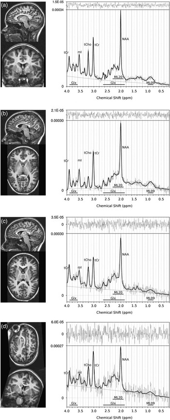
The MRS measurements were performed on a Gyroscan Achieva 1.5T release 2.6 (Philips Medical systems, Eindhoven, the Netherlands) with the transmit/receive head coil of bird cage type from the same vendor. The subject’s head was firmly supported with a vacuum cushion. MR imaging was performed for careful localisation of MRS volumes. Following a survey scan, a three-dimensional (3D) T1-weighted (T1W) turbo field echo sequence was used for imaging of the brain with nearly isotropic voxels (1.18×1.18×1.25 mm3). Single voxel MRS was performed at four different locations (Fig. 1) using the PRESS pulse sequence (echo time=30 ms, repetition time=2000 ms, CHESS water suppression).
Magnetic resonance spectroscopy (MRS) volume position (left column) and typical 1H magnetic resonance spectrum (right column) in (a) cingulate gyrus and sulcus, (b) occipital cortex, (c) thalamus and (d) right caudate nucleus. The MRS volume is shown in (a) upper left panel: sagittal view. Lower left panel: coronal view; (b) and (c) upper left panel: sagittal view. Lower left panel: transaxial view. (d) Upper left panel: long axis view of the caudate nucleus. Lower left panel: short axis view of the caudate nucleus; lower part of the spectrum panel: thick black line shows fitted spectrum, thin line shows acquired data, smooth thin line shows baseline and horizontal thick lines indicate chemical shift ranges where the spectral complexes of glutamate+glutamine (Glx), macromolecules and lipids ML09 and ML20 appear. Upper spectrum panel shows difference between fitted spectrum and acquired data. Cr, creatine; mI, myoinositol; NAA, N-acetylaspartate; T, thalamus; tCho, choline compounds.

Four VOIs for MRS were carefully positioned in consensus by the same two operators throughout the entire study. The position and size of the VOIs are shown in Fig. 1. The primary purpose of first VOI was optimisation of acquisition parameters such as water suppression, etc., however, as the region is found to be affected OCD the measurement results were included in the evaluation. The VOI was placed bilaterally above the mid body of corpus callosum and includes mainly a part of the cingulate gyrus and sulcus (CGS) and a smaller portion of corpus callosum itself, and is henceforth, denoted CGS. This VOI corresponds mainly to the Brodmann areas of 23 and 24, but includes also area 31, 32 and 33 to a lesser extent [anterior-posterior (AP)×left-right (LR)×feet-head (FH)=30×30×20 mm3=18 cm3, number of signals averaged (NSA)=32]. The three other VOIs were planned in grey matter (GM) with a focus on brain regions involved in the OCD pathophysiology, and kept small in order to minimise signal contribution from white matter (WM). The second VOI was placed bilaterally in the occipital cortex, denoted O, in parallel with and below the parieto-occipital sulcus (AP×LR×FH=20×14×15 mm3=4.2 cm3, NSA=256). The VOI consists mainly of the primary visual cortex (V1), but includes also to a lesser extent parts of V2 and V3. The third VOI was placed bilaterally in the medial, cranial thalamus (T) (AP×LR×FH=14×18×10 mm3=2.5 cm3, NSA=256). The last VOI was placed in the right CN, and included the superior part of the head and the anterior part of the body of the CN (AP×LR×FH=15×10×10 mm3=1.5 cm3, NSA=256). Subsequent multi-planar reformatting of the 3D T1W images were used to reconstruct long axis and short axis views of the right CN to facilitate a VOI location with minimal content of cerebrospinal fluid and maximal content of GM in the VOI, as described earlier (Reference Starck, Ljungberg and Nilsson12). A short 3D fast field echo MR imaging scan was performed before each of the MRS scans to check for head movements during the examination. If a shift >0.5 mm was detected in the feet–head direction between the 3D T1W scan and the short FFE scan before the caudate MRS measurement, the VOI position of the caudate VOI was corrected. Maximum adjustment needed in this study was 2 mm. Therefore, none of the acquired spectra was discarded due to movement during the examination. The total MR examination time was just below 60 min.
MRS quantification
LC Model, software version 6.2 (Reference Provencher21), was used for spectrum analysis and quantification. The basis set used for quantification was measured in house (Reference Bengtsson22) and included: Asp, creatine, gamma-aminobutyric acid, glucose (Glc), glutamine, glutamate, Glx, mI, lactate (Lac), NAA, NAAG, tNAA, scylloinositol (sI), taurine (Tau), guanine, tCho. Lipids and macromolecule resonances at 1.3 ppm (ML13), lipid resonances at 1.3 ppm (L13), lipids and macromolecule resonances at 0.9 ppm (ML09) and lipids and macromolecule resonances at 2 ppm (ML20) were simulated within LC Model. The spectral fitting was performed between 0.2 and 4.0 ppm. A non-water suppressed spectrum with NSA=16 was automatically measured in the beginning of each MRS scan and was used for eddy-current correction in the post-processing of the data. The unsuppressed water signal was also used as an internal reference. No correction for T1 relaxation was performed. Concentrations are given as institutional concentration units. The residual between the fitted and the acquired spectra were inspected carefully for artefacts, and all measured spectra were found to have a satisfactory spectrum quality. The full-width-at-half-maximum value of the NAA peak, as reported by LC Model, ranged between 0.02 and 0.09 ppm.
Tissue composition in VOI
The amount of GM, WM and cerebrospinal fluid (CSF) in each VOI was estimated with an in-house developed software program (Reference Montelius23), through segmentation within the MRS VOI in the 3D T1W image.
Questionnaire
Immediately, after completion of the MR examination the participants were given a questionnaire with two (for the controls) or four (for the patients) questions. All participants received the following questions: (1) How much anxiety did you experience during the investigation, and (2) How much anxiety did you experience during the investigation compared with what you normally experience? In addition, the patients received the following questions: (3) How much of obsessional thinking did you experience during the investigation?, and (4) How intensive were your obsessions during the investigation compared with what you normally experience? The five response alternatives to questions (1) and (3) were as follows (assigned score in parenthesis): None at all (1) – Modest (2) – Moderate (3) – Severe (4) – Unbearable (5). The five response alternatives to questions (2) and (4) were as follows: Considerably less (1) – Less (2) – Similar (3) – More (4) – Considerably more (5). One of the controls did not complete the questionnaire.
Statistical analyses
Multivariate analyses using SIMCA-P+, version 12.0 (Umetrics AB, Sweden) were performed on CY-BOCS scores and the concentration values determined with LC Model for the 20 metabolites in the four brain regions examined with MRS. Principal component analysis (PCA) and partial least square (PLS) projection to latent structures were applied. Any variable with >30% missing values was excluded from the analyses.
In PCA the original set of variables is reduced into a smaller set, commonly referred to as latent variables or principal components. The principal components are not correlated with each other and they show the significant relation between the original variables. PCA was performed to obtain an overview, detect trends in the data and to evaluate possible overall differences in metabolite concentrations between controls and the OCD subjects. The analysis was based on 74 metabolites and 24 subjects (controls and patients).
PLS analysis is a regression extension of PCA, where the relation between two blocks of variables, X and Y, is explored. PLS was performed with MRS data as X-variables (73 metabolites) and total CY-BOCS score as Y-variable. To further explore the relation to OCD symptoms, PLS was performed also separately with obsession and compulsion subscores, respectively, as Y-variable.
The results of the PCA and PLS analyses were presented as 2d projections, loading plots, where the relations between the variables are visualised. The loading plot shows how the MRS data are correlated with CY-BOCS scores. X-variables projected on the same side of origin as the Y-variable are positively correlated with Y and vice versa, and the greater the distance between the variable and origin the stronger the correlation.
Pre-processing of the data consisted of unit variance scaling and mean-centring. The significance of the components was evaluated by means of cross-validation (Reference Eastment and Krzanowski24) and only significant components are shown in figures. The goodness of fit of a model is given by R 2 and Q 2. The R 2 value states how much of the variation in the Y- and X-variables that can be explained by the model and Q 2 indicates the predictability of the model.
The Pearson’s correlation coefficient r was reported in the univariate correlation analyses. Mann–Whitney U-test was applied for group comparison within the questionnaire.
Results
Symptom severity for the patient group, given as CY-BOCS and it’s subscores, is given in Table 1.
The mean GM contribution was high in three of the four VOIs [T=89% (range 85–94%), O=73% (range 63–80%), CN=71% (range 61–83%) and CGS=53% (range 41–68%)]. The mean CSF contribution was low for all VOIs [T=8% (range 5–12%), O=6% (range 3–11%), CN=1% (range 0–6%) and CGS=4% (range 1–5%)].
PCA yielded a one-component model explaining (R 2) 12% and predicting (Q 2) 0.096% of the data variation. The model did not show any separation between controls and patients or between genders. Thus further analyses on potential group differences were not performed. Six measures of metabolites in specific VOIs were excluded due to missing values (Lac in all four VOIs and NAAG and Tau in the CN).
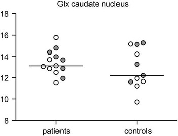
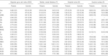
Mean metabolite concentrations for the patient and control group for all metabolites included in the analysis are given in Table 2. Furthermore, individual Glx values for patients and controls in the CN are shown in Fig. 2, which illustrates the substantial overlap between the patient and the control group.
Individual glutamate+glutamine (Glx) values for patients and controls in caudate nucleus. Median values for the groups are illustrated by solid horizontal lines. Females, unfilled circles; males, filled circles.

Metabolite concentrations, given as mean and standard deviation, for the patient and control group for all four measured volume-of-interest positions

Metabolites are as follows: Asp, aspartate; Cr, creatine; GABA, gamma-aminobutyric acid; Glc, glucose; Gln, glutamine; Glu, glutamate; Glx, Gln+Glu; Gua, guanine; L13, lipid resonances at 1.3 ppm; Lac, lactate; mI, myoinositol; ML09, lipids and macromolecule resonances at 0.9 ppm; ML13, lipids and macromolecule resonances at 1.3 ppm; ML20, lipids and macromolecule resonances at 2 ppm; NAA, N-acetylaspartate; NAAG, N-acetylaspartylglutamate; sI, scylloinositol; Tau, taurine; tCho, choline compounds; tNAA, NAA+NAAG.
Variables with >30% missing values were excluded from the analysis.
The concentrations are given as institutional concentration units (icu).
PLS resulted in a three-component model explaining
 $(R_{y}^{2} )$
99.8% and predicting (Q
2) 56.4% of the data variation when using total CY-BOCS score as Y-variable. An even stronger three-component model was obtained when metabolite concentrations were related to obsession subscores only, explaining
 $(R_{y}^{2} )$
99.8% of the data, and with a prediction value (Q
2) of 61.2%. Modelling compulsion subscores against metabolite concentrations yielded a two component model with a similar pattern, but with a substantially lower predictability (Q
2=13.2%). Seven metabolites were excluded from the PLS analyses due to missing values (Lac in all four VOIs and NAAG, Tau and Glc in the CN).
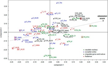
The loading plot for the obsession scores as Y-variable is shown in Fig. 3. Obsession scores show the strongest positive correlation to aspartate and tCho in the CN, and sI and ML09 in the CGS. The strongest negative correlation with obsession scores is seen for ML09 and ML20 in the thalamus, and for Glx, tNAA and mI in the occipital cortex. Using obsession scores instead of total CY-BOCS as Y-variable resulted in increased correlation with especially ML09 in the CGS, Glx in the occipital cortex and aspartate in the CN. The influence from CGS sI decreased (data not shown).
Loading plot from partial least square model of magnetic resonance spectroscopy data in relation to obsession score. Metabolites with the strongest impact on the model are marked by squares (variable influence on projection value>1.5). The loading plot shows that aspartate (Asp) and choline compounds (tCho) in the caudate nucleus (CN), and to scylloinositol (sI) and ML09 in the cingulate gyrus and sulcus (CGS) display the strongest positive correlation to obsession score. The strongest negative correlation to obsessions is seen for ML09 and ML20 in the thalamus (T), and for glutamate+glutamine (Glx), tNAA (NAA+NAAG) and myo-inosytol (mI) in the occipital cortex (O). Asp, aspartate; Cr, creatine; GABA, gamma-aminobutyric acid; Glc, glucose; Gln, glutamine; Glu, glutamate; Gua, guanine; L13, lipid resonances at 1.3 ppm; Lac, lactate; ML09, lipids and macromolecule resonances at 0.9 ppm; ML13, lipids and macromolecule resonances at 1.3 ppm; ML20, lipids and macromolecule resonances at 2 ppm; NAA, N-acetylaspartate; NAAG, N-acetylaspartylglutamate; Tau, taurine.

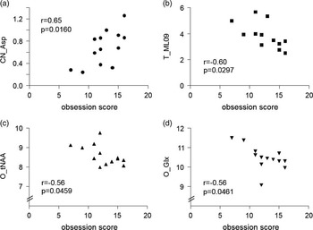
Individual correlation plots for the four metabolites with the strongest relationship to the obsession score, as recognised by the PLS analysis, are shown in Fig. 4.
Scatter plots of the four metabolites that exhibited the strongest correlations with the obsession score; that is, these metabolites are the four most distant from origin in the partial least square plot in Fig. 3. The Pearson’s correlation coefficient r and p-values are shown in each plot (a) aspartate (Asp) in the volume-of-interest (VOI) in the caudate nucleus (denoted CN_Asp) r=0.65; p=0.0160, (b) lipids and macromolecule resonances at 0.9 ppm (ML09) in the VOI in thalamus (T_ML09) r=−0.60; p=0.0297, (c) N-acetylaspartate+N-acetylaspartylglutamate (tNAA) in the VOI in occipital cortex (O_tNAA) r=−0.56; p=0.0459 and (d) glutamate+glutamine (Glx) in the VOI in occipital cortex (O_Glx) r=−0.56; p=0.0461.

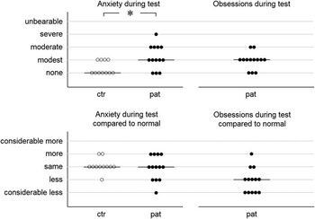
Results from the questionnaire are shown in Fig. 5. Anxiety during the MR examination was more pronounced in patients than in controls (p=0.02). Neither patients nor controls had a median anxiety level different from what they normally experience during the MR examination, but patients experienced less obsessions than usual (sign test: p=0.01).
Result of the questionnaire regarding the level of anxiety and obsessions during the magnetic resonance examination. Median values for the groups are illustrated by solid horizontal lines. *p=0.02 using Mann–Whitney U-test. ctr, Control group; pat, patient group.

Discussion
In the present, 1H MRS study in paediatric OCD patients we observed significant correlations between brain metabolite concentrations and symptom severity. Metabolites related to total CY-BOCS scores resulted in a significant PLS model, but an even stronger model was obtained when metabolites were correlated with the CY-BOCS obsessions subscores only. In contrast, the PLS model where metabolite levels were related to the CY-BOCS compulsions subscores was weaker. The present finding of a significant relationship between metabolite concentrations and OCD symptoms is in agreement with the results from our former study on adults (Reference Starck, Ljungberg and Nilsson12), but in that study the PLS model was not improved by correlating metabolites to Y-BOCS obsessions (or compulsions) subscores.
We did not observe any significant group differences between paediatric OCD subjects and healthy controls in Glx concentration or any other metabolite concentration in any of the investigated brain regions (right CN, thalamus, occipital cortex, CGS). This result is in accordance with our previous 1H MRS study performed on adult OCD subjects (on medication or with a history of other treatments) who were not found to differ significantly from a group of healthy controls with respect to MRS metabolite concentrations in the right CN, the occipital cortex or in the anterior cingulate gyrus (Reference Starck, Ljungberg and Nilsson12). This lack of group differences is also in agreement with the review of Brennan (Reference Brennan, Rauch, Jensen and Pope11).
The most important metabolites for the PLS model in the present study were CN Asp and tCho (positively correlated with obsessions), and thalamic ML09 and ML20 and occipital Glx, tNAA and mI (negatively correlated with obsessions). Three of these metabolites also showed up clearly in our previous study on adults (Reference Starck, Ljungberg and Nilsson12): in agreement with the present study, caudate tCho was positively correlated and occipital Glx was negatively correlated with OCD symptoms, and there was also a tendency for a negative correlation between occipital tNAA and symptom severity. On the other hand, occipital mI was in the previous study positively correlated with OCD symptoms, whereas, in the present study there was a negative correlation with CY-BOCS. The metabolic maturation of the brain was explored by Bluml et al. (Reference Bluml, Wisnowski and Nelson25) – even though this process continues in some regions up to 18 years of age, they found that mI in the parietal/occipital lobe was stable after about 6 months of age. Therefore, normal maturation of the brain is probably not the reason for the different occipital mI correlation pattern in this study compared with the adult study. A perhaps more plausible reason for the discrepancy regarding occipital mI could be the fact that the former investigation was performed in patients who had received various kinds of treatments in the past, as they were adults, with some even on medication at the time of the study, whereas the present study was carried out in drug-naive patients. Interesting in this context, Szeszko et al. (Reference Szeszko, MacMillan and McMeniman26), using diffusion tensor imaging, observed WM aberrations in, for example, the lingual gyrus of the left occipital lobe in OCD, possibly reflecting abnormalities in the myelin sheath. Inositol treatment has in two studies been reported to have beneficial effects in OCD (Reference Kim, McGrath and Silverstone27).
A comparison of the PLS loading plots from our two OCD studies discloses another interesting similarity. Although the pattern was more distinct in the adult study, there was a tendency in both studies for occipital variables to aggregate to the left of origin along the first component (i.e. tended to display negative correlations with symptom severity), whereas the reverse seemed to be true for caudate variables, which in both studies aggregated more to the right (i.e. tended to display positive correlations with symptom severity). These results suggest that there may be similarities between adult and paediatric OCD biochemistry, even though two variables, occipital and caudate mI, had a clearly deviant position along the first component in the present compared with the adult study.
The results from the questionnaire agree fairly well with our previous study. Similarly to the adult patients (Reference Starck, Ljungberg and Nilsson12), the paediatric subjects with OCD in the present investigation had a higher degree of anxiety than controls during the MR measurements. Moreover, in agreement with our former study, most of the paediatric OCD patients experienced less obsessions than usual during the MR examination. In the adult study most patients also reported lower anxiety levels than usual during the MR measurements but this was not the case among the paediatric OCD subjects. We cannot exclude the possibility that the lower than normal obsession intensity could have influenced the observed metabolite pattern.
We regard the finding of the negative correlation between occipital Glx and OCD symptoms observed in both studies particularly interesting. The occipital region has earlier been used as a control area without special interest for OCD pathophysiology and symptomatology. However, as we have pointed out in our former paper (Reference Starck, Ljungberg and Nilsson12), decreased metabolic activity in the parieto-occipital region has been observed in patients with OCD based on fluorodeoxyglucose (18F-FDG) positron emission tomography (PET) data (Reference Nordahl, Benkelfat, Semple, Gross, King and Cohen28). Interesting in this context is the fairly consistent neuropsychological finding of a poor performance on visuospatial tests in OCD, and that a negative correlation between visuospatial performance and Y-BOCS scores has been observed (Reference Lacerda, Dalgalarrondo, Caetano, Haas, Camargo and Keshavan29,Reference Boldrini, Del Pace and Placidi30). Visuospatial processing involves the occipito-parietal regions. In the present study, a large portion of the occipital VOI consists of the primary visual cortex (V1), but there is also a substantial contribution from visual association cortex (mainly V2 and V3).
Unfortunately, it is methodologically difficult to obtain reliable MRS measurements in the prefrontal cortex and such data are therefore lacking in the present study, but with other brain imaging techniques this region has fairly consistently been observed to be hyperactive in OCD [(Reference Carlsson7)and refs therein]. Interestingly, a recent review and meta-analysis of diffusion tensor imaging studies in OCD (Reference Piras, Piras, Chiapponi, Girardi, Caltagirone and Spalletta31) describes hypoconnectivity of the inferior fronto-occipital fasciculus, which connects the ventrolateral prefrontal cortex and medial orbitofrontal cortex to posterior parietal and occipital associative cortices which would cause a disturbed balance in activity and communication between anterior and posterior parts of the cortex. This anterior–posterior hypoconnectivity is interesting in the light of our observation in both adults and paediatric subjects with OCD that even though we found no significant group difference in occipital Glx, there was a negative correlation between occipital Glx and symptom severity. In other words, a stronger glutamatergic projection from the prefrontal cortex to the occipital cortex may serve to mitigate OCD symptoms related to a presumably hyperactive prefrontal cortex.
In the present study on paediatric subjects, we also observed a negative correlation between occipital tNAA and symptom severity and a similar tendency was seen in the study of adults, suggesting an important role of occipital cortex neuronal viability in this regard. A striking example of a disorder with an altered anterior–posterior cortical balance is Williams syndrome: these patients who display severe visuospatial deficits and enhanced emotionality have been found to have markedly reduced occipital lobe GM volumes, but increased volume and GM density in, for example, orbital and medial prefrontal cortices (as well as in the amygdala) (Reference Reiss, Eckert and Rose32). Szeszko et al. (Reference Szeszko, Christian and Macmaster33) found a similar pattern in OCD, that is, more GM in, for example, the orbital frontal cortex and less GM in the occipital cortex. Further, reduced perfusion in right visual association cortex, increased frontal perfusion but no difference in the perfusion of the CN was found in OCD subjects compared with healthy controls in a single-photon emission computed tomography (SPECT) study (Reference Harris, Hoehn-Saric, Lewis, Pearlson and Streeter34). A similar anterior–posterior SPECT pattern with, for example, increased frontal lobe and decreased occipital lobe perfusion was observed in a subgroup of OCD subjects in a more recent study (Reference Wen, Cheng, Cheng, Yue and Wang35). Moreover, PET studies by Nordahl et al. (Reference Nordahl, Benkelfat, Semple, Gross, King and Cohen28) and Kwon et al. (Reference Kwon, Kim and Lee36) confirm this anterior–posterior pattern in OCD.
In an attempt to further increase the reliability of the statistical analysis, we excluded metabolites with potentially uncertain concentration estimates (SD>100%) as reported by the LC Models software for spectrum quantification. We also performed corrections of partial volumes for GM/WM and CSF. However, neither of these measures resulted in increased predictability of the PCA and PLS models.
In conclusion, we did not find any separation between controls and patients in the PCA analysis, which is consistent with most other 1H MRS studies on OCD subjects (Reference Brennan, Rauch, Jensen and Pope11). However, we did find a correlation between metabolite concentrations and symptom severity, particularly with the CY-BOCS obsession subscore. Metabolites with the strongest impact on the PLS model in the present study were caudate Asp and tCho (positively correlated with obsessions) and thalamic ML09 and ML20, and occipital Glx, tNAA and mI (negatively correlated with obsessions). Particularly, interesting is the finding of the negative correlation between occipital Glx and OCD symptoms, an observation we also made in the previous study in adults.
Acknowledgement
The authors thank Mikael Montelius, MSc for the segmentation programme for partial volume corrections, Henric Rydén, MSc for Matlab-programming for efficient data-handling and Philip Healthcare Clinical Science Group for support. Authors’ Contribution: All authors took part in the study design; K.H. and T.I. performed the clinical evaluation; M.L., Å.C. and G.S. acquired the MR-data. M.L. performed the MR- and MRS-data analysis and M.N. performed the statistical evaluation. All authors took part in the interpretation of the data. M.L., M.N., M.C. and T.I. wrote the first draft of the manuscript and all authors took part in revising the manuscript to final version. All authors have approved the final manuscript.
Financial Support
This research was supported by the Swedish Research Council (14039), ‘Västra Götalandsregionen’, ‘Grants from the state under the LUA/ALF agreement’, ‘Frimurare Barnhusdirektionen in Göteborg’, ‘The Arvid Carlsson Foundation’ and ‘Wilhelm and Martina Lundgrens research fund’.
Conflicts of Interest
The authors declare that they have no potential conflicts of interest.
Ethical Standards
The authors assert that all procedures contributing to this work comply with the ethical standards of the relevant national and institutional committees on human experimentation and with the Helsinki Declaration of 1975, as revised in 2008.