Syncope is the sudden loss of consciousness and postural tone, from which recovery is spontaneous.Reference Kapoor1 Syncope is common, affecting approximately 15% of all children, and results in 1 out of every 1200 visits to a paediatric emergency department.Reference Lewis and Dhala2, Reference Pratt and Fleisher3 It is encountered commonly by emergency medicine or urgent care physicians due to a concern for serious cardiac or neurologic pathology. However, actual cardiac pathology is typically found in less than 5% of syncopal episodes in the paediatric population.Reference Driscoll and Edwards4 Potential worrisome syncopal events include those that occur with exertion, those that occur with concurrent chest pain, dyspnoea or palpitations, and those with focal or diffuse neurologic changes.Reference Lewis and Dhala2, Reference Pratt and Fleisher3, Reference Johnson, Mack, Kuntz, Brands, Porter and Fischer5
Here, we describe a 16-year-old female with syncope, diagnosed as neurocardiogenic syncope and postural orthostatic tachycardia syndrome, who after an extensive evaluation at several institutions was ultimately found to have a single coronary artery, which was felt to be clinically important.
Case report
The patient was the product of an uncomplicated pregnancy, labour, and delivery. She had her first syncopal episode at the age of 2 years, collapsing while brushing her teeth. At the age of 3 years, she had another syncopal episode with urinary incontinence. An electroencephalogram at that time was normal. She was diagnosed as having benign vasovagal syncope. She continued to have infrequent syncopal spells between the ages of 3 and 10 years, and then went without a syncopal episode between the ages of 10 and 13 years.
At the age of 14 years, the patient had loss of consciousness while running in a track meet. She fell to the ground suddenly, but was awake immediately after the fall. She was reported to be pale, diaphoretic, and disoriented at the time. Over the next month, she had similar exercise-induced events, occurring 1–2 times weekly in a similar manner to the first event. A month later, she began to have a right upper extremity tremor lasting 5–30 minutes after each spell. She then began having these right upper extremity tremors each time she exercised. After a month, the patient began to have spells occurring without exercise, including syncopal events while standing and walking between classes.
She was referred to paediatric cardiology at an outside institution at that time. As per the records, standard two-dimensional echocardiography was performed and was interpreted as being normal. A treadmill exercise test was performed with no evidence of exercise-induced ischaemia. She experienced mild lightheadedness after the test, but no other symptoms. After a further recurrence of her symptoms, she was referred to a paediatric electrophysiologist. A tilt table test was performed. After being tilted to 80 degrees for 24 minutes, she complained of blurry vision. She reported a feeling of her heart “trying to beat faster, but something was pressing really hard” on her chest. She then passed out, with a concurrent asystolic pause of 12.5 seconds. The patient was noted to then have a tonic clonic seizure with incontinence of urine. She was returned to the supine position, where she regained consciousness. She complained of a headache on waking. After this test, the patient was started on midodrine, and recommended to increase her fluid and salt intake.
Her spells persisted, and 1 month after the tilt table test, she had a Reveal loop recorder placed (Medtronic Corp, Minneapolis, Minnesota, United States of America). She had multiple syncopal spells while the recorder was in place; however, recorder interrogations showed her to be in sinus rhythm during each spell. A treadmill exercise test was repeated at that time, at the end of which she collapsed, appearing to trip on her feet. She remained at the end of the treadmill for 30 seconds, at which point she stood up when asked. There was no seizure-like activity or post-ictal confusion/fatigue. Her blood pressure and heart rate were reportedly normal despite the collapse. The possibility of a conversion disorder was entertained at that time, for which she underwent therapy. The patient not only continued to participate in sports after this workup, but also experienced syncopal events during practices and games. She never was injured as a result of her many falls.
The patient was then referred to our institution at the age of 15 years. She was given a trial of beta-blockade with propranolol, after which she had a period of 3 months with no events. However, the events returned, including weekly syncopal spells with exercise. She also began having different types of events, including absence-like events where she would not remember time, occurring once every 1–2 weeks. She also described episodes of emesis without a specific trigger occurring 3–4 times weekly. The patient was referred to neurology, both for management of her spells and for chronic headache. She had a normal repeat electroencephalogram and normal head magnetic resonance imaging, and was placed on an inpatient epilepsy monitoring unit for 48 hours. During this stay, she had one typical spell while riding an exercise bicycle, where she became nauseated and lightheaded. Her heart rate increased to 120 beats per minute during the spell, and then decreased to 41 beats per minute afterwards. Her blood pressure remained stable during the spell. Her electroencephalographic rhythm was normal throughout the period of the spell. She was discharged with a diagnosis of convulsive syncope, with possible postural orthostatic tachycardia syndrome.
After 6 months, at the age of 16 years, the patient continued to have chronic daily headache and intermittent spells. Her mother called, reporting that she had experienced a severe spell while at school walking with friends. She reportedly suddenly passed out, and a first responder could not feel pulses or see signs of active breathing. Cardiopulmonary resuscitation was started and continued for 1 minute, after which she woke up. Evaluation by emergency services was unremarkable, and she was discharged home. She returned to the cardiology clinic at that time.
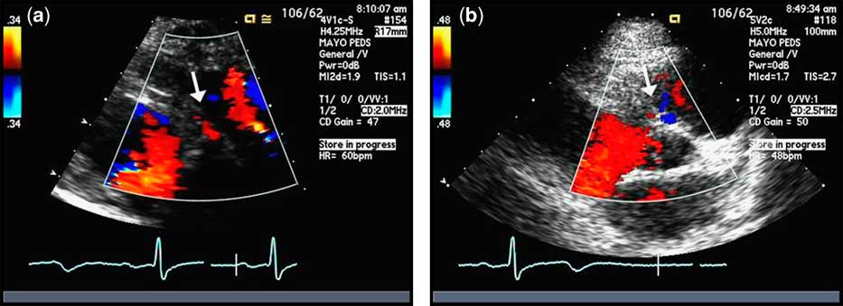
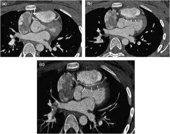
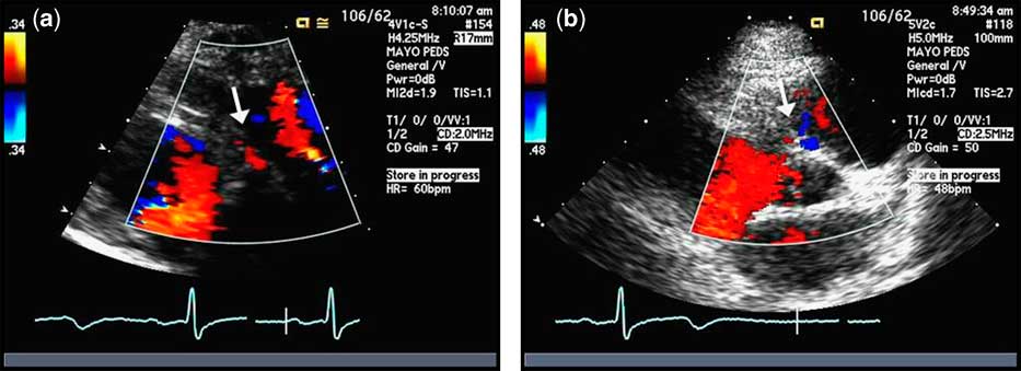
A repeat electrocardiogram was performed, and demonstrated normal sinus rhythm with a normal corrected QT interval. Chest X-ray showed a normal cardiac silhouette and normal pulmonary vascularity. An echocardiogram was performed, which revealed normal chamber sizes and function. The origin of the right coronary artery was normal with evidence of normal diastolic flow; however, the left coronary artery origin could not be confidently established. In addition, there was an abnormal diastolic flow noted between the aorta and pulmonary artery, suggesting anomalous coronary flow (Fig 1). She was thus referred for a computed tomography angiogram. This revealed a single coronary artery originating from the right aortic sinus, with the left coronary artery arising from the proximal trunk. The left coronary maintained an initial interarterial course, followed by an intraseptal course in the infundibular septum below the level of the pulmonary artery (Fig 2). On emerging from the septum, the left coronary then divided into a small left anterior descending and left circumflex coronary arteries. There was evidence of mild coronary narrowing along its intraseptal course.
(a) Still frame echocardiographic image demonstrating normal diastolic flow (arrow), indicating what appears to be the normal origin of the right coronary artery. (b) Still frame echocardiographic image demonstrating abnormal diastolic flow (arrow) between the aorta and pulmonary artery, suspicious of abnormal coronary flow.
Two axial slice images from a computed tomography angiogram, demonstrating (a) the left coronary artery originating from a single coronary artery trunk (white arrow), and (b) running with an intraseptal course in the infundibular septum (white arrows) below the level of the pulmonary artery. A thick-slice reconstructed image (c) of the entire course of the left main coronary artery is also shown.

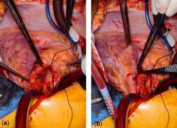
The patient was referred for surgical repair. After confirmation of the intramyocardial, posterior outflow tract course and location inferior to the pulmonary valve (Fig 3), it was felt that it would not be technically possible to mobilise the left coronary artery out of the right ventricular outflow tract muscle. Therefore, the left internal mammary artery was grafted to the left anterior descending coronary artery. However, flow probe studies demonstrated that the native left coronary artery flow was preventing competitive flow from the graft. To prevent the possibility of the graft not developing appropriately, a suture was placed around the left main coronary artery to partially restrict native flow and allow graft maturation.
Intraoperative photography demonstrating (a) the anomalous left coronary artery diving into the infundibulum after take-off from the proximal right coronary artery, and (b) the coronary emerging laterally.

The patient had an uncomplicated recovery. She was discharged home with aspirin monotherapy. She largely did well; however, she had an episode of chest pain that occurred 2 months post-operatively. Angiography was performed, which revealed a widely patent right coronary artery and left internal mammary artery graft. There was no flow through the native left main coronary artery, with the graft being the sole supply for the left coronary artery system. A month later, she underwent an exercise echocardiogram, which was negative for ischaemia. A treadmill exercise test was also negative for electrocardiographic evidence of ischaemia at that time.
Since surgery, our patient has experienced a dramatic improvement. In the year following her surgery, she had no syncope. She experienced a single episode of lightheadedness on standing, and a single episode of shortness of breath and chest pain with exercise. She was to return to athletic activities. A dobutamine stress echocardiogram performed 1 year after surgery showed normal exercise response with no evidence of myocardial ischaemia.
Discussion
The incidence of potentially fatal coronary anomalies is estimated in the published literature to be between 0.1% and 1%.Reference Angelini6–Reference Davis, Cecchin, Jones and Portman8 Patients with coronary artery anomalies are often asymptomatic at the time of diagnosis. If symptomatic, syncope and chest pain are the most common presenting complaints encountered. However, sudden death may be the first symptom. Here, we describe a 16-year-old female with a history of syncope, diagnosed as neurocardiogenic syncope and postural orthostatic tachycardia syndrome, who ultimately was found to have a single coronary artery origin. Her left coronary artery anomalously arose from the proximal single coronary trunk with an initially interarterial and subsequently intraseptal course.
A single coronary artery origin itself is an extremely rare entity. Akcay et al reported a series of greater than 70,000 coronary angiograms performed at several institutions, of whom only 10 patients (0.024%) were found to have a single coronary artery.Reference Akcay, Tuncer and Batyraliev9 Of these 10 patients, only one had a single coronary artery arising from the right aortic sinus with a left coronary artery arising from the proximal trunk with an interarterial course. No patients had an intraseptal course. Single coronary artery is reported in several case reports of sudden death in infants, children, and adults.Reference Ohshima, Lin and Sato10–Reference Moore and Byard12
It is important to distinguish an anomalous coronary artery with an intraseptal course, as seen in this case, from the “intramural” coronary artery. In the intramural variety, the proximal anomalous coronary artery courses completely within the wall of the aorta. This high-risk coronary anomaly also typically exhibits a slit-like orifice and acute angle of take-off from the aortic sinus, thus increasing the likelihood of potential compression. It can be treated by unroofing the coronary artery, opening up the orifice to a less acute angle.Reference Angelini6, Reference Davies, Burkhart and Dearani13 Intramural coronaries that course below the level of the commissure can be difficult, but often can be at least partially unroofed on the opposite side of the commissure.Reference Gulati, Reddy and Culbertson14, Reference Frommelt, Frommelt, Tweddell and Jaquiss15 In contrast, an anomalous coronary artery with an intraseptal course runs through the septal infundibulum. These coronary anomalies typically do not have a slit-like orifice or acute angle of take-off at their origin. Surgical treatment of these anomalies, if required, is more difficult, and the use of grafting is often necessary.Reference Davies, Burkhart and Dearani13 In addition, our patient had a single coronary origin from the right aortic sinus, with the intraseptal coronary artery arising from the opposite coronary artery, making it a fundamentally different lesion.
Our patient required grafting with the left internal mammary artery to provide consistent flow to the left coronary system. In the majority of patients with single coronary artery or anomalous origin of a coronary artery, the native coronary artery provides adequate flow during most situations, that is, it is a dynamic lesion. As such, competitive flow is often problematic, and can keep the implanted mammary artery graft from maturing.Reference Davies, Burkhart and Dearani13, Reference Fedoruk, Kern, Peeler and Kron16, Reference Tavaf-Motamen, Bannister, Corcoran, Stewart, Mulligan and DeVries17 A study of five patients treated with a right internal mammary artery grafting for anomalous origin of the right coronary artery found that two of five (40%) patients developed graft occlusion.Reference Fedoruk, Kern, Peeler and Kron16 This risk is confirmed by several other reports.Reference Davies, Burkhart and Dearani13, Reference Tavaf-Motamen, Bannister, Corcoran, Stewart, Mulligan and DeVries17 This is the reason for partially restricting native left coronary artery flow in our patient. An unroofing procedure is the treatment of choice when possible; however, this was clearly not a viable option in our case due to the intraseptal course. There are potential longevity concerns for our patient's graft, and long-term monitoring will be required.
Central to this patient's discussion is the misdiagnosis early in her clinical course. The incorrect initial echocardiogram reading led the diagnostic strategy to the realm of neurocardiogenic syncope and, ultimately, to postural orthostatic tachycardia syndrome. She most likely had neurocardiogenic syncope concurrently with her anomalous coronary artery, as evidenced by the asystolic pause seen on tilt testing and the presence of events unrelated to exercise. Currently, echocardiography has not been recommended for screening in all patients with postural orthostatic tachycardia syndrome or neurocardiogenic syncope.Reference Johnson, Mack, Kuntz, Brands, Porter and Fischer5, Reference Kanjwal, Kosinski and Grubb18 There are many known clinical mimickers of syncope and postural orthostatic tachycardia syndrome. These include all secondary causes of orthostatism, such as diabetes mellitus, chemotherapy, systemic lupus erythematosus, and Sjögren's syndrome.Reference Johnson, Mack, Kuntz, Brands, Porter and Fischer5, Reference Kanjwal, Kosinski and Grubb18 In addition, potentially fatal causes of syncope including Long QT Syndrome, Brugada Syndrome, and coronary anomalies are critical to rule out.Reference Johnson and Ackerman19, Reference Basso, Maron, Corrado and Thiene20 Coronary anomalies can be difficult to diagnose in some patients via echocardiography. A high index of suspicion is required to ensure that coronary anomalies are excluded when possible on screening echocardiography. If clinical suspicion exists despite an equivocal echocardiogram, consideration for further imaging including computed tomography angiography is warranted.Reference Hejmadi and Sahn21
Conclusions
We present a 16-year-old female with a previous diagnosis of neurocardiogenic syncope and postural orthostatic tachycardia syndrome, who was found to harbour a single coronary origin with an anomalous intraseptal left coronary artery. This case highlights the high index of suspicion that must be present when evaluating any patient with syncope, both clinically and via echocardiography.