Introduction
Bacillus thuringiensis (Bt), an aerobic, gram-positive spore forming bacterium commonly found in soil, produces parasporal crystal (Cry) proteins with insecticidal activity against a wide range of pests. The structure and function of these toxins are well reviewed (Jenkins et al., Reference Jenkins, Lee, Valaitis, Curtiss and Dean2000; Bravo et al., Reference Bravo, Gill and Soberon2007). The domain I of the toxin portion consists of seven α-helices involved in membrane insertion and pore formation (Schnepf et al., Reference Schnepf, Crickmore, Van Rie, Lereclus, Baum, Feitelson, Zeigler and Dean1998). Domain II is also called β-prism, with a threefold symmetry consisting of three β-sheets having a Greek key conformation. Domain III consists of two antiparallel β-sheets in a jelly-roll formation. Domains II and III are both involved in receptor recognition and binding (Schnepf et al., Reference Schnepf, Crickmore, Van Rie, Lereclus, Baum, Feitelson, Zeigler and Dean1998; Burton et al., Reference Burton, Ellar, Li and Derbyshire1999; Lee et al., Reference Lee, You, Gould and Dean1999; Jenkins et al., Reference Jenkins, Lee, Valaitis, Curtiss and Dean2000). Additionally, a role for domain III in pore function has been found (Chen et al., Reference Chen, Lee and Dean1993; Wolfersberger et al., Reference Wolfersberger, Chen and Dean1996; Schwartz et al., Reference Schwartz, Potvin, Chen, Brousseau, Laprade and Dean1997; Bravo et al., Reference Bravo, Gill and Soberon2007).
The structural similarity of all members of the family of three-domain Bt toxins and the separate roles of the domains in the processes of receptor binding and channel formation suggest that combining domains from different proteins could generate active toxins with novel specificities (Saraswathy and Kumar, Reference Saraswathy and Kumar2004; Gatehouse, Reference Gatehouse2008). Several examples of protein engineering of Bt Cry toxins have demonstrated enhanced activity in toxins that already expressed some level of activity (Bosch et al., Reference Bosch, Schipper, van der Kleij, de Maagd and Stiekema1994; de Maagd et al., Reference de Maagd, Kwa, van der Klei, Yamamoto, Schipper, Vlak, Stiekema and Bosch1996; Naimov et al., Reference Naimov, Hendriks, Dukiandjiev and De Maagd2001).
The old world bollworm Helicoverpa armigera (Hübner) (Lepidoptera: Noctuidae) is a major threat to the present day intensive agriculture in several parts of the world, because the larvae can feed on a wide range of economically important crops including cotton, maize, tomato, legumes and tobacco (King, Reference King, Mathews and Turnstall1994; Shanower and Romeis, Reference Shanower and Romeis1999). It causes an annual loss of >$2 billion in the semiarid tropics despite application of insecticides costing >$500 million annually (Sharma, Reference Sharma2005).
Hence it is necessary to investigate the insecticidal potency of two parental Cry1 and chimeric toxins, in which domain substitutions were carried out against neonate larvae of H. armigera.
Materials and methods
Bacterial strains and plasmids
Plasmids pET-29a and pKK-233 were used for cloning and overexpression of parental and chimeric genes. The DH5α strain was used for cloning, whereas BL21(DE3) and JM105 were used for protein expression.
Insect population
Nucleus culture of the test insect H. armigera was collected from cotton and chickpea fields at the Indian Agricultural Research Institute (New Delhi, India) and maintained on an artificial diet (Gupta et al., Reference Gupta, Birah and Rani2004) in an environmentally controlled growth chamber with an optimum temperature of 27 ± 1 °C, relative humidity 65 ± 5% and photoperiod 16 h:8 h scoto/photo regime conditions.
Construction and expression of chimeric genes
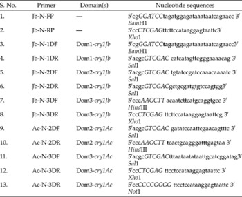
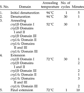
We constructed three chimeric genes by exchanging domain-coding fragments between cry1Jb and cry1Ac. Parental genes cry1Jb and cry1Ac were named as Jb1D-Jb2D-Jb3D and Ac1D-Ac2D-Ac3D to represent domains I, II and III of cry genes, respectively. The chimeric gene constructed by swapping the domains I and II of cry1Jb with domain III of cry1Ac was named as Jb1D-Jb2D-Ac3D, and the hybrid made by exchanging domain I of cry1Jb with domains II and III of cry1Ac was named as Jb1D-Ac2D-Ac3D. Another hybrid was constructed by exchanging the domain II of cry1Jb with that of cry1Ac and was named as Jb1D-Ac2D-Jb3D. Primer sequences were designed at the domain junctions (Table 1). To facilitate cloning of the domain fragments, restriction sites were created without altering the amino acid sequences. Polymerase chain reaction (PCR) conditions standardized for the amplification of the domain(s) are given in Table 2.
Primers used for the amplification of full-length cry1Jb from native strain EG 5092 and domain(s) from cry1Jb and cry1Ac native genes

PCR conditions standardized for the amplification of the domain(s)

The cry1Jb fragment in coding domains I and II was amplified (1350 bp) using domain-specific primers. This was further purified and digested with BamHI and Sal1, and ligated into the pET-29a vector. The amplified PCR product containing domain III of cry1Ac (2.18 kb) was digested with Sal1 and Not1 and cloned into pET-29a::Jb1D-Jb2D, which was restricted with the same enzymes to get the final construct of pET-29a::Jb1D-Jb2D-Ac3D. The chimeric gene construct of pET-29a::Jb1D-Jb2D-Ac3D was confirmed by restriction analysis.
Domain I encoded by cry1Jb was amplified with domain-specific primers and resolved in a 1% agarose gel. The amplified product of 750 bp was obtained and purified. The purified product was restricted with BamH1 and Sal1 and ligated into pET-29a vector. Domain II of cry1Ac gene was amplified using pKK233::cry1Ac as template. The amplified product (720 bp) was purified and restricted with Sal1 and HindIII, and cloned into the Sal1 and HindIII sites of pET-29a vector carrying the domain I of cry1Jb. Furthermore, domain III of cry1Jb was amplified using specific primers. The purified product (2.1 kb) was restricted with HindIII and Not1 and cloned into pET-29a::Jb1D-Ac2D, which was restricted with the same enzyme. The construct pET-29a::Jb1D-Ac2D-Jb3D was confirmed by restriction analysis.
The gene construct pET-29a::Jb1D-Ac2D-Ac3D was developed by PCR amplification of the domain I of cry1Jb with specific primers. The amplified product showing a size of 750 bp was purified and restricted with BamH1 and Sal1, and ligated into pET-29a. Domains II and III of cry1Ac (2.79 kb) were amplified using domain-specific primers, purified, restricted with Sal1 and Not1, and ligated in Sal1 and Not I sites of pET-29a::Jb1D. The final construct pET-29a::Jb1D-Ac2D-Ac3D was confirmed by restriction analysis.
All parental and chimeric genes were transformed into BL21(DE3) cells. A single colony was inoculated in 5 ml LB medium and grown overnight at 37 °C, at 220 rpm. The overnight grown culture was subcultured into 100 ml Luria Bertani medium (LB) and incubated at 37 °C, 220 rpm until the OD600 reached 0.6. Protein expression was induced by 1 mm Isopropyl-beta-thio galactopyranoside (IPTG), and cells were pelleted after 4 h. δ-Endotoxins were purified from Escherichia coli overexpressing clones as described by Ge et al. (Reference Ge, Shivarova and Dean1990).
Solubilization and activation of protoxins
The pellet was resuspended in an appropriate quantity of Na2CO3 buffer (pH 10.5) and kept for solubilization at 37 °C for 3–4 h with gentle shaking. After centrifugation at 8000 rpm for 10 min, the supernatant was recovered and stored at − 20 °C for further use. The total protein concentration was estimated by the Bradford assay (Bradford, Reference Bradford1976). Trypsin (10 mg/ml stock) was added (1:50 trypsin to protein w/w) and incubated at 37 °C for 1–2 h.
Analysis of proteins by sodium dodecyl sulphate (SDS)-polyacrylamide gel electrophoresis (PAGE) and immunoblotting
Total proteins from E. coli cells expressing Bt genes and trypsinized protein were resolved on a 12% SDS-PAGE. The amount of Bt protein in total protein was estimated by densitometric observation of the trypsin-resistant core on a SDS-PAGE and known bovine serum albumin concentration. The proteins were transferred onto polyvinylidene fluoride membrane (Millipore Immobilon-P) for immunoblotting and probed with rabbit polyclonal antibodies raised against Cry1Jb and Cry1Ac proteins.
Determination of toxicity
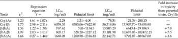
Bioassays were performed using the surface contamination assay. Toxin preparations were serially diluted in seven different toxin concentrations (as shown in Table 3) and tested by using 30 neonate larvae of H. armigera, Earias vittella F. and Spodoptera litura (Fab.) (both Lepidoptera: Noctuidae) per concentration. Toxin samples (75 μl) were applied uniformly into each vial with an artificial nutritive preparation (Gupta et al., Reference Gupta, Birah and Rani2004) and then allowed to dry. Larvae were placed on the nutritive surface, and reared at a temperature of 27 ± 1 °C, relative humidity 65 ± 5% and photoperiod 16 h:8 h scoto/photo regime. Mortality was assessed after 7 days. The effective dose (50% lethal concentration of the toxins, LC50) was calculated using probit analysis of the results obtained from five independent experiments.
Toxicity of parental and chimeric proteins against Helicoverpa armigera

Results
Selection of the parental toxins
A total of 200 different genes encoding crystal proteins have been cloned to date from Bt and two other species (a full list with further links can be found at: http://www.biols.susx.ac.uk/home/neil_crickmore/Bt/). We selected 50 cry genes that are specific to lepidopteran insects. Separate dendrograms were constructed for the whole toxins and individual domains using the amino acid sequences. Phylogenetic relationship revealed that Cry1Jb and Cry1Ac are widely separated, which in turn differs in their sensitivity to Cry1Ac and Cry1Jb against H. armigera. Multiple alignments of the amino acid sequences and superposition of 3D structures of the Cry1Jb and Cry1Ac toxins showed considerable sequence differences in the loops of domains II and III. Based on the above-mentioned results, Cry1Jb and Cry1Ac were selected for domain swapping.
Cloning and expression of parental and chimeric toxins
cry1Jb gene-specific primers were designed (Table 1), and PCR was carried out to amplify the gene sequence. The amplified PCR product was purified and cloned into pET-29a vector, and transformed into BL21(DE3) for overexpression. pKK233::cry1Ac construct was provided by Donald Dean (Ohio State University, Athens, OH, USA). All three chimeric genes were cloned and overexpressed. Results of 12% SDS-PAGE analysis of E. coli expressed parental and chimeric toxins showed a protoxin of 130 kDa. Trypsin activation yields a product of expected size of ≈ 60 kDa.
Insecticidal activity of Cry1Ac and Cry1Jb
Due to the considerable amino acid sequence differences between Cry1Ac and Cry1Jb, their biological activities were compared to illustrate the relationships between the sequences and activity. Cry1Ac and Cry1Jb were expressed in E. coli for biological assays. Cry1Ac is highly active against H. armigera (Table 3) and E. vittella with LC50 of 2.29 and 14.8 ng/ml, respectively. The activity assay with S. litura revealed no effect even at higher doses. Similarly, Cry1Jb was tested against all three insects. The LC50 of Cry1Jb towards H. armigera was found to be 6039 ng/ml, whereas no toxicity was observed against S. litura and E. vittella, even at the discriminative doses of 100 μg/ml.
Insecticidal activities of chimeric proteins generated from domain swapping between Cry1Jb and Cry1Ac
To evaluate the role of individual domain(s) of both Cry proteins in their toxicity and host specificity, and to explore the possibility of toxin improvement by domain swapping, chimeric genes of Jb-Jb-Ac, Jb-Ac-Jb and Jb-Ac-Ac were created and expressed in E. coli. All chimeric toxins were biologically active against H. armigera. Jb-Jb-Ac was 7.8-fold more toxic than Cry1Jb, followed by Jb-Ac-Jb and Jb-Ac-Ac with a 7.5- and 3.6-fold increase in toxicity against H. armigera, respectively (Table 3). No toxicity was recorded for chimeric toxins against S. litura even at a very high dose of 100 μg/ml. The only detectable effect was the reduced growth rate of the larvae.
Discussion
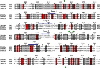
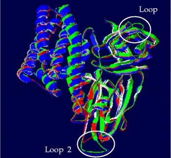
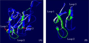
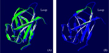
Multiple sequence alignment was carried out for the domains II and III of Cry1Ac and Cry1Jb, which showed considerable differences in the loop regions (Fig. 1). Difference in the loops of both domains of the toxin was further confirmed by superpositioning of 3D structures. Figure 2 shows the superpositioning of the whole toxin, and Figs 3 and 4 show domains and loops of II and III, respectively. These differences may partially account for the observed differences in the toxicity. Studies have shown that loops of domain II are the least homologous parts and extend beyond the β-sheet core surface (Grochulski et al., Reference Grochulski, Masson, Borisova, Pusztai-Carey, Schwartz, Brousseau and Cygler1995). Introduced changes/mutations in these loops affect toxin binding and specificity (Lee et al., Reference Lee, You, Gould and Dean1999; Jurat-Fuentes and Adang, Reference Jurat-Fuentes and Adang2001).
Amino acid sequence alignment for loops and adjacent regions of domains II and III (star indicates the start and end points of domains II and III) of Cry1Ac and Cry1Jb. Alignments were produced with the CLUSTAL program. Potential loops (shown by solid blue lines) were identified with Cry1Aa as a reference point. The order in which the toxins are listed corresponds to the order on the dendrogram based on levels of amino acid sequence similarity of domains II and III. Amino acids in the loops of domain II show sequence difference, which may affect the receptor binding. Amino acids from 503 to 511 in Cry1Jb show sequence difference with that of Cry1Ac, which may result in structural difference in the loops of domain III. (A colour version of this figure can be found online at: journals.cambridge.org/jti).

Superposition of 3D structure of Cry1Aa (blue), Cry1Ac (green) and Cry1Jb (red) toxins. The main differences in the three toxins are located in the loops of domain II and domain III, which is implicated in receptor recognition and binding. (A colour version of this figure can be found online at: journals.cambridge.org/jti).

The predicted domain II structure of Cry1Jb (green) is superimposed on that of Cry1Ac (blue). A and B, comparison of the structures adopted by the three surface-exposed loops of Cry1Ac and Cry1Jb at the extremity of the domain II. The locations of the loops are indicated, and close-up (B) highlights the structural differences in these putative receptor-binding regions. (A colour version of this figure can be found online at: journals.cambridge.org/jti).

The predicted domain III structure of Cry1Jb (green) is superimposed on that of Cry1Ac (blue). A and B, comparison of the structures adopted by the surface-exposed loops of Cry1Ac and Cry1Jb at the extremity of the domain III. The location of the loops is indicated, and close-up (B) highlights the structural differences (from 503 to 511 amino acid) in these putative receptor-binding regions. (A colour version of this figure can be found online at: journals.cambridge.org/jti).

The genes of Cry1 family are toxic to lepidopteran insects, which include some of the most devastating crop pests. The Cry1Ac has been found to be more toxic among the different Cry proteins known so far towards H. armigera, a polyphagous pest of many important crops (Chakrabarti et al., Reference Chakrabarti, Mandaokar, Kumar and Sharma1998). Cry1Ac with its known tertiary structure and relatively well-characterized receptor-binding regions is an ideal candidate for the domain swapping to alternate the specificity. Cry1Jb and Cry1Ac are presumed to share a similar mode of action but target different insect species.
The absence of toxicity of Cry1Jb protein against certain insects such as S. litura, E. vittella and H. armigera suggests that Cry toxins are highly specific in their action. Specificity in case of Cry toxins has been attributed to toxin–receptor interaction (Van Rie et al., Reference Van Rie, Jansens, Höfte, Degheele and Van Mellaert1990). Cry1Ac displayed very high toxicity against H. armigera and E. vittella. These results are in agreement with the previous bioassays, which showed low LC50 values on H. armigera and E. vittella (Van Rie et al., Reference Van Rie, Jansens, Höfte, Degheele and Van Mellaert1989; Bohorova et al., Reference Bohorova, Cabrera, Abarca, Quintero, Maciel, Brito, Hoisington and Bravo1997; Chakrabarti et al., Reference Chakrabarti, Mandaokar, Kumar and Sharma1998; Jurat-Fuentes and Adang, Reference Jurat-Fuentes and Adang2001). The variation in LC50 values observed in various laboratories has been attributed to the nature and mode of application of the toxins, the method used to estimate protein concentration, the temperature at which bioassays were conducted and the origin of the insects (Avilla et al., Reference Avilla, Osunav, Cabrera, Ferre and Zamoraj2005; Pardo et al., Reference Pardo, Cabreral, Parla and Rodriguez2006).
A specific site, only present in the domain III of Cry1Ac (amino acid residues 503–525), is responsible for the interaction with the N-acetylgalactosamine moiety of the aminopeptidase N (APN) receptor from H. armigera (Jenkins et al., Reference Jenkins, Lee, Valaitis, Curtiss and Dean2000). This may be the reason for the increased toxicity of Cry1Ac than that of Cry1Jb against H. armigera. Mutations or differences in the structural loops of the Cry1Ac domain II as well as domain III swapping combinations demonstrated their involvement in specific Cry1Ac membrane receptor/complex interaction (Luo et al., Reference Luo, McLachlin, Brown and Adang1999; Rang et al., Reference Rang, Vachon, de Maagd, Villalon, Schwartz, Bosch, Frutos and Laprade1999; de Maagd et al., Reference de Maagd, Weemen-Hendriks, Stiekema and Bosch2000; de Maagd et al., Reference de Maagd, Bravo and Crickmore2001).
Hybrids derived from toxins with different specificities are powerful tools in determining the role of the primary amino acid sequences in specificity. Earlier results showed that exchanging domains of Cry proteins might improve toxicity to a particular target insect. This suggested that domain swapping could become a valuable tool in the modification of existing toxins, thereby increasing the repertoire of available toxins for use in agriculture (de Maagd et al., Reference de Maagd, Kwa, van der Klei, Yamamoto, Schipper, Vlak, Stiekema and Bosch1996). Several studies have demonstrated that the creation of hybrid Cry toxins can result in substantially improved toxins in terms of toxicity or target spectrum (de Maagd et al., Reference de Maagd, Bravo and Crickmore2001).
Domains of Cry1Jb and Cry1Ac were swapped to create three chimeric combinations of Jb-Jb-Ac, Jb-Ac-Jb and Jb-Ac-Ac. Protoxin-specific C-terminal half was retained, as earlier reports suggested that it enhances the toxicity of the proteins (de Maagd et al., Reference de Maagd, Bravo and Crickmore2001). The chimeric toxins were stable, and a trypsin-resistant core was obtained after trypsin digestion, showing that the chimeric proteins have folded properly and may be finally active. All the chimeric toxins had no significant activity against S. litura but showed considerable difference in growth with respect to the control. However, there was an increase in the toxicity of all the three chimeric toxins to H. armigera.
The effect of domain III changes is dependent on the origin of domains I and II as well (de Maagd et al., Reference de Maagd, Bakker, Masson, Adang, Sangadala, Stiekema and Bosch1999; Karlova et al., Reference Karlova, Weemen-Hendriks, Naimov, Ceron, Dukiandjiev and de Maagd2005). The Jb-Jb-Ac chimeric toxin is 7.6 times more toxic than Cry1Jb. This finding indicates that domains I and II of Cry1Jb are important determinants of the higher activity of Jb-Jb-Ac. However, the combinations with domain III and the protoxin-specific C-terminal part of Cry1Ac render the resulting hybrid toxins to be more toxic than Cry1Jb. The hypotheses that domains II and III can function separately in a two-step mechanism of binding of Cry1Ac to APN (Jenkins et al., Reference Jenkins, Lee, Valaitis, Curtiss and Dean2000) and that Cry1Ab binds to a cadherin-like protein as well as to APN (Bravo et al., Reference Bravo, Gomez, Conde, Munoz-Garay, Sanchez, Zhuang, Gill and Soberon2004) could help explain why novel combinations of domains II and III can have altered toxicity.
Bioassay results showed a higher toxicity of the Jb-Jb-Ac chimeric toxin, which was obtained by the substitution of domain III involved in high affinity binding. All hybrid toxins containing various domains I and II of Cry1 linked to domain III of Cry1Ac were more active against lepidopteran insects than the original Cry1 toxins but were less toxic than Cry1Ac. Domain III of Cry1Ac interacts specifically with H. armigera brush-border membrane vesicles (BBMV), and domain II can compete with domain III binding suggesting that the initial specific interaction is mediated by domain III as was demonstrated by domain swapping studies (de Maagd et al., Reference de Maagd, Weemen-Hendriks, Stiekema and Bosch2000, Reference de Maagd, Bravo and Crickmore2001; Avisar et al., Reference Avisar, Keller, Gazit, Prudovsky, Sneh and Zilberstein2004). This may be one of the reasons for the increased toxicity of the Jb-Jb-Ac chimeric toxin.
Domain III binding may also be rate limiting, which may be the cause of increased toxicity (Lee et al., Reference Lee, You, Gould and Dean1999). If the domain II-mediated binding/oligomerization step was rate limiting/absent, little/no increase in toxicity would occur even with domain III of Cry1Ac. The increase in toxicity of Jb-Jb-Ac may also be due to the replacement of amino acids 450–612 on Cry1Jb by amino acid from Cry1Ac, which was found to be more toxic against H. armigera than Cry1Jb parent (Schnepf et al., Reference Schnepf, Tomczak, Ortega and Whiteley1990; Caramori et al., Reference Caramori, Albertiai and Galizzi1991; Ge et al., Reference Ge, Rivers, Milne and Dean1991; Karlova et al., Reference Karlova, Weemen-Hendriks, Naimov, Ceron, Dukiandjiev and de Maagd2005). Formation of hybrids by substituting domain III is possibly an evolutionary strategy for Bt itself to modify toxicity.
The domain II substitution gave Jb-Ac-Jb a 7.5-fold increased toxicity compared with Cry1Jb against H. armigera. The present investigation not only identifies domain II of Cry1Ac as the most important determinant for the activity against H. armigera but also emphasizes the point that the domain II function of Cry1Ac is required in an insect-specific manner for the oligomer formation, which probably precedes/coincides with membrane insertion of the toxin (Herrero et al., Reference Herrero, Gonzalez-Cabrera, Ferre, Bakker and de Maagd2004). New combinations of the receptor-binding domains II and III resulted in increased activities of hybrids for several lepidopterans (Ge et al., Reference Ge, Rivers, Milne and Dean1991; de Maagd et al., Reference de Maagd, Kwa, van der Klei, Yamamoto, Schipper, Vlak, Stiekema and Bosch1996). The molecular mechanisms underlying this effect are well understood, although studies of Cry1Ac binding to a putative receptor Lymantria dispar APN suggested that domains II and III confer two separate steps in binding to this protein in a two-step model, and that one step may be rate limiting for that binding (Jenkins et al., Reference Jenkins, Lee, Valaitis, Curtiss and Dean2000). Following this line of reasoning, one could speculate that both Cry1Jb and Cry1Ac bind to the same receptor (both the toxins bind to a 110 kDa receptor in ligand blotting experiments) in H. armigera, but those different steps are rate limiting for the two toxins. Hence the optimal combinations of domains II and III may optimize both binding steps and thus increase activity. This may be the reason for the increased toxicity of Jb-Ac-Jb compared with that of Jb-Ac-Ac.
Bioassays conducted on insect larvae showed that the chimeric toxin Jb-Ac-Ac was active against H. armigera. The reduction in the toxicity of the Jb-Ac-Ac protein in vivo indicates that although it is able to recognize a binding site, make pores and kill the larvae like the parental Cry1Ac toxin, this protein cannot generate a toxic effect on insects similar to that of the Cry1Ac toxin. Reduced toxicity of Jb-Ac-Ac may be due to modification in the affinity (less strict and less tight association of domains II and III) for the receptor, leading to its lower binding capability and reduced toxicity.
Exchanging domain I results in the reduction in toxicity even when domains II and III from an active toxin are present. Swapping of domain I without creating mutations within the structural domains of the toxin or within the hinge region separating domains I and II was shown to influence the pore-forming ability of the activated toxin. These properties result from interactions between domain I and domains II and/or III. In addition, the structural domains must be compatible to yield a toxin capable of membrane permeation (Rang et al., Reference Rang, Vachon, de Maagd, Villalon, Schwartz, Bosch, Frutos and Laprade1999).
Exchange of domains within Cry proteins influences toxicity, level of protoxin production, protoxin stability and crystal formation, most likely by influencing the structure and folding of both the active toxin and the protoxin. Domain I is linked to domain II by salt bridges, four of which were identified in Cry1Aa (Grochulski et al., Reference Grochulski, Masson, Borisova, Pusztai-Carey, Schwartz, Brousseau and Cygler1995). Associating domain I of Cry1Jb with domain II of Cry1Ac protein may result in an improper presentation of the charged amino acids involved in the formation of these salt bridges. This may, in turn, generate an improperly folded toxin with altered toxicity. Further analysis on the role of the interdomain salt bridges and the interactions between the structural domains of Jb-Ac-Ac is needed to better understand the structure–function relationships in the Cry toxins. However, the three-dimensional structure of the parental and chimeric toxins (Jb-Jb-Ac, Jb-Ac-Jb and Jb-Ac-Ac) is still unknown, and it is therefore not possible to determine whether salt bridges are also involved in toxin–protoxin interactions. Analysing the structure–function relationships in the protoxin as a whole therefore will be valuable to better understand the mode of action of the toxins, a key step towards improved toxicity and resistance management (Rang et al., Reference Rang, Vachon, Coux, Carret, Moar, Brousseau, Schwartz, Laprade and Frutos2001). This may be the reason for the lower toxicity of the chimeric Jb-Ac-Ac compared with Jb-Ac-Jb and Jb-Jb-Ac. Thus, it is concluded that hybrid toxins resulting from domain swapping may have improved toxicity.
Acknowledgements
The work was supported by a financial grant from the Department of Biotechnology, Ministry of Science and Technology. Anuradha Chelliah is grateful to the Council of Scientific and Industrial Research, Government of India, for her Senior Research Fellowship.