INTRODUCTION
Hermaphroditism is widespread and takes on many forms among teleosts, having been reported for more than 350 species in 34 families and eight orders (Kuwamura & Nakashima, Reference Kuwamura and Nakashima1998). The most common form is the sequential hermaphroditism (mainly protogyny). At the other end of the spectrum there are simultaneous hermaphrodites in which individuals maintain both gonad tissue types throughout their life (Zorica et al., Reference Zorica, Sinovčić, Pallaoro and Čikeš Keč2006).
In the deep-sea environment, hermaphroditism is known only within some families. In particular, in addition to the protandrous type well known and restricted to a few species of two genera Cyclothone and Gonostoma of Gonostomatidae (Kawaguchi & Marumo, Reference Kawaguchi and Marumo1967; Badcock, Reference Badcock1986; Miya & Nemoto, Reference Miya and Nemoto1987), simultaneous hermaphroditism is a common sex allocation pattern within many Aulopiformes, among the Chlorophthalmidae (Chlorophthalmus agassizi, C. albatrossis, C. brasiliensis and Parasudis truculentus; Mead, Reference Mead1960; Hirakawa et al., Reference Hirakawa, Suzuki, Narimatsu, Saruwatari and Ohno2007) and Ipnopidae family (Bathypterois sp.; Mead, Reference Mead1960, Mead et al., Reference Mead, Bertelsen and Cohen1964 and Bathytyphlops sp.; Merrett, Reference Merrett1980).
The hermaphrodite Mediterranean tripodfish Bathypterois mediterraneus Bauchot, 1962 is a bathydemersal Ipnopidae, endemic to the Mediterranean Sea (Quignard & Tomasini, Reference Quignard and Tomasini2000) and not commercially important. The species is distributed both in the western basin (Catalan Sea (Stefanescu et al., Reference Stefanescu, Lloris and Recubado1992a), Balearic Islands (Moranta et al., Reference Moranta, Stefanescu, Massutí, Morales-Nin and Lloris1998; D'Onghia et al., Reference D'Onghia, Lloris, Poulitou, Sion and Dokos2004b), off the Gulf of Genoa (Tortonese & Relini-Orsi, Reference Tortonese and Relini-Orsi1970) and Sardinian Channel (Follesa et al., Reference Follesa, Mulas, Murenu, Sabatini and Cau2005)) where it has been found between 700 and 2800 m depth, and in the eastern (along the coast of Israel (Galil & Goren, Reference Galil and Goren1994), Cretan Sea (Papaconstantinou, Reference Papaconstantinou1988; Klausewitz, Reference Klausewitz1989; Kallianotis et al., Reference Kallianotis, Sophronidis, Vidoris and Tselepides2000), Levantine Sea (Galil, Reference Galil2004) and Ionian Sea (D'Onghia et al., Reference D'Onghia, Lloris, Poulitou, Sion and Dokos2004b)) between 800 and 3300 m.
The knowledge of the biology and ecology of B. mediterraneus is rather fragmentary, although this species is one of the dominant and most characteristic fish in deep-sea communities of the Mediterranean Sea. In general, abundance, biomass, bathymetric distribution, population structure and growth patterns have been studied in the western (Stefanescu et al., Reference Stefanescu, Lloris and Recubado1992a,Reference Stefanescu, Recubado and Llorisb, Reference Stefanescu, Lloris and Recubado1993, Reference Stefanescu, Morales-Nin and Massutí1994; Morales-Nin, Reference Morales-Nin1990, Reference Morales-Nin2001; Morales-Nin et al., Reference Morales-Nin, Massutí and Stefanescu1996; Cartes et al., Reference Cartes, Maynou, Moranta, Massutí, Lloris and Morales-Nin2004; Moranta et al., Reference Moranta, Palmer, Massutí, Stefanescu and Morales-Nin2004) and eastern Mediterranean (D'Onghia et al., Reference D'Onghia, Lloris, Sion, Capezzuto and Labropoulou2004a). Also, few aspects regarding the feeding ecology (Carrassón & Matallanas, Reference Carrassón and Matallanas1990, Reference Carrassón and Matallanas2001), morphological characteristics of the digestive tract (Carrassón & Matallanas, Reference Carrassón and Matallanas1994) and trophic relationships among the fish assemblages have been studied in the Catalan Sea (Carrassón & Cartes, Reference Carrassón and Cartes2002) and south-western Balearic Islands (Polunin et al., Reference Polunin, Morales-Nin, Pawsey, Cartes, Pinnegar and Moranta2001). Only Fishelson & Galil (Reference Fishelson and Galil2001) and D'Onghia et al. (Reference D'Onghia, Lloris, Sion, Capezzuto and Labropoulou2004a) reported information about the gonad hermaphrodite structure and reproductive cycle for the eastern Mediterranean.
As the data of the reproductive biology of hermaphroditic bathyal species are generally scanty, the aim of our work is to provide further information on the reproductive characteristics (sexual cycle, maturation, spawning period, hermaphroditism and length at first sexual maturity) exhibited by B. mediterraneus caught in the south-eastern Sardinian Sea (Sardinian Channel, central-western Mediterranean) at depths of about 800 to 1600 m.
MATERIALS AND METHODS
A total of 348 specimens of B. mediterraneus were caught during seasonal experimental trawl surveys carried out between 817 and 1598 m depth on compact mud bottoms off the south eastern Sardinian Sea (Sardinian Channel, central-western Mediterranean).
For each specimen the standard length (SL) was measured to the nearest mm. Total weight (TW) was recorded to the nearest g, and gonads (GW) and liver (LW) weights were noted to the nearest 0.01 g.
The fish were dissected and the gonads removed, and examined macroscopically according to the Fishelson & Galil (Reference Fishelson and Galil2001) criteria modified ad hoc by the authors of this work. A piece of tissue from the middle region of the gonad was cut and preserved in Carnoy's fixative and subsequently processed histologically to enable the observation of the process of gonadal development. Transverse sections (3 µm) were stained with sodium iodoeosine and toluidine blue (Dominici's method) (Mazzi, Reference Mazzi1977). Oocyte development stages were identified according to the scale proposed by Forberg (Reference Forberg1982), whereas the development stages of the testicular germinal cells were identified based on the spermatogenic differentiation developed by Grier (Reference Grier1981), both with the use of an optic microscope (Laborlux 12) at 40–250 × magnification. Oocytes of a defined number of microscopic fields were measured and counted by stage and only oocytes sectioned through the nucleus were measured with the image analysis program, tpsDig2 (Rohlf, Reference Rohlf2005).
The reproductive period was established by analysing the values of the gonadosomatic index (GSI = GW × 100/TW) (Anderson & Gutreuter, Reference Anderson, Gutreuter, Nielsen and Johnson1983) and the temporal evolution in the per cent frequency of the maturity stages of female and male components. The hepatosomatic index (HIS = LW × 100/TW) and the condition factor (K = TW × 100/SL3) were also calculated. The indices were calculated separating the specimens according to month of capture. Monthly values of all indices were compared using a one-way ANOVA test completed by a multiple sample comparison of means (Dagnélie, Reference Dagnélie and Duculot1970). Size at sexual maturation was determined by expressing the proportion of reproductively active fish (Stages III and IV) collected during the spawning season as a percentage of the total number of fish in each size-class. The length at first maturity (L50) was determined as the proportion of reproductively active fish in each size-class (macroscopic Stages III and IV) and by fitting a logistic ogive:
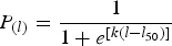
where P is the percentage of mature fish at length l; l50 the length at first maturity and k the model parameter.
RESULTS
The annual length distribution of B. mediterraneus, obtained from monthly sampling, showed a range in size between 47 and 150 mm SL with a dominant mode between 110 and 120 mm and a mean of 104±19 (mm±SD) (Figure 1).
Annual length distribution of Bathypterois mediterraneus sampled in the south-eastern Sardinian Sea between 817 and 1598 m. N, number of specimens.

Macroscopic analysis of the gonads
The gonads of B. mediterraneus were of an elongated and subcylindrical shape ranging from the liver region over the median genital papilla (Tortonese & Relini-Orsi, Reference Tortonese and Relini-Orsi1970). The criteria of Fishelson & Galil (Reference Fishelson and Galil2001) were used to classify macroscopically the gonads of the species with the difference that one stage was added after the Stage II and other one before the Stage III.
In each stage, the ovary was the most evident component of the ovotestis while the testis appeared as a colourless filament varying in thickness which maturity stage was never discriminated macroscopically.
The macroscopic stages of the female component were as follows:
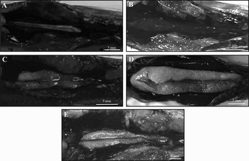
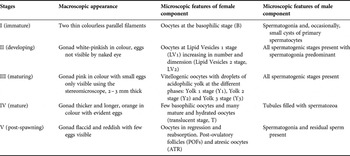
Stage I, immature: the gonad consists of two thin colourless parallel filaments (Figure 2A);
Stage II, developing: the ovary appears to be white-pinkish in colour, eggs not visible by naked eye (Figure 2B);
Stage III, maturing: the gonad appears to be 2–3 mm thick, pink in colour with small eggs only visible using the stereomicroscope (Figure 2C);
Stage VI, mature: the gonad appears to be thicker and longer, orange in colour with evident eggs (Figure 2D);
Stage V, post-spawning: the gonad appears flaccid and reddish with few eggs visible (Figure 2E).
Macroscopic developmental stages of Bathypterois mediterraneus ovotestis. (A) Stage I, immature; (B) Stage II, developing; (C) Stage III, maturing; (D) Stage IV, mature; (E) Stage V, post-spawning.

The histological analysis revealed the presence of an ovotestis in which both female and male tissue occurred contemporaneously and clearly subdivided into two zones by a connective tissue (Figure 4E).
Microscopic analysis of female component
The macroscopic scale adopted for this species was confirmed by the histological observation of oogenesis. On the basis of the oocyte stages, five different ovarian developmental phases were distinguished:
Immature ovary: the gonad showed mainly oocytes at early perinucleolus stage (Forberg et al., 1982) with a high nucleus to cytoplasm ratio, small size and lightly basophilic cytoplasm (B). Oocyte diameter 24–166 µm; mean diameter 74.57±1.30 (μm±SE) (Figure 3A);
Developing ovary: the pre-vitellogenetic ovary contained oocytes with a less basophilic cytoplasm due to the presence of few lipid vesicles (Lipid Vesicles 1 stage (LV1)) that subsequently increase in number and dimension (Lipid Vesicles 2 stage (LV2)). LV1 oocyte diameter 93–244 µm; mean diameter 135,02±5.28 (μm±SE); LV2 oocyte diameter 116–380 µm; mean diameter 189.28±25.76 (μm±SE) (Figure 3B);
Maturing ovary: the ovary was characterized by few basophilic oocytes (B), the previtellogenetic oocytes (LV2) and by vitellogenic oocytes with droplets of acidophilic yolk at the different phases: Yolk 1 stage (Y1) with droplets of yolk in the periphery of the cytoplasm, Yolk 2 stage (Y2) with more and larger yolk droplets and Yolk 3 stage (Y3) with compacting and successive fusion of the yolk droplets together with a progressive migration of nucleus towards the animal pole. Y1 oocyte diameter 210–332 µm, mean diameter 291.04±21.85 (μm±SE); Y2 oocyte diameter 392–618 µm, mean diameter 601.87±41.10 (μm±SE); Y3 oocyte diameter 502–702 µm, mean diameter 642.69±28.16 (μm±SE) (Figure 3C);
Mature ovary: the ovary showed a few basophilic oocytes and many mature and hydrated oocytes (translucent stage (T)) that reach maximum dimensions owing to the clarification of the yolk. Oocyte diameter 600–1053 µm, mean diameter 837.95±47.18 (μm±SE) (Figure 3D).
Post-spawning ovary: the female portion was characterized by oocytes in regression and reabsorption. Post-ovulatory follicles (POFs) and atresic oocytes (ATR) were present (Figure 3E).
Transverse sections of Bathypterois mediterraneus female component illustrating oogenesis. (A) Immature stage containing: B, basophil oocytes; (B) developing stage containing: B, basophil oocytes, LV1, Lipid Vesicles 1, LV2, Lipid Vesicles 2; (C) maturing stage showing Yolk1 (Y1) and (Y3) oocytes; (D) mature stage with translucent oocytes (T); (E) post-spawning stage with atresic oocyte (ATR) and post-ovulatory follicle (POF).

Microscopic analysis of male component
Testis tissue consisted of a convoluted seminiferous tubule of the lobular type (Nagahama, Reference Nagahama, Hoar, Randall and Donaldson1983); it is also called unrestricted spermatogonial testis-type (Grier, Reference Grier1981), because spermatogonia are distributed along the entire length of the tubule and not only restricted to the distal terminus.
We described five different developmental stages on the basis of germinal cells proportions (Figure 4):
Stage I, immature testis: the testis contained spermatogonia and, occasionally, small cysts of primary spermatocytes (Figure 4A);
Stage II, developing testis: spermatogonia predominated, but all spermatogenic stages were present. Rare groups of sperm could be seen attached to the lobular wall (Figure 4B);
Stage III, maturing testis: all spermatogenic stages were present. Spermatozoa detached from lobular wall filled the lumen of seminiferous lobules (Figure 4C);
Stage IV, mature testis: tubules were filled with spermatozoa (Figure 4D);
Stage V, post-spawning testis: the spermatogenic activity was very limited with spermatogonia and residual sperm (Figure 4E).
Transverse sections of Bathypterois mediterraneus male component illustrating spermatogenesis. (A) Immature stage (I) containing: SPG, spermatogonia and SP I primary spermatocytes; (B) developing stage (II), an intense spermatogenic activity is observed, SPG, spermatogonia and SP I and II, primary and secondary spermatocytes, SPZ, spermatozoa; (C) maturing stage (III); (D) mature stage (IV) with tubules filled with spermatozoa; (E) post-spawning stage (V) with spermatogenic activity limited.

Table 1 shows the summary of macroscopic and microscopic scale of B. mediterraneus gonads.
Macroscopic scale of Bathypterois mediterraneus gonad with histological features of female and male components.

Oocyte size–frequency
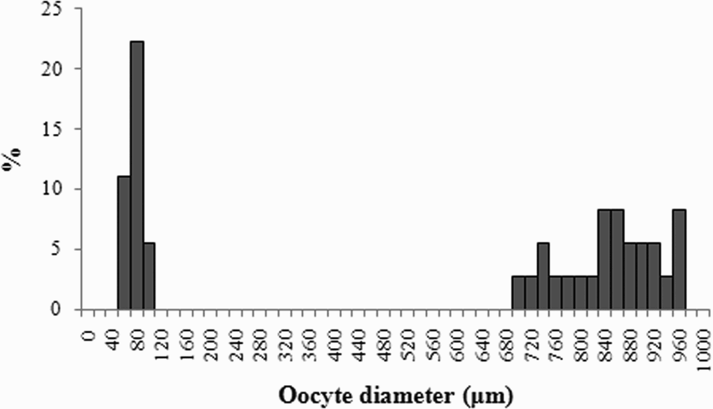
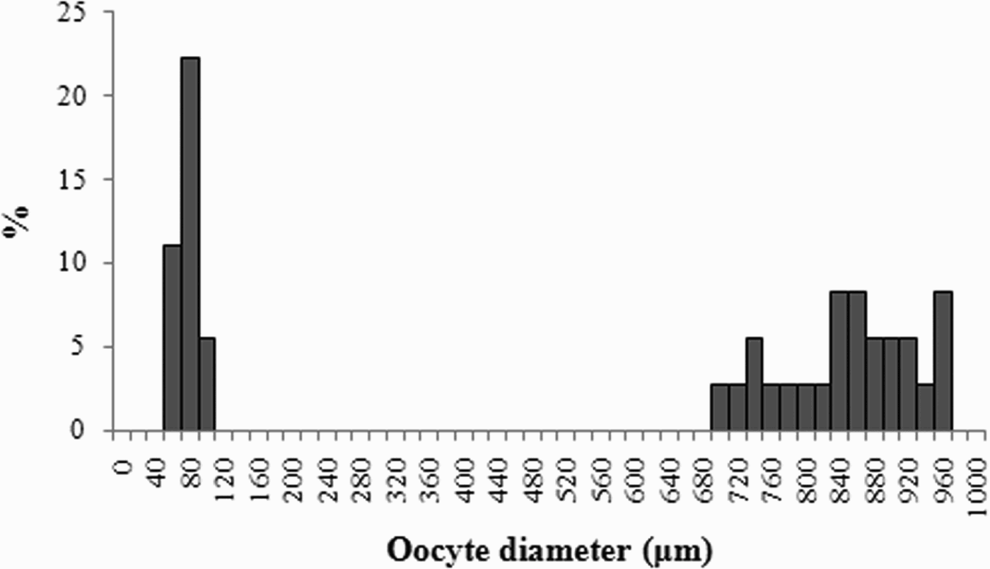
The oocyte size–frequency histograms determined for some mature ovaries' sections of tripodfish is shown in Figure 5. Oocytes of two stages of development were present: basophil oocytes (60–100 µm) and translucent oocytes (700–960 µm). All these features indicated that the species exhibited a ‘group-synchronous’ ovarian development organization visible also in Figure 3D.
Size–frequency distribution of oocytes determined from histological sections for some mature ovaries of Bathypterois mediterraneus.
Reproductive cycle
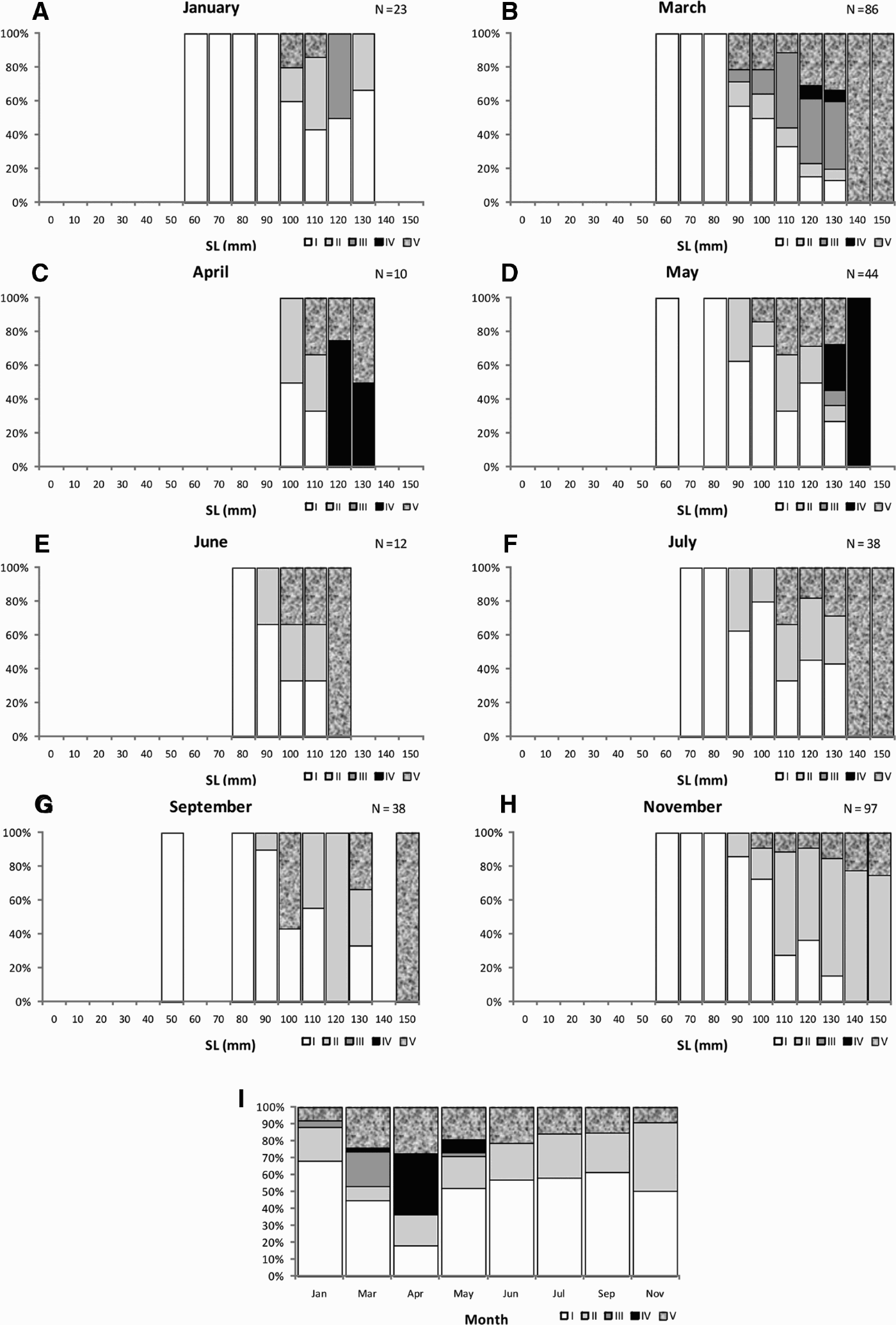
Monthly distributions of the different maturity stages for female component, showed a clear reproductive seasonality from March to May with a peak in April (Figure 6I).
Length–frequency distribution by months and stages (A–H) and monthly distributions of various maturity stages (I) of female component for Bathypterois mediterraneus (Stage I, immature; Stage II, developing; Stage III, maturing; Stage IV, mature; Stage V, post-spawning).
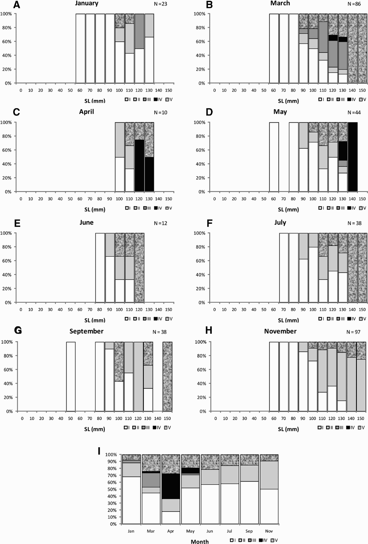
The specimens of size ranged between 50 and 80 mm SL were found to be immature (Stage I) throughout the year. A single specimen almost mature (Stage III; SL 111 mm) was found in January (Figure 6A), while the specimens with a maturing female component (minimum size-class 90 mm SL) appeared only in March with a value of 20.7% (Figure 6B). The occurrence of mature female portion (Stage IV) was detected from March (2.3%, size-classes 120 and 130 mm) to May (7.7%, size-classes 130 and 140 mm) with a peak in April (four out ten specimens; 120 mm and 130 mm size-classes, Figure 6C). Post-spawning Stage V females (from 100 mm SL) were found during all reproductive periods up to November (Figure 6A–H).
The smallest mature specimen (111 mm SL) was observed in March, while the largest (136 mm SL) was observed in May. The size at 50% maturity (L50) was attained at 119 mm SL (Figure 7).
Sexual maturity ogive for Bathypterois mediterraneus female component. n1 is the number of data used in the estimation of the equation, and n2 the total number of individuals used to estimate the percentage of mature fish.

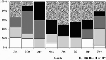
The percentage of the different male stages over the study period indicated a more protracted reproductive period compared to the female component (Figure 8).
Monthly distributions of maturity stages of Bathypterois mediterraneus for male component of the gonad (Stage I, immature; Stage II, developing; Stage III, maturing; Stage IV, mature; Stage V, post-spawning).

Inactive males (Stages I and II) were found in all sample months. Maturing males (Stage III) appeared mainly from March (50%) to April (40%), while mature males (Stage IV) are dominant from March to November with a peak (40%) in April. Post-spawning males (Stage V) were found throughout the year (except April) with higher proportions from May to September (40%) (Figure 8).
Trends in GSI, HIS and K
As defined by the monthly distributions of different maturity stages of female component, the highest gonadosomatic index (GSI) values were observed between March and May. The GSI peak was observed in April. The differences of GSI during the sampled months were highly significant (one-way ANOVA, P < 0.05). Particularly, multiple sample comparisons of means showed that GSI values of March, April and May differed statistically from values of the other months (June–January) and were different between them (P < 0.05) (Figure 9A).
Monthly changes in the gonadosomatic index (A, GSI), hepatosomatic index (B, HSI) and the condition factor (C, K) of Bathypterois mediterraneus. Each value represents the mean±SE (boxes) based on the number of fish sampled at each month; the vertical line represents the maximal and the minimal values.

Hepatosomatic index (HIS) showed a clear seasonal pattern, highlighting significant variation across months (one-way ANOVA; P < 0.05). In the peak of the reproductive period (April) the individuals showed the highest HIS median value, which remained high until May. Furthermore, multiple sample comparisons of means showed that HSI values of April and May differed statistically from HSI registered between June and November (P < 0.05) (Figure 9B).
The monthly profile of the condition factor (K) was not marked, with only a slight increase in April. However, April values appeared different from other months (multiple sample comparisons of means, P < 0.05) (Figure 9C).
DISCUSSION
This paper represents the first study regarding the reproductive biology of B. mediterraneus in the central western Mediterranean Sea.
Macroscopic investigation of B. mediterraneus gonads showed that all individuals maintained male and female gonad tissue with the ovarian component predominant on the testis. By histological analysis, the two gonadal portions were clearly distinct from each other and simultaneously functional, highlighting the presence of a simultaneous hermaphroditism as already confirmed by other studies (Mead, Reference Mead1960; Fishelson & Galil, Reference Fishelson and Galil2001; D'Onghia et al., Reference D'Onghia, Lloris, Sion, Capezzuto and Labropoulou2004a). This aspect appears to be a feature common to other species of the genus (B. longipes and B. dubius) (Sulak, Reference Sulak1977) and family (Bathytyphlops sewelli) (Merrett, Reference Merrett1980, Reference Merrett1994).
Histological analysis is also useful to identify the type of reproductive organization, emphasizing the type of development of both gonadal components. For the female component seven stages of oocytes development that characterize the different stages of maturity were identified.
The oocyte size (larger than those found by Fishelson & Galil, Reference Fishelson and Galil2001) frequency distributions in mature component indicated that the tripodfish exhibits a ‘synchronous-group’ ovary (Wallace & Selman, Reference Wallace and Selman1981) characterized by the presence of two contemporaneous populations of oocytes: one at the translucent stage and another, more heterogeneous consisting of small immature oocytes. This dynamic organization and the absence of post-ovulatory follicles (POF) in the ripe component highlighted a type of monocyclic ovary characterized by deposition in a single batch of eggs per year (total spawner) as confirmed by Fishelson & Galil (Reference Fishelson and Galil2001) in the eastern Mediterranean.
The testis appeared as a colourless filament never discriminated macroscopically. The histological structure common to many other teleosts was of the ‘unrestricted’ type with spermatogonia along the entire length of the tubules, other more mature germinal cells near the lumen and spermatozoa in the centre (Grier, Reference Grier1981).
The species showed a late size at first maturity (L50) of 119 mm SL. The tripodfish reaches a maximum SL of 160 mm while the smallest mature specimen was 110 mm SL. In the eastern Mediterranean, Fishelson & Galil (Reference Fishelson and Galil2001) reported a different result with mature individuals of greater size (140–158 mm SL). The delayed reproduction is a common characteristic in other deep-sea species such as Nezumia sclerorhynchus and Coelorhynchus coelorhynchus (D'Onghia et al., Reference D'Onghia, Basanisi and Tursi2000).
Bathypterois mediterraneus showed a typical seasonal reproductive pattern. A seasonal spawning is known in other Mediterranean fish species such as Phycis blennoides (Rotllant et al., Reference Rotllant, Moranta, Massutí, Sardà and Morales-Nin2002) distributed in the first part of the tripodfish depth-range. Through macroscopic and microscopic gonad analysis and monthly variation of GSI, for female component, a definite reproductive period in spring, between March and May with a peak in April was identified. From June to November, however, the individuals were immature or in post-spawning. The male component showed, instead, the large amount of mature germ cells in sperm ducts all years more or less showing a prolonged in time. This longer spawning period of component male is common also in many simultaneous hermaphroditic species such as Chlorophthalmus agassizi (Follesa et al., Reference Follesa, Cabiddu, Davini, Porcu and Cau2004; Anastasopoulou et al., Reference Anastasopoulou, Yiannopoulos, Megalofonou and Papaconstantinou2006) probably guaranteeing continuous spermatogenesis at any time of year.
The hepatosomatic index (HIS) and the condition factor (K) were high during the spawning season, assuming that the energy reserves used for the reproductive act are mainly composed of liver reserves and that, in this particular period, the species shows a more active trophism.
The spawning period of the species appears to be concordant with the only data reported in literature for the central Mediterranean (Gulf of Genova) by Tortonese & Relini-Orsi (Reference Tortonese and Relini-Orsi1970), but differs totally from the finding of Fishelson & Galil (Reference Fishelson and Galil2001) for the eastern Mediterranean, where the spawning period was identified between September and November.
The timing and duration of spawning in fish is generally accepted to coincide with periods in which environmental conditions are favourable for larval survival and growth. Furthermore, in deep-sea species seasonal reproduction species seem also to be mainly regulated by food supply (Gage & Tyler, Reference Gage and Tyler1991). The most plausible explanation of the discrepancy in spawning data of B. mediterraneus from the two sides of the Mediterranean basin (eastern and western) could be due to different availability of food. The oligotrophic Mediterranean waters are affected, in fact, by two annual peaks of primary production, i.e. spring and autumn. This periodicity has given rise to seasonal food availability, not only in shallow waters, but also in deeper ones. In the western Mediterranean Polunin et al. (Reference Polunin, Morales-Nin, Pawsey, Cartes, Pinnegar and Moranta2001) found a greater metabolic activity in the May samples of B. mediterraneus than October ones (on the basis of stable isotope data), highlighting the differences in function of primary production which appears highest in spring, increasing the downward flow of particles in the water column. In the eastern Mediterranean, instead, the presence of a greater concentration of meioplancton in late summer (Weikert et al., Reference Weikert, Koppelmann and Wiegratz2001), with a large amount of copepods, preferential preys of the diet of B. mediterraneus (Carrassón & Matallanas, Reference Carrassón and Matallanas2001) could justify the autumn spawning period found by Fishelson & Galil (Reference Fishelson and Galil2001).
The spawning period in a season of maximum concentration of organic matter could ensure, in populations with low density where there is little probability that individuals will encounter a mate (Ghiselin, Reference Ghiselin1969; Merrett, Reference Merrett1994), an increase of reproductive and offspring survival chances (Coggan et al., Reference Coggan, Gordon and Merrett1998).
In summary, the deep-sea fish B. mediterraneus provides a good example of a species adaptation to an oligotrophic environment by means of reproductive strategies. This species presented reproductive features such as simultaneous hermaphrotiditism, delayed sexual maturity, spawning period limited to one season (monocyclic ovary) and small eggs that can be seen as adaptations to life in a poor environment. This conclusion is further supported by other data in literature that show, for this species, a low fecundity (2000–2400 eggs in a clutch probably developed close the bottom; Sulak, Reference Sulak1977; Fishelson & Galil, Reference Fishelson and Galil2001) and a slow growth rate (Morales-Nin et al., Reference Morales-Nin, Massutí and Stefanescu1996, Morales-Nin, Reference Morales-Nin2001; D'Onghia et al., Reference D'Onghia, Lloris, Sion, Capezzuto and Labropoulou2004a).