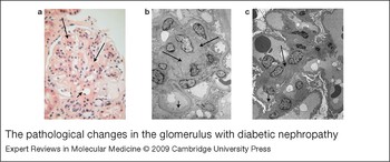
Why do approximately a third of patients with diabetes develop nephropathy irrespective of glycaemic control? Hyperglycaemia is known to play a critical role in the pathogenesis of diabetic nephropathy via activation of genes regulating extracellular matrix (ECM) synthesis and degradation. The accumulation of ECM accounts for the prominent pathological changes of diabetic nephropathy, as seen on the kidney biopsy in Figure 1. While a hyperglycaemic environment is necessary, it is not sufficient alone to cause diabetic nephropathy (Ref. Reference Marre1). Marked ethnic variation and familial predisposition is also observed, suggesting a significant genetic component (Refs Reference Chowdhury2, Reference Granier3). The pathogenesis of diabetic nephropathy likely results from a complex interaction between both metabolic and haemodynamic factors that depend on genetic predeterminants within a specific environment (Ref. Reference Wada, Makino and Kanwar4). The current proposed metabolic mechanisms mediating diabetic kidney disease include accumulation of advanced glycation endproducts in the kidney, oxidation of renal glycoproteins by reactive oxygen species (ROS), intracellular accumulation of sorbitol via the reduction of glucose by aldose reductase, and activation of protein kinase C (PKC), the hexosamine biosynthetic pathway (HBP) and the mitogen-activated protein kinase (MAPK) cascade – ultimately culminating in production of profibrotic growth factors, such as transforming growth factor β1 (TGF-β1) (Refs Reference Wada, Makino and Kanwar4, Reference Zhu, Usui and Sharma5).
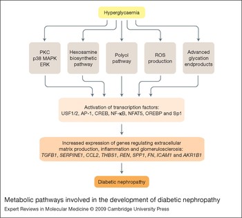
This review focuses on a select group of transcription factors that have been implicated in the regulation of TGF-β1, as well as other downstream genes that are involved with the progression of diabetic kidney disease (Fig. 2). Investigating transcription factors such as the upstream stimulatory factors (USF1 and 2), activator protein 1 (AP-1), cAMP-response-element-binding protein (CREB), nuclear factor (NF)-κB, nuclear factor of activated T cells (NFAT), and stimulating protein 1 (Sp1) may help elucidate the translation of a high-glucose signal into the production of mediators of progressive nephropathy.
The pathological changes in the glomerulus with diabetic nephropathy. (a) Light microscopy of a human glomerulus with diabetic nephropathy. Long arrows show areas of diffuse mesangial expansion. Short arrow depicts arteriolar hyalinosis. (b) Electron microscopy of diabetic glomerulus. Long arrows depict diffuse mesangial matrix expansion. Note that the mesangial matrix does not have clear borders. The short arrow points to a thickened glomerular basement membrane. (c) Electron microscopy of a normal glomerulus. The long arrow points to normal mesangial matrix. The short arrow shows the thickness of a normal glomerular basement membrane.

Metabolic pathways involved in the development of diabetic nephropathy. Hyperglycaemia leads to intracellular activation of the PKC, MAPK, HBP and polyol pathways, and also leads to accumulation of ROS and production of advanced glycation endproducts. These pathways lead to activation of the transcription factors USF1/2, AP-1, CREB, NF-κB, NFAT5, OREBP and Sp1. These transcription factors have been demonstrated to increase the expression of genes that ultimately culminate in extracellular matrix accumulation, inflammation and glomerulosclerosis in the diabetic kidney. Abbreviations: AKR1B1, aldo-keto reductase family 1, member B1; AP-1, activator protein 1; CCL2, chemokine (C-C motif) ligand 2; CREB, cAMP-response-element-binding protein; ERK, extracellular-signal-regulated kinase; FN, fibronectin; HBP, hexosamine biosynthetic pathway; ICAM1, intercellular adhesion molecule 1; MAPK, mitogen-activated protein kinase; NF-κB, nuclear factor κB; OREBP, osmotic-response-element-binding protein; PKC, protein kinase C; REN, renin; ROS, reactive oxygen species; SERPINE1, serpin peptidase inhibitor, clade E, member 1; Sp1, stimulating protein 1; SPP1, secreted phosphoprotein 1; TGFB1, transforming growth factor β1; THBS1, thrombospondin 1; USF, upstream stimulatory factor.

Overview of transcription factor activation and classification
Transcription factors are proteins that bind to promoter regions of genes and activate or silence their transcription into mRNA (Ref. Reference Levine and Tjian6). In addition to availability of the DNA-binding site, activity of a transcription factor often requires post-translational modification and binding with nuclear-localisation proteins (Ref. Reference Frith7). Thus, transient signals such as high glucose can be converted into long-term changes in gene expression by activating various signal transduction pathways within the cell that result in transcription factor phosphorylation and glycosylation (Refs Reference Han and Kudlow8, Reference Weigert9, Reference Kreisberg, Radnik and Kreisberg10).
Most eukaryotic genes are packed into chromatin structures, comprising DNA and histone proteins, that require chromatin remodelling to facilitate transcription (Ref. Reference Miao11). Aberrant histone modifications may lead to changes in chromatin structure, allowing for dysregulated gene transcription and disease progression. Epigenetics is a term used to describe heritable alterations in gene expression by mechanisms other than changes in the underlying DNA sequence (Ref. Reference Miao12). Current interest in the epigenetics of diabetic complications has included evaluating the impact of high glucose on histone lysine acetylation and methylation patterns in vascular smooth muscle cells derived from type 2 diabetic mice and in human monocytes (Refs Reference Miao12, Reference Villeneuve13). These modifications can result in a heritable change in gene expression known as cellular or metabolic memory, which may explain why patients with diabetes continue to develop inflammation and vascular complications even after achieving glycaemic control (Refs Reference Liu, Li and Tollefsbol11, Reference Villeneuve13). However, the emerging field of epigenetics is not a major aspect of this review as we primarily focus on data demonstrating the role of specific transcription factors in diabetic kidney disease.
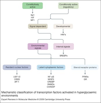
Transcription factors can be classified by their structural motif or mechanistically, such as constitutively active and regulatory transcription factors (Refs Reference Brivanlou and Darnell14, Reference Pabo and Sauer15) (Fig. 3). The constitutively active transcription factors facilitate the expression of genes that are always transcribed, such as structural proteins and crucial metabolic enzymes, and include the ubiquitously expressed transcription factor Sp1 (Ref. Reference Brivanlou and Darnell14). Regulatory transcription factors are either developmental transcription factors important in embryonic cell type differentiation or signal-dependent transcription factors, which are largely inactive until exposed to the appropriate intra- or extracellular signal (Ref. Reference Brivanlou and Darnell14). The signal-dependent transcription factors can be further categorised into those that are activated by internal stimuli (sterol-regulatory-element-binding proteins; SREBPs) and transcription factors activated by environmental stimuli. The transcription factors that are activated by environmental stimuli can be further subcategorised into resident nuclear factors (CREB, AP-1 and USF1/2), which reside in the nucleus but must be activated to affect transcription; latent cytoplasmic factors (NF-κB and NFAT proteins), which exist in the cytoplasm in an inactive form and then translocate to the nucleus upon activation; and the steroid receptor superfamily (e.g. the peroxisome-proliferator-activated receptors and the glucocorticoid receptor), which can reside in the nucleus or cytoplasm and function as transcription factors once activated (Refs Reference Levine and Tjian6, Reference Brivanlou and Darnell14) (Fig. 3).
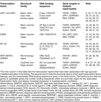
The characteristics of the transcription factors discussed in this review are summarised in Table 1.
Mechanistic classification of transcription factors activated in hyperglycaemic environments. This diagram shows a classification schema for transcription factors based on their functional characteristics. Overall, transcription factors are either constitutively active or conditionally active. Included in this table are transcription factors that are either activated in the diabetic state or important in glucose homeostasis. This review focuses on signal-dependent transcription factors. Signals can arise from the intracellular environment, or from an environmental stimulus, such as glucose. Glucose has been shown to activate transcription factors that reside in the nucleus as well as those that reside in the cytoplasm and translocate to the nucleus in order to affect gene transcription. The superfamily of steroid receptor proteins are transcription factors that reside in the cytoplasm or nucleus and have the ability to directly bind DNA once activated. Abbreviations: AP-1, activator protein 1; ChREBP, carbohydrate-responsive-element-binding protein; CREB, cAMP-response-element-binding protein; GR, glucocorticoid receptor; HIF, hypoxia-inducible factor; HNFs, hepatocyte nuclear factors; NF1, nuclear factor 1; NFAT5, nuclear factor of activated T cells 5; NF-κB, nuclear factor κB; OREBP, osmotic-response-element-binding protein; PPARs, peroxisome-proliferator-activated receptors; Sp1, stimulating protein 1; SREBPs, sterol-regulatory-element-binding proteins; USF, upstream stimulatory factor.

Characteristics of transcription factors activated by high glucose and potential gene targets in models of diabetic kidney diseasea

aThe first column in this table lists the transcription factors that have been associated with high-glucose-mediated activation in the kidney. The second column lists the structural class to which each transcription factor belongs, and the characteristic DNA binding sequence is given in the third column. The fourth column lists the genes or candidate genes that contain binding sequences within the promoter region for the associated transcription factor and display increased expression in hyperglycaemic conditions.
Abbreviations: ADIPOQ, adiponectin; AKR1B1, aldo-keto reductase family 1, member B1; AP-1, activator protein 1; CCL2, chemokine (C-C motif) ligand 2; CRE, cAMP-response element; CREB, cAMP-response-element-binding protein; DCN, decorin; FN, fibronectin; ICAM1, intercellular adhesion molecule 1; MIOX, myo-inositol oxygenase; NFAT, nuclear factor of activated T cells; NF-κB, nuclear factor κB; ORE, osmotic-response element; OREBP, osmotic-response-element-binding protein; PTGS2, prostaglandin-endoperoxide synthase 2; REN, renin; RSOR, renal-specific oxidoreductase; SERPINE1, serpin peptidase inhibitor, clade E, member 1; Sp1, stimulating protein 1; SPP1, secreted phosphoprotein 1; TGFB, transforming growth factor β; THBS1, thrombospondin 1; TonE, tonicity-response element; USF, upstream stimulatory factor.
Transcription factors involved in development of diabetic nephropathy
Upstream stimulatory factors 1 and 2
USF1 and USF2 are ubiquitously expressed and belong to the Myc family of transcription factors that are characterised by a basic–helix–loop-helix leucine-zipper motif (bHLH–zip) (Refs Reference Atchley and Fitch16, Reference Henrion17). USF1 and USF2 are encoded by two separate genes but share the highly conserved C-terminal domain that includes the basic domain for DNA binding and the leucine zipper for dimerisation (Refs Reference Kreisberg, Radnik and Kreisberg10, Reference Pabo and Sauer15, Reference Atchley and Fitch16, Reference Luo and Sawadogo18). Factors containing the basic binding domain must form dimers in order to bind DNA, and the USF proteins function primarily as a USF1–USF2 heterodimer or a USF1 homodimer (Refs Reference Atchley and Fitch16, Reference Latchman19). The DNA-binding basic region recognises the E-box (CACGTG) or E-box motif (CANNTG), which for the purposes of this review will be regarded as the glucose-responsive element (GRE) and part of the carbohydrate-response element (ChoRE), the latter consisting of two E-box motifs separated by 5 bps (Refs Reference Shi20, Reference Liu, Shi and Wang21, Reference Zhu22, Reference Corre and Galibert23).
In response to high glucose, activation of PKC appears to be a critical signalling pathway affecting USF1 levels, while USF2 appears to be regulated by several signalling pathways including p38 MAPK, ERK1/2 (extracellular-signal-regulated kinases 1/2) and HBP, as well as PKC (Refs Reference Zhu22, Reference Kutz24, Reference Wang25). USF1 and USF2 have been linked to a variety of glucose-regulated genes in mesangial cells, hepatocytes, epithelial cells and smooth muscle cells, and the relative amounts of each protein vary among cell types (Refs Reference Zhu22, Reference Vallet26, Reference Vallet27). Mice lacking USF2 show growth delay and increased postnatal death, whereas USF1-knockout mice are phenotypically normal, possibly due to compensation from USF2 (Refs Reference Vallet26, Reference Vallet27). In streptozotocin-induced diabetic rats, only USF1, and not USF2, is increased in glomeruli after two weeks of diabetes (Ref. Reference Zhu22). However, overexpression of USF2 in transgenic mice leads to albuminuria in nondiabetic mice by 6 months of age, which is further accelerated by inducing diabetes (Ref. Reference Liu, Shi and Wang21). The role of USF1/2 in diabetic nephropathy likely involves transcriptional upregulation of the genes for TGF-β1 (TGFB1), thrombospondin 1 (THBS1), and osteopontin (SPP1) in response to high glucose, although binding sites have also been reported in the promoters of the genes for plasminogen activator inhibitor 1 (PAI-1; SERPINE1) and renin (REN) (Refs Reference Weigert9, Reference Liu, Shi and Wang21, Reference Zhu22, Reference Kutz24, Reference Wang25, Reference Pan28, Reference Bidder29).
Transforming growth factor β1
The profibrotic cytokine TGF-β1 has been implicated in the development of diabetic kidney disease as a result of its role in excess ECM accumulation in a high-glucose environment (Ref. Reference Zhu22). The TGFB1 gene contains an E-box located within the murine promoter at −641 to −636 and within the human promoter at −1013 to −1002 (Refs Reference Weigert9, Reference Zhu22). Murine mesangial cells and porcine mesangial cells transfected with the human TGFB1 promoter demonstrate increased USF1 homodimer and USF1–USF2 heterodimer binding activity to the TGFB1 GRE, although in murine mesangial cells there is preferential binding of USF1 (Refs Reference Weigert9, Reference Zhu22). USF2 homodimers in vivo are scarce, and require USF1 as a heterodimer to regulate promoter activity (Refs Reference Zhu22, Reference Sirito30). Overexpression of USF1 or USF2 in transfected porcine mesangial cells increases the TGFB1 promoter activity twofold, which is prevented by a mutation in the GRE (Ref. Reference Weigert9). In murine mesangial cells and aortic smooth muscle cells, USF1 protein levels increase with sequential elevations in the glucose concentration, even within the high-normal range, while there have been conflicting reports of high-glucose-enhanced USF2 expression (Refs Reference Shi20, Reference Zhu22, Reference Bidder29). Overexpression of USF1 or USF2 in human embryonic kidney cells increases TGFB1 promoter activity; however, only the USF1-transfected cells exhibited increased secretion of TGF-β1 protein (Ref. Reference Zhu22).
Renal TGF-β1 expression has been studied under fasting and carbohydrate refeeding conditions in USF1-knockout and USF2-knockout mice (Ref. Reference Zhu22). In mice lacking USF1, renal TGF-β1 mRNA is not stimulated in response to high carbohydrate intake, whereas mice lacking USF2 have the same degree of stimulation of renal TGF-β1 as wild-type mice (Ref. Reference Zhu22). USF2-expressing mesangial cells as well as the kidneys of USF2-transgenic mice display increased active TGF-β levels although in mesangial cells total TGF-β levels are not affected (Refs Reference Liu, Shi and Wang21, Reference Wang25). The data thus far suggest that USF1 is necessary and sufficient for stimulation of the renal TGFB1 gene in states of high intake of carbohydrate (Ref. Reference Zhu22).
TGF-β2, along with TGF-β1, has been implicated in diabetic kidney disease; however, there are limited data on the regulation of TGFB2 by high glucose. An E-box has been identified in the TGFB2 promoter that binds USF1/2, as well as a binding site for CREB, and a mutation in either binding site results in a 60–80% reduction in TGF-β2 expression (Ref. Reference Scholtz, Kingsley-Kallesen and Rizzino31). No studies have yet been conducted evaluating glucose-induced binding of USF1/2 or CREB to the TGFB2 promoter.
Thrombospondin 1
Thrombospondin 1 (TSP1) converts latent TGF-β1 to the active form and therefore is an important factor in the development of diabetic nephropathy (Refs Reference Shi20, Reference Wang25). USF1/2 binds an E-box motif (CAGATG) in the human THBS1 promoter located at −924 to −919 that regulates high-glucose-induced TSP1 expression in mesangial cells (Ref. Reference Wang25). Mutations in this GRE inhibit glucose-induced USF binding and THBS1 promoter activity (Ref. Reference Wang25). USF2 overexpression in transfected rat mesangial cells significantly augments THBS1 promoter activity and TSP1 protein levels under normal glucose conditions (Ref. Reference Wang25). USF2-transgenic mice with streptozotocin-induced diabetes exhibit increased TSP1 compared with wild-type controls (Ref. Reference Liu, Shi and Wang21). The role of USF2 in regulation of TGF-β1 may be predominantly via increased TSP1 expression leading to active TGF-β formation.
Osteopontin
Osteopontin is a bone matrix protein and pro-inflammatory cytokine produced in response to biological stressors, and increased expression strongly correlates with albuminuria and glomerulosclerosis in human diabetic kidneys (Refs Reference Bidder29, Reference Lorenzen32, Reference Hsieh33, Reference Junaid and Amara34). Osteopontin-knockout mice are protected from diabetes-induced albuminuria and mesangial expansion (Refs Reference Lorenzen32, Reference Hsieh33, Reference Fischer35). The promoter for the rat osteopontin gene (SPP1; secreted phosphoprotein 1) contains a CCTCATGAC motif at −80 to −72 relative to the initiation site, to which USF proteins and AP-1 bind as a complex (Ref. Reference Bidder29). In rat aortic vascular smooth muscle cells, transient expression of either USF1 or AP-1 upregulates SPP1 promoter activity (Ref. Reference Bidder29). Glucose upregulates both AP-1 and USF binding activities, and selectively upregulates protein levels of USF1 and the AP-1 component FOS (Ref. Reference Bidder29).
Activator protein 1
AP-1 belongs to the basic–leucine-zipper family of transcription factors, which contain a basic region responsible for DNA binding and a leucine zipper that mediates dimer formation (Refs Reference Kreisberg, Radnik and Kreisberg10, Reference Brivanlou and Darnell14, Reference Pabo and Sauer15). The most common form of AP-1 is a dimer of JUN and FOS that binds the sequences TGACTCT and TGTCTCA (Ref. Reference Wu36). AP-1 is activated by high glucose, oxidative stress, angiotensin II, low-density lipoprotein (LDL) and oxidised LDL (Refs Reference Wu36, Reference Weigert37, Reference Weigert38). PKC appears to be the key regulator for AP-1 via post-translational phosphorylation of JUN and FOS, but the stress-activated kinases JNK and p38 MAPK also regulate the transcriptional activity of AP-1 proteins (Refs Reference Wu36, Reference Weigert37, Reference Weigert38, Reference Wilmer and Cosio39, Reference Haneda40). In studies of vascular smooth muscle cells, glucose-mediated AP-1 binding has been demonstrated in the promoters for SPP1 and the aldose reductase gene (AKR1B1), but further studies are needed in models of diabetic kidney disease (Refs Reference Bidder29, Reference Ramana41). The fibronectin gene (FN) promoter contains an AP-1-binding site that displays increased angiotensin-II-induced transcriptional activation, but there are limited data regarding glucose-induced increases in transcriptional activity in the kidney (Refs Reference Chen42, Reference Chen43, Reference Weigert44). In models of diabetic kidney disease, glucose-mediated activation of AP-1 has been demonstrated to increase expression of TGF-β1 and PAI-1 (Refs Reference Weigert37, Reference Huang45).
Transforming growth factor β1
The human TGFB1 promoter region contains two binding sequences for AP-1, designated AP-1 box A (TGACTCT) and box B (TGTCTCA), which mediate the upregulation of promoter activity via a PKC-dependent pathway after exposure of cells to a high-glucose environment (Refs Reference Weigert37, Reference Weigert38). Increased binding activity of AP-1 is observed in peripheral blood mononuclear cells (PBMCs) from humans with diabetic nephropathy compared with control PBMCs (Ref. Reference Nam46). Glucose markedly increases binding activity of nuclear AP-1 proteins JUND and FOS in mesangial cells to box B (Ref. Reference Weigert37). A mutation in AP-1 box A, box B or in both boxes prevents high-glucose-mediated increases in TGFB1 promoter activity, with the greatest effect when both sites are mutated and lowest effect with the AP-1 box B mutation (Ref. Reference Weigert37). Addition of the AP-1 inhibitor curcumin, a yellow pigment found in the spice turmeric, obliterates glucose- and oxidised-LDL-mediated increases in TGFB1 transcription (Refs Reference Wu36, Reference Weigert37). In the murine model, the regulation of glucose responsiveness of the TGFB1 promoter cannot be fully explained by AP-1 sites, as the glucose-responsive region of the mouse promoter was localised between −835 and −406 upstream of the first transcriptional start site, a region lacking AP-1-binding sites (Ref. Reference Weigert37).
Plasminogen activator inhibitor 1
Elevated PAI-1 blood levels are observed in diabetes, and increased expression of its gene, SERPINE1 (serpin peptidase inhibitor clade E, member 1) appears to be involved in many of the complications of diabetes including glomerulosclerosis and tubulointerstitial fibrosis, presumably due to decreased plasmin degradation of ECM (Refs Reference Huang45, Reference Goldberg, Scholey and Fantus47, Reference Goldberg48, Reference Vulin and Stanley49, Reference Nicholas50). PAI-1 deficiency has been shown to be protective against diabetic nephropathy in experimental animal models of both type 1 and type 2 diabetes (Refs Reference Huang45, Reference Nicholas50). The promoter of the SERPINE1 gene contains four AP-1-binding sites, as well as an oxidant-response element to which AP-1 binds (Refs Reference Vulin and Stanley49, Reference Ahn51). Glucose upregulates SERPINE1 gene expression in AP-1-activated vascular smooth muscle cells via activation of MAPK and PKC (Ref. Reference Suzuki, Akimoto and Hattori52). Oxidative stress increases JUN mRNA, phosphorylated nuclear JUN, activation of MAPK, and AP-1 DNA-binding activity, which ultimately results in increased SERPINE1 transcription (Ref. Reference Vulin and Stanley49). A mutation in the AP-1-binding site and use of the antioxidant glutathione abolishes the effect of oxidative stress on increased PAI-1 expression (Ref. Reference Vulin and Stanley49).
cAMP-response-element-binding protein
CREB belongs to the basic–leucine-zipper family of transcription factors and binds the 8 bp palindromic sequence TGACGTCA known as the c-AMP-response element (CRE) (Refs Reference Jung53, Reference Bowlus, McQuillan and Dean54). Tissue-specific regulation of CREB-targeted genes depends on recruitment of a coactivator known as CREB-binding protein (CBP) (Ref. Reference Barrera and Ren55). CREB must be phosphorylated by kinases such as PKA, PKC and MAPK for dimerisation, binding of CBP, and increased affinity for the CRE (Refs Reference Kreisberg, Radnik and Kreisberg10, Reference Brivanlou and Darnell14, Reference Latchman19, Reference Bowlus, McQuillan and Dean54). The role of CREB in the development of diabetic nephropathy includes transcriptional upregulation of FN, USF2 and decorin (DCN) genes; although binding sites have been identified in the REN and TGFB2 gene promoters, there are limited data in regards to glucose-mediated activation in the kidney (Refs Reference Shi20, Reference Pan28, Reference Jung53, Reference Wahab56).
Fibronectin
Fibronectin is a major component of the ECM and is overexpressed in target organs of diabetic complications (Ref. Reference Chen42). The 5′ flanking region of the human FN gene contains three functional CREs that vary in sequence, to which activated CREB binds leading to increased fibronectin mRNA expression (Refs Reference Chen43, Reference Bowlus, McQuillan and Dean54, Reference Singh57). Treatment of mesangial cells with high glucose increases fibronectin synthesis, and phosphorylation of CREB is mediated largely via HBP, although activation via PKC and p38 MAPK pathways by high glucose has also been demonstrated (Refs Reference Jung53, Reference Singh57, Reference Kim58). The enzyme 12/15-lipoxygenase (12/15-LO) has been implicated in diabetic nephropathy, as lipoxygenase products induce cellular hypertrophy and ECM deposition (Ref. Reference Kim58). Murine mesangial cells derived from 12/15-LO-knockout mice display slower growth rates, reduced activation of MAPK, attenuated CREB and AP-1 DNA-binding activity, and lower amounts of fibronectin production (Ref. Reference Kim58). A p38 MAPK inhibitor has been shown to ameliorate CREB activation in high-glucose-stimulated mesangial cells (Ref. Reference Jung53).
Upstream stimulatory factor 2
High-glucose treatment in rat mesangial cells increases CREB binding to a CRE located at −1740 to −1620 in the USF2 gene promoter and increases USF2 gene promoter activity (Ref. Reference Shi20). Mutations in the CREB-binding site, and small interfering (si)RNA-mediated CREB knockdown, abolish glucose-induced USF2 promoter activity and protein expression (Ref. Reference Shi20). Activation of the HBP has been shown to increase USF2 mRNA, which may be due to O-glycosylation of other transcription factors such as Sp1, as a GC-rich region in the USF2 promoter has also been reported (Ref. Reference Weigert9).
Decorin
Decorin is a small proteoglycan component of the ECM and inhibits active TGF-β, and appears to be a protective factor against the development of diabetic nephropathy (Refs Reference Wahab56, Reference Williams59). Diabetic mice deficient in the DCN gene exhibit increased glomerular active TGF-β and matrix accumulation (Refs Reference Williams59, Reference Sharma, Kulkarni and Chopra60). In advanced diabetic nephropathy, decorin deposition is found in fibrotic regions and is colocalised with deposits of collagen type I (Ref. Reference Sharma, Kulkarni and Chopra60). Decorin may modulate progression of TGF-β-mediated fibrosis through formation of complexes of decorin, type I collagen and TGF-β (Ref. Reference Sharma, Kulkarni and Chopra60). Both high glucose and TGF-β upregulate decorin in mesangial cells; however, a region in the DCN promoter, known as the TGF-β inhibitor element, has also been identified that mediates downregulation of decorin expression in response to TGF-β (Ref. Reference Wahab56). It appears that DCN escapes the negative regulation of TGF-β via increased CREB binding to a CRE-like sequence (TGACGTCA) in the promoter (Ref. Reference Wahab56).
Nuclear factor κB
NF-κB belongs to the Rel family of transcription factors (NF-κB, NFAT5, OREBP) which is defined by the highly conserved Rel-homology region (RHR); the DNA-binding loop is in the RHR-N domain and dimerisation occurs at the RHR-C domain (Refs Reference Aramburu61, Reference Macian, Lopez-Rodriguez and Rao62, Reference Hoffmann, Natoli and Ghosh63). The RHR-N domain recognises the DNA consensus sequence (κB site) GGGRNNYYCC, where R is purine, Y is pyrimidine, and N is any base (Refs Reference Zabel, Schreck and Baeuerle64, Reference Mezzano65, Reference Guijarro and Egido66). The major form of NF-κB is the p50–p65 heterodimer, but it can also be a homo- or heterodimer composed of variable subunits: c-Rel, p65 (RelA), RelB, p50 and p52 (Refs Reference Hoffmann, Natoli and Ghosh63, Reference Nagarajan67, Reference Ha68). NF-κB exists in the cytoplasm in an inactive state bound to the inhibitory protein IκB (Ref. Reference Gilmore69). NF-κB is activated by a large variety of stimuli including hyperglycaemia, oxidative stress or ROS, advanced glycation endproducts, elevated free fatty acids, and pro-inflammatory cytokines (Refs Reference Nam46, Reference Yang70, Reference Hofmann71, Reference Rovin72, Reference Brownlee73). High glucose induces phosphorylation of IκB kinase (IKK), which then phosphorylates IκB leading to its degradation, allowing NF-κB to translocate to the nucleus and induce expression of target genes (Ref. Reference Ramana41). High glucose increases expression of the p65 subunit of NF-κB via histone methylation, and this effect persists up to six days in cultured aortic endothelial cells, despite a return to physiological glucose levels (Ref. Reference El-Osta74). The PKC pathway has also been shown to induce increased NF-κB binding activity and ROS generation in high-glucose-treated mesangial cells (Ref. Reference Ha68).
High glucose leads to NF-κB activation in endothelial cells, retina, heart and kidneys of diabetic rats, with the most pronounced activation in the retina (Ref. Reference Chen42). The activation of NF-κB in both human PBMCs and kidney biopsies has been shown to correlate with severity of proteinuria and degree of glycaemic control in diabetic nephropathy (Refs Reference Nam46, Reference Mezzano65, Reference Hofmann71, Reference Hofmann75, Reference Schmid76). NF-κB binds to the promoter regions of several genes thought to be important in the pathogenesis of diabetic nephropathy: TGFB1, AKR1B1, CCL2 (encoding C-C motif chemokine 2; also known as monocyte chemoattractant protein, MCP-1) and ICAM1 (encoding intercellular adhesion molecule 1) (Refs Reference Nam46, Reference Nagarajan67, Reference Ha68, Reference Yang70, Reference Park77). Mutating the NF-κB-binding sites of the promoter of the cyclooxygenase 2 gene (PTGS2) in human mesangial cells cultured in high glucose abrogates PTGS2 promoter activation as well as prostaglandin synthesis, which may be involved in the glomerular hyperfiltration observed in early diabetes (Ref. Reference Kiritoshi78). Several potential NF-κB-binding sites have also been identified in the FN promoter, and high-glucose-induced fibronectin mRNA synthesis in cultured endothelial cells is blocked by NF-κB inhibitors, but this relationship has not been investigated in mesangial cells or animal models of diabetic nephropathy (Refs Reference Chen42, Reference Chen43).
Transforming growth factor β1
NF-κB appears to have an indirect regulatory role in glucose-induced TGF-β1 expression. The regulatory protein SMAD7 is synthesised in response to TGF-β1, and inhibits TGF-β1 signalling in a negative feedback loop (Ref. Reference Zhang, Fraser and Phillips79). The activity of the SMAD7 promoter is inhibited by NF-κB and enhanced by SMAD3 (Ref. Reference Nagarajan67). The competitive interaction between the SMAD proteins and NF-κB is mediated by the transcriptional coactivator CBP (Ref. Reference Nagarajan67). NF-κB mediates disruption of the SMAD–CBP interaction and may block SMAD activity; therefore, NF-κB may not directly affect the TGFB1 promoter itself but decrease the expression of the inhibitory SMAD7 (Ref. Reference Nagarajan67). This is supported by models of downregulated SMAD7 that show a progressive fibrosis in obstructive nephropathy (Ref. Reference Zhang, Fraser and Phillips79). Both urinary TGF-β1 excretion and the TGF-β1 level in PBMCs significantly correlate with NF-κB and AP-1 DNA-binding activity in PBMCs of patients with diabetic nephropathy (Ref. Reference Nam46).
Aldose reductase
The AKR1B1 (aldo-keto reductase family 1, member B1) gene product, aldose reductase, is the first and rate-limiting enzyme in the polyol pathway, and abnormal activation of this pathway in diabetes leads to osmotic and oxidative stress that results in chronic tissue injury (Refs Reference Ramana41, Reference Yang70, Reference Jiang80, Reference Olmos81, Reference Yang82). Aldose reductase mRNA and protein levels are increased in patients with diabetic microvascular complications compared with those without diabetic complications, and it has been found to be one of the TGF-β1-responsive genes (Refs Reference Yang82, Reference Jiang83). The AKR1B1 gene is activated by osmotic stress via osmotic-response elements (OREs) and is largely regulated by the transcription factor NFAT5; however, NF-κB and AP-1 may play an important regulatory role as well (Refs Reference Yang70, Reference Jiang80). NF-κB shares a binding site with NFAT5 at the ORE designated OREC, and hyperglycaemia-induced flux through the polyol pathway leads to activation of NF-κB (Ref. Reference Yang70). In patients with type 1 diabetes and nephropathy, PBMCs cultured under high-glucose conditions have significantly increased DNA-binding activities of NF-κB to the κB motif of the AKR1B1 gene compared with patients who have uncomplicated diabetes (Ref. Reference Yang70). Levels of the aldose reductase protein are also significantly increased in the PBMCs of patients with diabetic nephropathy cultured under high-glucose conditions, compared with patients who have uncomplicated type 1 diabetes (Ref. Reference Yang70). In vascular smooth muscle cells cultured under high-glucose conditions, the presence of siRNA targeting NF-κB subunit p65 reduces transcription of aldose reductase (Ref. Reference Ramana41).
CCL2
CCL2 is a potentially important mediator of glomerular monocyte/macrophage infiltration, and urinary CCL2 levels correlate with the degree of albuminuria in type 1 diabetes (Refs Reference Rovin72, Reference Zhang84, Reference Giunti85, Reference Kanamori86, Reference Tesch87, Reference Park88, Reference Banba89). The promoter region of the human CCL2 gene has a binding site for NF-κB located between −2612 and −2603, and in the mouse there are two binding sites for NF-κB, at −2374 and −2348 (Refs Reference Rovin72, Reference Zhang84). High glucose induces NF-κB activation in murine mesangial cells, and leads to increased NF-κB binding to the promoter region of CCL2, with resultant increased production of CCL2 mRNA (Ref. Reference Ha68). When human mesangial cells are cultured in the presence of glycated albumin or high glucose, CCL2 gene and CCL2 protein expression are upregulated, and this effect is abolished by use of PDTC, an NF-κB inhibitor (Ref. Reference Banba89). In human monocytes, aortic endothelial cells, and vascular smooth muscle cells, histone modifications have been shown to be enriched at inflammatory gene promoters under diabetic conditions, as well as regulate NF-κB activity at pro-inflammatory genes such as CCL2, TNF-α (encoding tumour necrosis factor α) and VCAM1 (encoding vascular cell adhesion molecule 1) (Refs Reference El-Osta74, Reference Li90).
Intercellular adhesion molecule 1
During the process of glomerulosclerosis, adhesion of circulating monocytes to mesangial cells and endothelial cells is thought to be an early event, mediated by adhesion molecules such as ICAM-1, VCAM-1 and E-selectin (Ref. Reference Park77). Cloning of the human ICAM1 gene revealed a sequence that closely resembles the consensus sequence for NF-κB at position −540, and there are binding sequences for AP-1 and Sp1 as well (Ref. Reference Voraberger, Schafer and Stratowa91). During the early stages of diabetes, the expression of ICAM-1 mRNA and protein is upregulated and promotes recruitment of mononuclear cells in rat glomeruli through glomerular hyperfiltration (Ref. Reference Park77). In cultured murine mesangial cells, high glucose increases mesangial cell proliferation and ICAM-1 expression, but not VCAM-1 expression (Ref. Reference Park77). In human aortic endothelial cells transiently exposed to high glucose, increased expression of ICAM-1 is preserved up to six days after return to normoglycaemia, possibly via histone modifications and increased NF-κB activity (Ref. Reference El-Osta74).
Nuclear factor of activated T cells
The NFAT proteins belong to the RHR family of transcription factors and include ORE-binding protein (OREBP) and NFAT5 (also known as tonicity-response-element-binding protein; TonEBP) (Refs Reference Macian, Lopez-Rodriguez and Rao62, Reference Ko92). OREBP shares significant homology with NFAT5, but NFAT5 is the largest of the Rel proteins and differs from OREBP at the N-terminus (Refs Reference Aramburu61, Reference Prabhu93). The DNA-binding domain of NFAT5 is thought to have evolved from NF-κB; however, the rest of the protein differs considerably from other Rel transcription factors (Ref. Reference Aramburu61). NFAT5 has at least four spliced isoforms, which bind with varying affinities to the ORE TGGAAA(C/A/T) (Refs Reference Aramburu61, Reference Yang82, Reference Nayak94). NFAT5 exists in the cytoplasm heavily phosphorylated, but is activated by hyperosmolar stress in which an influx of calcium activates calcineurin (a type 2b phosphatase), which dephosphorylates NFAT5 and allows for nuclear translocation (Refs Reference Brivanlou and Darnell14, Reference Ko92, Reference Prabhu93). NFAT proteins have a potential role in the pathogenesis of diabetic nephropathy via glucose-mediated activation of the gene AKR1B1 (Ref. Reference Yang82). NFAT proteins also regulate expression of myo-inositol oxygenase (MIOX), which is an enzyme important in cellular adaptation to a hypertonic environment, and renal-specific oxidoreductase (RSOR) (Refs Reference Ko92, Reference Prabhu93, Reference Nayak94). Expression of RSOR is remarkably increased in parallel to the degree of hyperglycaemia in mouse models of diabetic nephropathy and during embryonic tubulogenesis, and exposure to high glucose has a differential effect on renal RSOR expression (Refs Reference Nayak94, Reference Yang95, Reference Kanwar96, Reference Kanwar97). Further investigation is warranted of the role of NFAT proteins in glucose-mediated MIOX and RSOR expression in models of diabetic kidney disease.
Aldose reductase
The expression of AKR1B1 is tightly regulated by NFAT5 binding the three OREs (OREA, OREB and OREC), which are also known as the tonicity-response element (TonE) (Refs Reference Yang70, Reference Jiang80, Reference Yang82). High glucose increases the binding activity of NFAT5 to the OREs of AKR1B1, and this increase is significantly higher in patients with type 1 diabetes and nephropathy than in those without diabetic complications (Ref. Reference Yang70). In PBMCs and human mesangial cells cultured under normal conditions, NFAT5 binding to OREA is the highest in patients with uncomplicated type 1 diabetes (Ref. Reference Yang82). PBMCs and human mesangial cells from those with diabetic nephropathy cultured in high-glucose conditions display increased DNA-binding activity of NFAT5 to all three OREs, although OREB and OREC binding is greater in diabetic nephropathy than controls (Ref. Reference Yang82). Thus, OREB and OREC may have a greater role in contributing to stress conditions than OREA. Silencing NFAT5 with siRNA reduces high-glucose-stimulated increases in AKR1B1 transcription (Ref. Reference Yang82).
Stimulating protein 1
Sp1 belongs to the two-cysteine–two-histidine zinc-finger (Cys2His2 zinc-finger) family of ubiquitous transcription factors that are mainly involved in basal promoter activity and cell cycle regulation (Refs Reference Goldberg, Scholey and Fantus47, Reference Kang98). Sp1 exists in a variety of isoforms that bind with varying affinities to GC-rich promoters at sequences designated as Sp1 sites (5′-GGGGCGGGGC-3′) in order to regulate the expression of a wide variety of genes, including many ECM genes (Ref. Reference Kang98). Flux through the HBP leads to O-linked glycosylation of Sp1, which is crucial for Sp1 transcriptional activation (Refs Reference Nam46, Reference Goldberg48). This process is dynamic, reversible and responsive to extracellular stimuli (Ref. Reference Goldberg48). Sp1 often cooperates with other transcription factors or proteins such as AP-1 and SMADs in order to regulate transcription (Ref. Reference Goldberg, Scholey and Fantus47). Sp1 is activated by glucose and has been implicated in diabetic nephropathy via the upregulation of TGFB1 and SERPINE1 (Refs Reference Weigert37, Reference Goldberg, Scholey and Fantus47, Reference Goldberg48). Binding sites for Sp1 have been identified in the promoter regions of the genes ADIPOQ (encoding adiponectin), USF2, ICAM1 and RSOR, although the role of Sp1 in glucose-mediated gene expression in the kidney has not been defined (Refs Reference Weigert9, Reference Voraberger, Schafer and Stratowa91, Reference Nayak94, Reference Zhang99).
TGF-β1
Sp1 has a high-affinity GC-box binding site on the TGFB1 promoter, and thus has been considered to be one of the transcription factors involved in the development of diabetic nephropathy (Refs Reference Weigert37, Reference Chae100). However, in human studies, binding activity of Sp1 does not correlate with urine or PBMC levels of TGF-β1 or with 24 h urinary albumin excretion in patients with or without diabetic nephropathy, unlike NF-κB and AP-1 (Ref. Reference Nam46). In porcine mesangial cells transfected with the human TGFB1 promoter, a mutation in the Sp1-binding site decreases the activity of the TGFB1 promoter in both normo- and hyperglycaemic media, likely due to Sp1-mediated changes on basal activity as hyperglycaemia still significantly increases promoter activity (Ref. Reference Weigert37). Additionally, introduction of mithramycin, an inhibitor of Sp1, does not abolish the high-glucose effect on the TGFB1 promoter (Ref. Reference Weigert37). Mutation of the binding site for Sp1 in the human TGFB1 promoter also does not change angiotensin-II-mediated increases in TGFB1 promoter activity (Ref. Reference Weigert38). Although activation of Sp1 by glucose has been demonstrated, it does not appear to have a significant effect on glucose-induced TGFB1 expression.
PAI-1
The human SERPINE1 promoter has two Sp1-binding sites located at −85 to −63 that are required for glucose-induced activation in mesangial cells (Refs Reference Goldberg, Scholey and Fantus47, Reference Goldberg48). Disruption at the two putative Sp1-binding sites blocks induction of SERPINE1 promoter activity by glucose- or glucosamine-induced HBP flux (Ref. Reference Goldberg, Scholey and Fantus47). O-linked glycosylation is crucial for Sp1 transcriptional activation, SERPINE1 promoter activation, and high-glucose-induced SERPINE1 gene expression (Ref. Reference Goldberg48). Suppression of O-linked glycosylation in mesangial cells prevents high-glucose-induced increases in PAI-1 mRNA levels and SERPINE1 promoter activity (Ref. Reference Goldberg48).
Clinical implications and applications
Identifying the molecular mechanisms by which diabetic nephropathy occurs has important therapeutic implications, as drug therapies can then be tailored to target those at risk. Single-nucleotide polymorphisms (SNPs) in the genes encoding certain transcription factors have been linked to development of diabetic nephropathy, perhaps by altering the way these transcription factors interact with target genes under high-glucose conditions. Recently, a SNP in the Sp1-binding site of the ADIPOQ promoter was found to be associated with diabetic nephropathy among patients with type 1 diabetes (Ref. Reference Zhang99). Polymorphisms in the USF1 gene have been linked to incident type 2 diabetes in some populations, as well as the metabolic syndrome, which is associated with modest fluctuations in blood glucose levels (Refs Reference Auro101, Reference Ng102, Reference Holzapfel103). Additionally, polymorphisms in the promoter regions of AKR1B1, CCL2 and SERPINE1 are associated with diabetic nephropathy in certain populations (Refs Reference Gosek104, Reference Moon105, Reference Wong106).
Interfering with transcription factor binding to a promoter region has shown a reduction in progression of diabetic nephropathy in various models of diabetes, which may have important therapeutic implications in the treatment of patients with uncontrolled diabetes. Several strategies have been employed to interfere with upregulation of target genes by blocking the action of transcription factors. Synthetic double-stranded oligodeoxynucleotides (ODNs) containing a consensus sequence for a specific transcription factor have been developed that downregulate expression of the targeted gene (Ref. Reference Ahn107). Both an AP-1 and Sp1 decoy ODN have been shown to diminish TGF-β1 and PAI-1 expression, as well as synthesis of other ECM proteins in mesangial cell cultures and animal models of diabetes (Refs Reference Ahn51, Reference Kang98, Reference Chae100, Reference Ahn107). Pyrrole-imidazole polyamides are small synthetic nuclease-resistant compounds that recognise and bind specific nucleotide sequences in the minor groove of double-helical DNA with high affinity and block binding of specific proteins, and can be used to target transcription-factor-binding elements (Ref. Reference Matsuda108). In vivo studies using a polyamide targeting the AP-1-binding site of the rat TGFB1 promoter result in decreased expression of TGF-β1, connective tissue growth factor, collagen type Iα1 and fibronectin mRNA, resulting in a reduction of urinary protein and albumin in rats fed a high-salt diet (Ref. Reference Matsuda108). Use of polyamides and ODNs can be particularly helpful as regulatory sequences that bind specific transcription factors can be targeted while still maintaining the baseline expression of the candidate gene (Ref. Reference Matsuda108).
Another therapeutic approach is to inhibit pathways of transcription factor activation. Use of the PKC inhibitor calphostin C reduces NF-κB-mediated increases in CCL2 and ICAM-1 expression in cultured mesangial cells (Refs Reference Ha68, Reference Park77). Diabetic rats treated for three months with a p38 MAPK inhibitor display reduced CREB activation, reduced fibronectin expression, and decreased apoptosis in glomeruli and mesangial cells (Ref. Reference Jung53). 1,25-dihydroxyvitamin D3 stabilises the inhibitory protein IκB, thereby reducing nuclear translocation of NF-κB, and blocks high-glucose-induced CCL2 expression in mesangial cells and may reduce hyperglycaemia-induced renal injury (Ref. Reference Zhang84). Aldose reductase inhibitors and curcumin have both been shown to prevent hyperglycaemia-induced NF-κB activation via suppression of IKK activation, thereby preventing degradation of IκB (Refs Reference Ramana41, Reference Sharma, Kulkarni and Chopra60, Reference Yang70, Reference Shishodia, Singh and Chaturvedi109). Curcumin also inhibits AP-1 via direct interaction with the AP-1-binding motif, and has been shown to ameliorate renal disease in diabetic rats and block high-glucose-induced fibronectin mRNA accumulation in cultured endothelial cells (Refs Reference Chen43, Reference Sharma, Kulkarni and Chopra60, Reference Shishodia, Singh and Chaturvedi109). Treatment with the antioxidant α-lipoic acid decreases NF-κB binding activity in PBMCs of patients with diabetic nephropathy, and the antioxidants pyrrolidine dithiocarbamate (PDTC), N-acteylcysteine (NAC) and trolox abolish NF-κB-mediated increases in CCL2 protein secretion in murine mesangial cells cultured in high glucose (Refs Reference Ha68, Reference Hofmann75). Pirfenidone, an antifibrotic, is a promising agent being studied in diabetic nephropathy that also likely acts via reduced activation of NF-κB (Refs Reference McGowan, Zhu and Sharma110, Reference Nakanishi111).
Recent interest in the prevention and treatment of diabetic nephropathy has also centred on histone modifications, which are believed to be a core process behind the phenomenon of metabolic memory. Resveratrol, a polyphenol found in red wine, administered to steptozotocin-induced diabetic rats decreases plasma creatinine, lowers blood pressure, and prevents activation of p53 and dephosphorylation of histone H3, possibly preventing cell death (Ref. Reference Tikoo112). Resveratrol has also been demonstrated to lower albuminuria and markers of cardiovascular disease in ageing mice fed a high-calorie diet (Ref. Reference Pearson113). Curcumin treatment in diabetic rats also affects post-translational modifications of histone H3, expression of MAPK p38, and HSP27 (heat shock protein 27) and appears to protect against diabetic nephropathy (Ref. Reference Tikoo114). Histone deacetylase inhibitors are currently being investigated as novel agents in the treatment of diabetic nephropathy (Ref. Reference Lee115).
Research in progress and outstanding research questions
In the quest for personalised medicine, the identification of transcription factors that are activated in response to a hyperglycaemic environment may lead to more effective strategies for identifying and treating patients at risk for diabetic nephropathy. Further studies to delineate clearly the role of each transcription factor individually or in co-operation with other transcription factors will allow for a systems biology understanding of the pathogenesis of diabetic nephropathy. These studies will hopefully lead to targeted therapies based on transcription factor biology to arrest nephropathy in the early stages of development.
Acknowledgements and funding
The authors acknowledge the NIDDK for grants R01DK053867, R01DK63017 and U01DK076133, and the American Diabetes Association. We thank Dr Noel Weidner (University of California San Diego Medical Center, CA, USA) for providing the electron microscopy image of a normal glomerulus, and the anonymous peer reviewers for their constructive comments on this article.