A previously healthy 9-year-old boy was referred for sports participation clearance after his primary care physician detected a heart murmur. He denied any exertional intolerance, chest pain, palpitations, syncope, or presyncope. His review of systems and medical history were negative. His family history was positive for hypertension in his maternal grandparents. However, he and his family denied any cardiac disease including congenital cardiac disease, sudden death, or coronary artery disease in young adults.
Cardiac examination revealed a regular rhythm with a normal S1 and S2. He had a grade 2 out of 6 continuous murmur noted at the left lower sternal border and an additional continuous murmur noted at the apex. There was no S3, S4, or click. He had 2 plus pulses bilaterally in both upper and lower extremities. He had no abdominal aortic bruit.
An electrocardiogram showed normal sinus rhythm with no conduction or repolarisation abnormalities.
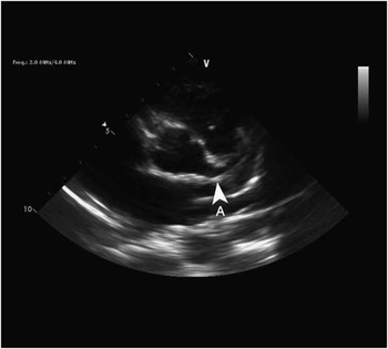
An echocardiogram showed normal cardiac connections with good biventricular function. There was no chamber dilation. The left coronary artery was significantly dilated and suggested the presence of a large coronary fistula draining to the pulmonary artery. There also appeared to be an additional fistula from the right coronary artery that drained to the right ventricular outflow tract.
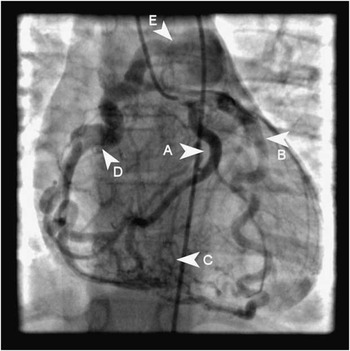
A cardiac catheterisation undertaken 3 weeks after the initial consultation showed an anomalous right coronary artery arising from the main pulmonary artery with retrograde flow via collaterals from the left coronary system.
Two months later, surgical repair was performed with reimplantation of the right coronary artery to the aorta. Post-operative and follow-up echocardiograms showed prograde flow in the right coronary artery.
Discussion
An anomalous origin of a coronary artery from the pulmonary artery, usually involving the left coronary artery, is a well-known congenital coronary anomaly. Origin of the right coronary artery from the pulmonary trunk is much less common and can be isolated or associated with other congenital cardiac diseases, such as aortopulmonary window (30%) or tetralogy of Fallot (19%), in up to 40% of patients.Reference Radke, Messmer, Haager and Klues1
In the past 20 years, there have been fewer than 10 reported cases of anomalous origin of the right coronary artery from the pulmonary artery in children. These patients ranged in age from 2 months to 7 years. Four were asymptomatic and one had congestive cardiac failure.Reference Vairo, Marina, De Simone and Marceletti2 One had an associated atrial septal defectReference Kuhn3 and three had murmurs warranting further investigation.Reference AL-Ammouri, Berkwits and Fahey4–Reference Kajihara, Asou, Takeda and Kosaka6 They had similar echocardiographic findings, and all showed improvement after surgical repair.
Coronary collateral circulation and pulmonary vascular resistance determines the myocardial perfusion and clinical course in a patient who has an abnormal origin of the right coronary artery from the pulmonary trunk. If an adequate collateral network has established in infancy, the area of the right coronary artery is perfused by collateral vessels from the left coronary artery. If the collateral circulation has not developed, then the area of the right coronary artery may be ischaemic. In addition, because of the low pressure in the pulmonary artery, the blood flow in the right coronary artery generally becomes reversed and coronary steal may develop.
Short axis view demonstrating the dilated left main coronary artery (A).

Selective left main coronary artery injection demonstrating the dilated left circumflex coronary artery (A), left anterior descending coronary artery (B), collateral vessels between the left and right coronary circulations (C), dilated right main coronary artery (D), and insertion of the right main coronary artery into the main pulmonary artery (E).

Physical examination is often normal except for a continuous murmur with diastolic accentuation. The presence of intercoronary collaterals within the ventricular septum is one of the most sensitive echocargiographic signs.Reference Yang, Nanda and Wang7 Diagnosis is mainly made by conventional coronary arteriography showing retrograde filling of the coronary artery from the collaterals. Recently, multislice computed tomography has been reported to be useful for the detection of an anomalous origin of the right coronary artery in the adult population.Reference Ichikawa, Sato, Komatsu, Hirayama, Kodama and Saito8
An anomalous right coronary artery arising from the pulmonary trunk was first described by Brooks during necropsy in 1885, and it was first showed by angiography in 1962 and by echocardiography in 1985.Reference Vijitbenjaronk, Glancy, Ferguson, Tan and Helmcke9 Approximately 80 cases of an anomalous origin of the right coronary artery from the pulmonary trunk have been documented in the literature.Reference Williams, Gersony and Hellenbrand10 Most patients (41%) without other associated cardiac lesions presented with an asymptomatic heart murmur. When present, symptoms include dyspnea (13%), congestive cardiac failure (30%), angina (17%), and cardiac arrest (17%).Reference Radke, Messmer, Haager and Klues1 Angina was rarely seen in those presenting before 20 years of age. Sudden death and cardiac arrest were documented in all ages. Therefore, surgical correction has been routinely advised upon diagnosis.Reference Collins, Davis and Stephens5
The goal of surgical therapy is the establishment of a bicoronary circulation.Reference Kuhn3 The first surgical treatment for an anomalous right coronary artery was reported by Tingelstad in 1972. Reimplantation of the anomalous right coronary artery to the aortic root is recommended.Reference Vijitbenjaronk, Glancy, Ferguson, Tan and Helmcke9 There is approximately a 50% regression in the diameter of the left anterior descending artery and left circumflex artery and a 17% regression in the diameter of the right coronary artery 14 weeks after surgical correction.Reference Radke, Messmer, Haager and Klues1