Introduction
Using genomic DNA extracted from meat, blood, hair or embryonic tissue for sex identification of those samples is an important tool in forensic science and genotypic analysis. Methods of sex identification include cytogenetic analysis, detection of H-Y antigen, measurement of X-linked enzymes before Barr body formation and Y-chromosome-specific probes (Bondioli, Reference Bondioli1992). Among sexing methods, polymerase chain reaction (PCR) amplification of sex-specific DNA sequences is relatively simple, rapid, inexpensive and highly accurate. Initial PCR-based systems for sex identification in various mammalian species involved amplification of the SRY gene, which has a Y-chromosome-specific sequence. By itself, however, the lack of an SRY-based band cannot be attributed definitively to a female sample rather than to experimental error. A gene present in both males and females (e.g., ZFX/Y) can be amplified as a positive control (Taberlet et al., Reference Taberlet, Mattock, Dubois-Paganon and Bouvet1993; Kohn et al., Reference Kohn, Knauer, Stoffella, Schroder and Paabo1995; Wasser et al., Reference Wasser, Houston, Koehler, Cadd and Fain1997), but doing so requires additional time and materials.
A recently developed PCR-based system for sex identification uses the amelogenin gene (AMEL) instead of SRY. In most mammals, copies of AMEL are present on both the X (AMELX) and Y (AMELY) chromosomes and differences in the introns of these two alleles enable them to be distinguished from each other. The X- and Y-chromosome-specific sequences for AMEL have been incorporated into methods of sex identification for cattle (Ennis et al., 1994), sheep (Pfeiffer & Brenig, Reference Pfeiffer and Brenig2005)) and humans (Sullivan et al., Reference Sullivan, Mannucci, Kimpton and Gill1993). The use of AMEL in PCR-based methods has simplified sex identification, because a single primer set can amplify both chromosome-specific forms of the gene.
In the present study, we compared the nucleotide sequences for porcine AMELX (EMBL/GenBank accession number, AB091791) and AMELY (AB091792) and designed a set of PCR primers that amplified both alleles in a single reaction. The amplicons demonstrated characteristic X–Y-specific length polymorphism among all breeds of pigs tested. We also successfully applied the AMEL-PCR method to sex identification of individual porcine embryos.
Materials and Methods
Extraction of DNA from porcine tissue samples
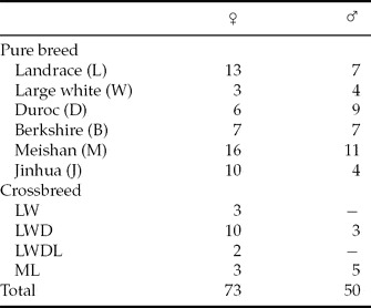
Genomic DNA was isolated from ear, tail or leukocyte samples from 123 pigs representing various breeds (Table 1). To prepare genomic DNA from leukocytes, samples of whole blood (10 ml) were combined with 10 ml Dulbecco's phosphate-buffered saline and centrifuged (1500 g, 15 min, 4 °C). After the supernatant was discarded, the pellet was resuspended in ACK buffer (0.16 M NH4Cl, 10 mM KHCO3, 0.1 mM EDTA·2Na) for hemolysis. After centrifugation (1500 g, 10 min, 4 °C), the pellet (leukocytes) was resuspended in lysis buffer (50 mM Tris–HCl [pH 8.0], 100 mM EDTA, 100 mM NaCl, 1% [w/v] sodium dodecyl sulfate, 0.5 mg/ml proteinase K) and incubated at 37 °C overnight. After centrifugation (1500 g, 10 min, 4 °C), the supernatant was extracted with an equal volume of phenol–chloroform. After centrifugation (1500 g, 10 min, 4 °C), the aqueous layer was combined with ethanol for precipitation of DNA. The precipitated DNA was pelleted and the DNA pellet was washed gently with 70% ethanol and then dissolved in TE buffer (10 mM Tris–HCl [pH 7.5], 0.1 mM EDTA). Tail and ear samples were minced and then lysed with proteinase K, extracted with phenol–chloroform, precipitated with ethanol and resuspended in TE buffer, as described for leukocytes.
Porcine breeds from which genomic DNA was extracted.

LW: Landrace (♀) × Large White (♂) crossbreed; LWD: LW (♀) × Duroc (♂) crossbreed; LWDL: LWD (♀) × Landrace (♂) crossbreed; ML: Meishan pig (♀) × Landrace (♂) crossbreed.
Extraction of DNA from porcine embryos
Cumulus–oocyte complexes were aspirated from 3 to 5 mm ovarian follicles in prepubertal gilts. The complexes were matured in modified NCSU-37 medium (Peters & Wells, Reference Peters and Wells1993) for 44 h, fertilized in vitro and then cultured in PZM5 (Yoshioka et al., Reference Yoshioka, Suzuki, Tanaka, Anas and Iwamura2002). The preimplantation embryos were sampled at 2 or 5 to 6 days after fertilization (day 0). Each day-2 embryo was treated in 5 μl of lysis solution (0.13 mg/ml proteinase K); the entire volume was used in the subsequent PCR amplification. Blastocysts recovered 5 to 6 days after fertilization each were treated in 20 μl of lysis solution and 5 μl was used for PCR. As the controls, genomic DNA from electroactivated embryos (EA) and germinal vesicle (GV)-stage oocytes was prepared for PCR as described for preimplantation embryos.
Polymerase chain reaction
Using published sequences for the porcine amelogenin genes (EMBL/GenBank accession numbers, AB091791 [AMELX] and AB091791 [AMELY]), we designed a set of degenerate PCR primers (AMEL-F, 5′-CRCMTTCATTGAYAATTCAC-3′; AMEL-R, 5′-CCAGAGGTTGTAACCTTACAG-3′; R = A or G; M = A or C; Y = C or T) to amplify a portion of intron 2 that was expected to yield PCR products of different sizes between AMELX and AMELY. For amplification of tissue samples, 50 ng of genomic DNA was used in each reaction; for embryos, 5 μl lysis solution containing genomic DNA was used. Amplification conditions were initial denaturation at 94 °C for 2 min followed by 40 cycles of denaturation at 98 °C for 10 s, primer annealing at 60 °C for 30 s and primer extension at 72 °C for 30 s (GeneAmp 9700, Applied Biosystems, Foster City, CA). PCR products were separated by electrophoresis in 1.5% agarose gels and visualized by ethidium bromide staining under UV illumination.
Sequence of porcine amelogenin gene
The male-specific fragment and that common to both male and female samples were excised from the gel and the DNA contained in the isolated gel slices was purified by using the QIAquick Gel Extraction Kit (Qiagen, Hilden, Germany) according to the manufacturer's instructions. The DNA fragments were subcloned into pBluescript KS(+) (Stratagene, La Jolla, CA); resulting plasmids were sequenced by using T3 and T7 primers and dye-termination methodology (ABI-Prism BigDye Terminator v3.1 Cycle Sequencing Kit, Applied Biosystems) on an automated sequencer (ABI Prism 310 Genetic Analyzer, Applied Biosystems) according to the manufacturer's instructions.
Statistical analysis
Statistical differences were analysed by the chi-squared test. A probability of less than 0.01 was considered significant.
Results
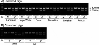
Figure 1a, b shows the amplification of male and female genomic DNA fragments by using primers for the porcine AMEL gene. Samples from male pigs were expected to yield two bands (at about 520 and 350 bp), whereas those from female pigs were anticipated to produce a single band (520 bp). Neither male nor female samples yielded non-specific amplification.
Gel electrophoresis showing amplicons of amelogenin gene fragments from various (a) purebred and (b) crossbred pigs. M, molecular weight markers (ΦX174 HincII digest).

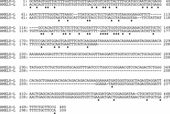
The nucleotide sequences of the 520 and 350 bp PCR-derived products matched those of AMELX and AMELY, respectively (data not shown). The amplified region of the AMELX allele in Chinese breeds (Meishan and Jinhua) has a single-nucleotide deletion relative to the sequences of European breeds. At these amplified region of AMEL genes, sequence differences containing insertions and deletions were frequent between AMELX and AMELY (Fig. 2). Among the breeds evaluated, the lengths of the PCR-amplified regions of porcine AMELX and AMELY differed by approximately 171 bp overall.
DNA sequence comparison between the X- and Y-specific amelogenin gene fragments of Landrace pigs. *, single-base substitution; –, deleted base.

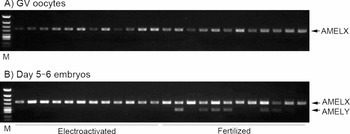
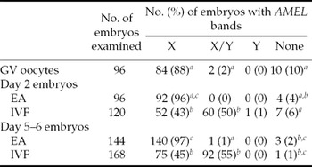
When genomic DNA from porcine blastocysts and GV-stage oocytes was used for PCR analysis, more than 88% of GV-stage oocytes and EA embryos yielded AMELX-derived bands only (Fig. 3, Table 2). In contrast, 50% of day 2 embryos (data not shown) and 55% of day 5–6 embryos generated both amplicons.
Gel electrophoresis showing amplicons of the amelogenin gene fragments of porcine (a) GV-stage oocytes and (b) embryos at 5 to 6 days after electroactivation or in vitro fertilization. M, molecular weight markers (ΦX174 HincII digest).

Detection of AMELX and AMELY in porcine embryos by AMEL-PCR.

EA: electroactivated; IVF: in vitro fertilized.
a–c Values with different superscripts in the same column differ significantly (p < 0.01, chi-squared analysis).
Discussion
In the present study, we developed a PCR-based method using the amelogenin gene for sex identification of pigs and porcine embryos. The amelogenin gene has been conserved during the evolution of vertebrates (Lyngstadaas et al., Reference Lyngstadaas, Risnes, Nordbo and Flones1990). In cattle (Gibson et al., Reference Gibson, Golub, Abrams, Shen, Ding and Rosenbloom1992, Yamamoto et al., Reference Yamamoto, Tsubota, Komatsu, Katayama, Murase, Kita and Kudo2002), sheep (Pfeiffer & Brenig, Reference Pfeiffer and Brenig2005) and humans (Salido et al., Reference Salido, Yen, Koprivnikar, Yu and Shapiro1992), copies of the amelogenin gene are found on both the X and Y chromosomes, whereas AMEL is only located on the X chromosome in mice (Chapman et al., Reference Chapman, Keitz, Disteche, Lau and Snead1991) and on autosomes in monotremes and marsupials (Watson et al., Reference Watson, Spencer, Graves, Snead and Lau1992). Like those in cattle, sheep and humans, the sequences of the X- and Y-specific genes in pigs differ. We therefore designed a set of primers that generated PCR products of different sizes between AMELX and AMELY. As expected, using these primers in PCR analysis of genomic DNA from male pigs yielded two bands (about 520 and 350 bp; derived from AMELX and AMELY, respectively), compared with the single band (520 bp; AMELX) from female pigs. Our PCR-based sexing method using porcine AMEL was 100% reliable (123 samples analysed; ♀: 73, ♂: 50) among the European breeds, Chinese breeds and crossbred pigs evaluated. Previous PCR-based methods for sex identification of pigs (Fajfar-Whetstone et al., Reference Fajfar-Whetstone, Rayburn, Schook and Weeler1993; Pomp et al., Reference Pomp, Good, Geisert, Corbin and Conley1995) involved the detection of SRY, a Y-chromosome-specific gene that determines sex. To our knowledge, the present report is the first to show that the AMEL gene can be used for PCR-based sex identification of pigs.
In the present study, we designed a set of primers that amplified a portion of intron 2 in both AMEL alleles. Comparing porcine AMELX and AMELY sequences revealed many differences (insertions and deletions were frequently found in intron 2; Fig. 2). In addition, PCR products obtained by using the primers in the present study showed that a significant difference (about 170 bp) in their lengths was observed between the X and Y chromosomes. A single AMEL-PCR primer pair that spans a length variation in AMEL intron 1 is used for sex determination in humans (Sullivan et al., Reference Sullivan, Mannucci, Kimpton and Gill1993), whereas a length variation in intron 5 is used in cattle (Chen et al., Reference Chen, Hu, Wang, Hung, Wu, Choo and Cheng1999). Still another sequence variation is used in the Japanese black bear (Yamamoto et al., Reference Yamamoto, Tsubota, Komatsu, Katayama, Murase, Kita and Kudo2002). In these reports, difference in lengths between AMELX and AMELY PCR fragments are 6–106 bp, which depend on the regions of AMEL sequences for PCR amplification. Our AMEL-PCR analysis can detect the differences of fragment sizes between AMELX and AMELY compared with the reports described above. As a difference of 170 bp is visually discriminable in the AMEL-PCR results, our PCR method using AMEL genes provides results that are easy to discern and reliable for sex identification.
Almost all GV-stage oocytes and EA embryos yielded the X amplicon only; given that GV-stage oocytes and EA embryos have only maternal genomes (that is, no Y chromosome), the present results are reasonable. In addition, about half of the day 2 and day 5–6 embryos produced both X and Y amplicons, suggesting that the AMEL-PCR analysis in the present study is reliable. However, several oocytes and EA and IVF embryos failed to yield any product at all, perhaps due to loss of genomic DNA during sample preparation or to errors in setting up the reaction tubes. Further, one day-2 IVF embryo yielded only an AMELY-derived band; the reason for this result is unknown, given that every embryo produced by EA or IVF must contain an X chromosome (and thus AMELX).
In conclusion, our findings show that the described PCR assay based on the AMEL gene is reliable for sex identification of porcine IVP embryos. A key advantage of this assay is the ability of identify the sex of a sample from a small amount of genomic DNA (e.g., a single embryo). Although techniques for in vitro fertilization and development of porcine oocytes and embryos are quite advanced, problems such as polyspermy and poor rate of development to term still plague current culture systems. Once such problems are solved, the described assay could be applied to sex identification of porcine embryo biopsies and execute sex control for livestock management.
Acknowledgements
We thank K. Iijima, N. Watanabe and T. Funakubo for technical assistance; the pig farm staff (National Institute of Livestock and Grassland Science) for the care and maintenance of the pigs and for assistance in handling the animals; and T. Nagai for proofreading of the manuscript.