Balloon dilatation of recoarctation of the aorta after repair by resection with end-to-end anastomosis is associated with good results in 82–94% of cases.Reference Brown, Ruzmetov, Hoyer, Rodefeld and Turrentine 1 Complications include recoarctation in 13–60%, rupture of the aortic wall in 2–8%, pseudoaneurysm formation in 4–10%, and thrombosis of the femoral artery in 3–7%.Reference Oliver, Gallego, Gonzalez, Aroca, Bret and Mesa 2 , Reference Rao 3 Nevertheless, the indications for balloon dilatation for recoarctation after surgical correction using an interposition dacron tube graft are not known. This case report documents the successful surgical repair of an aorto-bronchial fistula and a large aortic pseudoaneurysm at the proximal anastomosis of a dacron interposition tube graft that was balloon dilated for recurrent coarctation.
Clinical case
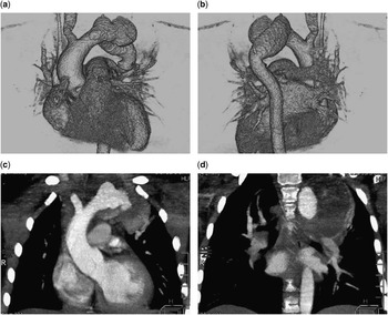
A 6-year-old girl was admitted with haemoptysis, shortness of breath, headaches, and anaemia. Her past history was remarkable for repair of coarctation of the aorta, at another institution at 4 years of age, using an 18-mm dacron interposition graft (Vascutek, Terumo, Renfrewshire, United Kingdom); 3 months after this initial surgery, a recoarctation of the proximal interposition graft anastomosis was diagnosed. This recoarctation was treated with balloon dilatation. Within 10 months of the balloon dilation, the patient began to complain of shortness of breath. Over the subsequent 2 months, she experienced several episodes of haemoptysis. On admission to our institution, the patient had an equal blood pressure of both the right arm and the legs (80/60 mmHg). An echocardiogram suggested the presence of a mass involving the proximal part of the interposition graft. An emergency CT scan with contrast (Fig 1) demonstrated a 58×56×49 mm pseudoaneurysm with parietal thrombosis. The neck of the pseudoaneurysm was 15 mm distal to the origin of the right-sided innominate artery and was 15 mm in diameter. There was left upper lobe atelectasis due to compression by the pseudoaneurysm.
CT scan of the aorta and pseudoaneurysm after balloon dilatation of post-operative recoarctation. ( a ) Anterior projection of a three-dimensional reconstruction of a CT scan showing the pseudoaneurysm at the proximal anastomosis of the interposition graft. ( b ) Posterior projection of a three-dimensional reconstruction of a CT scan showing the pseudoaneurysm projecting anterior to the interposition graft. ( c ) Contrast-enhanced coronal view showing the true lumen. ( d ) Contrast-enhanced coronal view showing pseudoaneurysm true lumen with parietal thrombosis.

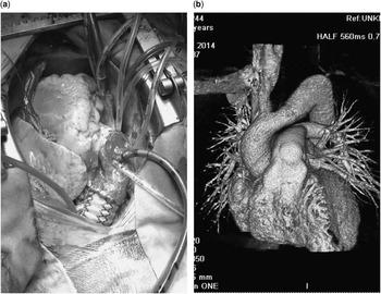
The patient underwent urgent surgical repair on the following day. A median sternotomy was performed and a 55-mm diameter pseudoaneurysm was observed. The left common carotid and subclavian arteries were not located during the surgical repair. After cannulation of the ascending aorta and the right atrium, cardiopulmonary bypass was initiated with cooling of the patient to 25°C. To isolate the false aneurysm, a proximal clamp was placed just distal to the right-sided innominate artery, but proximal to the neck of the pseudoaneurysm, and a distal clamp was placed distal to the pseudoaneurysm. Cardiopulmonary bypass flow was reduced while still maintaining a perfusion pressure of 40–50 mmHg in the right radial artery. Cardioplegia was not required because the myocardium was continually perfused by cardiopulmonary bypass. The pseudoaneurysm was opened. The proximal anastomosis between the dacron interposition graft and the aorta was disrupted up to two-thirds of the anastomotic diameter. The dacron graft, thrombi, and fibrous walls of the pseudoaneurysm were completely excised. The aorto-bronchial fistula was located between the false aneurysm and the left lower lobe bronchus. The bronchus was repaired by suturing lung tissue over it. A new 16-mm diameter dacron graft was interposed (Fig 2a). The aorta was clamped for 63 minutes. The patient was discharged home on the 8th post-operative day. A CT scan with contrast, performed 6 months later, confirmed a good result (Fig 2b).
Intra-operative photograph and CT scan of the surgical repair. ( a ) Intra-operative photograph of the implanted 16-mm interposition dacron vascular graft. ( b ) Anterior projection of a three-dimensional reconstruction of a CT scan 6 months post-operatively.

Discussion
Incorrect surgical approach for coarctation may lead to serious complications. In this case, recoarctation at the proximal anastomosis site of an oversized dacron interposition graft occurred within 3 months, possibly as a result of kinking or due to turbulent flow related to the size mismatch between the native aorta and the dacron graft. In response to the development of the recoarctation, the outside institution chose to perform balloon dilatation. This led to a partial disruption of the proximal interposition graft suture line. Although this complication can lead to fatal bleeding, in this case, a large pseudoaneurysm formed due to the presence of dense surrounding post-operative adhesions. This is a rare but serious complication.Reference Oliver, Gallego, Gonzalez, Aroca, Bret and Mesa 2 , Reference Rao 3 Conservative treatment of such a pseudoaneurysm leads to unpredictable results. Development of an aorto-bronchial fistula is yet another major complication (92%) in such cases.Reference Smayra, Otal and Soula 5 This aorto-bronchial fistula was caused by compression of the left lower lobe bronchus by the pseudoaneurysm. The main clinical symptom of the aorto-bronchial fistula was haemoptysis. In a situation such as this, the first episode of haemoptysis often leads to death, but sometimes a blood clot occludes the fistula and the haemoptysis stops transiently. Displacement or lysis of the thrombus may result in recurrent and often fatal, massive re-bleeding. Open surgery is the procedure of choice for pseudoaneurysms complicated by aorto-bronchial haemorrhage, although there are successful case reports treated by implantation of an endovascular stent graft.Reference Takawira, Sinyangwe and Mooloo 4 , Reference Smayra, Otal and Soula 5 We chose a more definitive single-stage surgical correction of the pseudoaneurysm and the aorto-bronchial fistula that allowed complete excision of the pathology and promoted an uncomplicated post-operative course.
Conclusion
This case demonstrates that balloon dilatation for recoarctation of the aorta after surgical repair with an oversized interposition dacron graft may result in severe complications. The use of an interposition graft in infants and children to repair coarctation of the aorta should be avoided, if at all possible, and if an interposition graft recoarctation is present the use of balloon dilation alone may be contraindicated.
Acknowledgement
None.
Financial Support
The authors received no financial support for the research, authorship, and/or publication of this article.
FConflicts of Interest
The authors declare no potential conflicts of interest with respect to the research, authorship, and/or publication of this article.