Introduction
Dicrocoeliasis is a worldwide parasitic disease caused mainly by Dicrocoelium dendriticum and also by D. hospes, D. suppereri and D. chinensis (Rojo-Vazquez et al., 2012). Dicrocoelium dendriticum is more frequently present throughout Europe, Asia, North Africa and North America, while other species have a limited distribution and are found in Africa, Asia and some focal points of Eastern Europe (Otranto & Traversa, Reference Otranto and Traversa2002). Adult worms live in the bile ducts and gall bladder of numerous mammalian species, mainly domestic and wild ruminants, but rabbits, pigs, dogs, horses and humans can also be infected (Otranto & Traversa, Reference Otranto and Traversa2003). To complete its life cycle, the parasite needs to develop in the body of land snails and ant species, as the first and second intermediate hosts, respectively (Campo et al., Reference Campo, Manga-Gonzalez and Gonzalez-Lanza2000). Various surveys have indicated that D. dendriticum is commonly found in ruminants throughout Iran (Ansari-Lari & Moazeni, Reference Ansari-Lari and Moazeni2006; Daryani et al., Reference Daryani, Alaei, Arab, Sharif, Dehghan and Ziaei2006; Movassaghi-Ghazvini et al., Reference Movassaghi-Ghazvini, Valilou, Ahmadzadeh, Karimi and Zirak2008; Ahmadi & Meshkehkar, Reference Ahmadi and Meshkehkar2010). The infection has been described in lowlands or mountain pastures at high altitudes. It appears that calcareous or alkaline soils are favourable biotopes for the survival and development of land snails and ants (Chartier & Reche, Reference Chartier and Reche1992). Accordingly, the highest rates of infection have been reported in the north and north-west of Iran (Daryani et al., Reference Daryani, Alaei, Arab, Sharif, Dehghan and Ziaei2006; Movassaghi-Ghazvini et al., Reference Movassaghi-Ghazvini, Valilou, Ahmadzadeh, Karimi and Zirak2008). In addition, human dicrocoeliasis has already been reported in Iran, in spite of the highly specific nature of this parasite's life-cycle (Farid, Reference Farid1971; Zali et al., Reference Zali, Jafa-Rimehr, Rezaian, Meamar, Vaziri and Mohraz2004). The most important economic and health aspects of dicrocoeliasis are due to confiscation of parasitized livers, reduced milk and meat production and the costs associated with anthelminthic treatments (Wolff et al., Reference Wolff, Hauser and Wild1984; Jithendran & Bhat, Reference Jithendran and Bhat1996).
Dicrocoelium species are readily distinguished at adult stage by morphological characteristics (Malek, Reference Malek1980; Taira et al., Reference Taira, Shirasaka, Taira, Ando and Adachi2006; Otranto et al., Reference Otranto, Rebien, Weigl, Cantacessi, Parisi, Paolo Lia, Meter and Olson2007). According to Rollinson et al. (Reference Rollinson, Walker and Simpson1986), morphologically similar helminths show marked differences in epidemiologically significant factors such as infectivity, pathogenicity, antigenicity and sensitivity to chemotherapeutic agents. Therefore, markers based on DNA sequences do differentiate between parasite isolates. Early studies have analysed variability and heterogeneity between D. dendriticum strains from cows, sheep and goats, using electrophoresis of isoenzymes in polyacrylamide gels (Leon-Ortega et al., Reference Leon-Ortega, Hermoso and Monteoliva1986, Reference Leon-Ortega, Monteoliva and Sanchez-Moreno1988, Reference Leon-Ortega, Monteoliva and Sanchez-Moreno1989; Campo et al., Reference Campo, Manga-Gonzalez, Gonzalez-Lanza, Rollinson and Sandoval1998). In addition, Sandoval et al. (1999) and Martinez-Ibeas et al. (Reference Martinez-Ibeas, Martinez-Valladares, Gonzalez-Lanza, Minambres and Manga-Gonzalez2011) applied random amplified polymorphic DNA analysis (RAPD) for the differentiation and estimation of genetic diversity among different populations of D. dendriticum. To date, several molecular studies, using mainly ribosomal DNA sequences (rDNA), have investigated the genetic diversity of this parasite (Maurelli et al., Reference Maurelli, Rinaldi, Capuano, Perugini, Veneziano and Cringoli2007; Otranto et al., Reference Otranto, Rebien, Weigl, Cantacessi, Parisi, Paolo Lia, Meter and Olson2007; Bazsalovicsova et al., Reference Bazsalovicsova, Kralova-Hromadova, Spakulova, Reblanova and Oberhauserovk2010; Arbabi et al., Reference Arbabi, Dalimi-Asl, Ghaffarifar and Foorozandeh-Moghadam2012).
To the best of our knowledge, no study has previously been conducted on the mitochondrial sequences of this trematode worldwide and ribosomal sequencing data of existing Iranian D. dendriticum strains are rare. Therefore, the present phenotypic and molecular study was designed to identify accurately the causative agent(s) of dicrocoeliasis in Iran and to assess the level of intraspecific and/or interspecific variation in sheep and cattle isolates using both ribosomal (internal transcribed spacer 2 (ITS-2)) and mitochondrial (NADH dehydrogenase gene (nad1)) sequences.
Materials and methods
Collection and examination of adult worms
Up to 44 adult worms of Dicrocoelium were collected from the liver and gall bladder of sheep and cattle slaughtered in various localities in Iran. These include, Tehran (35°41′N, 51°25′E), Fars (29°37′N, 52°31′E), Razavi Khorasan (36°17′N, 59°36′E), Gilan (37°16′N, 49°34′E) and Golestan (36°50′N, 54°26′E). Adult worms were carefully washed in physiological saline, maintained in 70% ethanol solution and frozen at − 20°C before DNA extraction. All standardized morphometric characteristics of adult worms including body length (BL), body width (BW), BL/BW, body area, body perimeter and length of vitelline glands were made according to the methods described previously for Dicrocoelium (Hinaidy, Reference Hinaidy1983; Taira et al., Reference Taira, Shirasaka, Taira, Ando and Adachi2006).
Molecular analysis
DNA of 28 adults of D. dendriticum from different hosts and geographical localities was extracted using a commercially available kit (Qiagen DNeasy Blood & Tissue Kit; Qiagen, Valencia, California, USA) according to the manufacturer's protocol. DNA samples were then kept at − 20°C until used for polymerase chain reaction (PCR). The amplification reactions were carried out in a 50 μl volume containing: 10 mm Tris-HCl (pH = 9.0), 50 mm KCl, 4 μl of DNA extracted as template, 1.5 mm MgCl2, 200 μm deoxynucleoside triphosphates (dNTPs), 20 pm of each primer and 2 U of Taq DNA polymerase (Fermentas, Hanover, Maryland, USA). The primers for the ITS-2 region have been described previously by Otranto et al. (Reference Otranto, Rebien, Weigl, Cantacessi, Parisi, Paolo Lia, Meter and Olson2007) as follows: forward, Dd58SF1 (5′-ATATTGCGGCCATGGGTTAG-3′; reverse, Dd28SR1 (5′-ACAAACAACCCGACTCCAAG-3′). Also, universal primers JB11 (5′-CAGAGGTTTGCTGATCTATTG-3′) and JB12 (5′-CTTTCAGCCTCAGCATAATCC-3′) were used for amplification of an approximately 520 bp fragment of the nad1 gene (Bowles & McManus, Reference Bowles and McManus1993; Herwerden et al., Reference Herwerden, Blair and Agatsuma2000). PCR reactions were performed under the following conditions: one 95°C cycle for 5 min for early denaturation, then 94°C for 45 s (denaturation), annealing (at 55°C and 58°C for nad1 and ITS-2, respectively) for 1 min, and extension at 72°C for 45 s. All three steps were repeated for 35 cycles and a final extension at 72°C for 5 min was applied. PCR products were analysed after electrophoresis in 1.5% (w/v) agarose gel and visualized in ethidium bromide (0.5 μg/ml) under ultraviolet illumination.
Data analysis
Morphometric data were tested using SPSS version 11.5 (SPSS Inc., Chicago, Illinois, USA). An independent Student's t-test was used to assess differences between morphological characteristics of all 44 isolates from sheep and cattle and P values of < 0.05 indicated significant differences.
Products from each assay were purified using a PCR kit (Bioneer, Korea) and sequenced directly using the capillary DNA analyser (ABI 3730; Applied Biosystems, Foster City, California, USA). Nucleotide sequence analysis was undertaken using the National Center for Biotechnology information BLAST (Basic Local Alignment Search Tool) programs and databases. Multiple sequence alignments and construction of a phylogenetic tree were made with the Clustal W and UPGMA methods using MEGA4 software (Tamura et al., Reference Tamura, Dudley, Nei and Kumar2007).
Results
Morphometrics
Morphometric data of the 44 Dicrocoelium spp. samples (table 1) indicated that all were D. dendriticum. The existence of minor phenotypic variation among adult flukes with different hosts was not statistically significant (P>0.05). Dicrocoelium dendriticum isolates exhibited two distinct morphological indices in comparison with those reported for D. hospes (Malek, Reference Malek1980) and D. chinensis (Otranto et al., Reference Otranto, Rebien, Weigl, Cantacessi, Parisi, Paolo Lia, Meter and Olson2007). First, testes lay tandem, and second, the position of the maximal body width was always behind the middle part of the body.
Comparison of morphometrics (mean±SD in mm) of adult Dicrocoelium from sheep and cows in Iran; body length (BL), body width (BW), body length/body width (BL/BW), body area (mm2), body perimeter and length of vitellaria.

Molecular analysis
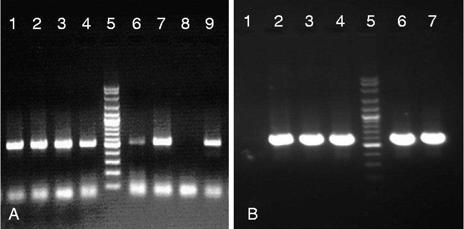
The partial nad1 (~520 bp) and the complete ribosomal ITS-2 fragment, with parts of the flanking 3′ end of the 5.8S and 5′ end of the 28S rDNA (561 bp) were amplified and sequenced for different hosts and geographical isolates (fig. 1). PCR amplification and direct sequencing were successfully performed on all of 28 isolates for both nad1 and ITS-2 genes, except for three isolates that did not produce an appropriate nad1 sharp band for sequencing and further analysis.
Agarose gel electrophoresis of nad1 (A) and ITS-2 (B) amplicons produced from Dicrocoelium dendriticum compared with the molecular weight marker 100 bp (lanes (A) 5, (B) 5) and negative controls (lanes (A) 8, (B) 1).

The sequence alignment based on ITS-2 data obtained from the five geographically distant D. dendriticum samples revealed only one nucleotide intraspecific substitution, with a diversity of 0–0.42%. The ITS-2 sequence data were deposited in GenBank under accession numbers, JQ966972 and JQ966973 as ITS-A and ITS-B haplotypes. Up to 17 of 28 isolates belonged to ITS-A haplotype. In ITS-2 complete sequence analysis, a heterogeneity (A/R) at position 109 was detected in 11 isolates of the ITS-B haplotype, which has not been reported previously. Finally, the nucleotide sequences obtained for the ITS-2 gene in Iran were compared with sequences of other related species in GenBank, i.e. D. dendriticum, accession numbers AB369980, AB369981, HM358027, HM026461, AB367789 and DQ379986; D. chinensis AB369982, AB367790 and EF547132; and D. hospes, EF102026.
A comparison of the ITS-2 sequences with those for other strains of Dicrocoelium spp. deposited in the GenBank database revealed additional variable positions (n= 41) in the sequence alignment. The interspecific variability in the ITS-2 region worldwide is 3.3–13.7% compared with 0–1.3% for intraspecific variability.
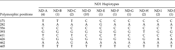
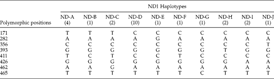
The nad1 sequences of 25 isolates were deposited in GenBank under accession numbers JX050110 to JX050134. Results obtained from this region showed that intraspecific variations within the population of all D. dendriticum isolates were detected at eight positions – 171, 282, 356, 393, 411, 426, 462 and 465 – in the range of 0–0.97% (table 2). However, a BLAST search of D. dendriticum nad1 sequences against those previously published for other trematodes (Fasciola hepatica, Fasciola gigantica, Opisthorchis viverrini, Clonorchis sinensis) revealed that the sequence homology ranges from 67 to 75%.
Intraspecific variation in the ND1 sequences of Dicrocoelium dendriticum from sheep and cattle in Iran; frequency of isolates are given in brackets.
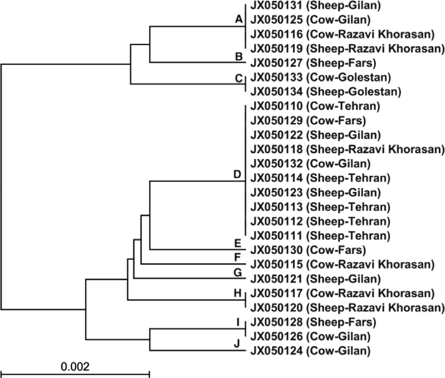
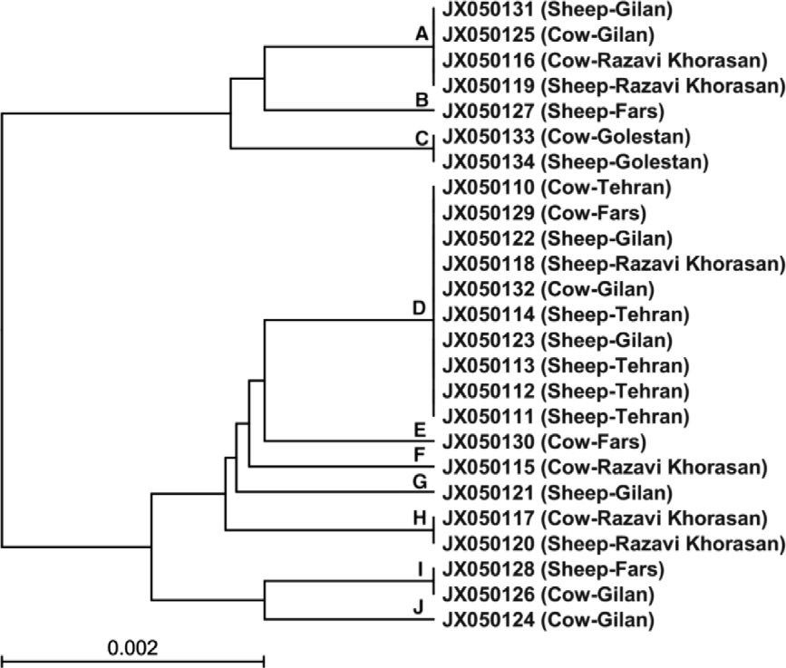
The UPGMA cluster analysis, based on these different polymorphic regions, grouped the Iranian isolates into ten distinct haplotypes of ND-A to ND-J (fig. 2). No obvious correlation was observed between D. dendriticum haplotypes and any host or geographical origin of the strains. The majority of strains, with 10 of 25 isolates, belonged to the ND-D haplotype.
The nad1 cluster analysis, constructed according to UPGMA (unweighted pair group method with arithmetic mean) method to show the position of Dicrocoelium isolates in sheep and cattle in various locations in Iran; A–J at nodes represent haplotypes.
For nad1, intraspecific nucleotide variation was mainly related to changes at the third codon position (n= 7), whereas only one change was detected at the second codon position. Comparison of the nad1 sequences represented seven transition types (C ↔ T or A ↔ G) and only one transversion (G ↔ T). However, in this gene there were two changes in amino acid sequences among isolates, in which the mutational change at positions 356 and 393 of the nad1 gene resulted in putative amino acid alteration of alanine (GCA) to valine (GTA) and leucine (TTG) to phenylalanine (TTT), respectively. The other mutations were silent.
Discussion
An accurate identification of the causative agents of dicrocoeliasis has important implications for the epidemiology, control and management of this disease (Saki et al., Reference Saki, Khademvatan and Yousefi2011). Phenotypic and genetic variation within species of Dicrocoelium, especially the nuclear DNA regions, has been reported previously (Hinaidy, Reference Hinaidy1983; Taira et al., Reference Taira, Shirasaka, Taira, Ando and Adachi2006; Maurelli et al., Reference Maurelli, Rinaldi, Capuano, Perugini, Veneziano and Cringoli2007; Otranto et al., Reference Otranto, Rebien, Weigl, Cantacessi, Parisi, Paolo Lia, Meter and Olson2007; Bazsalovicsova et al., Reference Bazsalovicsova, Kralova-Hromadova, Spakulova, Reblanova and Oberhauserovk2010; Martinez-Ibeas et al., Reference Martinez-Ibeas, Martinez-Valladares, Gonzalez-Lanza, Minambres and Manga-Gonzalez2011; Arbabi et al., Reference Arbabi, Dalimi-Asl, Ghaffarifar and Foorozandeh-Moghadam2012); however, data on phenotypic and genetic diversity of the parasite are limited.
Ribosomal DNA analysis is a tool to demonstrate intra- and interspecific variability of distinct parasite species because rDNA includes regions with varying rates of evolution, from highly conserved (18S, 5.8S and 28S) to highly variable regions (such as internal transcribed spacers 1 and 2 (ITS-1 and ITS-2)) (Maurelli et al., Reference Maurelli, Rinaldi, Capuano, Perugini, Veneziano and Cringoli2007). These molecular regions are the most frequently used markers for population genetic and evolutionary biology applications in digeneans (Hillis & Dixon, Reference Hillis and Dixon1991). Mitochondrial markers have been used extensively for phylogenetic studies and population differentiation because of their relatively rapid rate of evolution and importance in discrimination of closely related organisms; in addition, these markers are maternally inherited and do not undergo any recombination (McManus & Thompson, Reference McManus and Thompson2003). For example, NADH dehydrogenase subunit 1 gene (nad1) has been shown to be well suited for investigating the population genetics of trematodes, i.e. Schistosoma japonicum (Bowles et al., Reference Bowles, Hope, Tiu, Liu and McManus1993) and Opisthorchis viverrini (Saijuntha et al., Reference Saijuntha, Sithithaworn, Wongkham, Laha, Chilton, Petney, Barton and Andrews2008; Thaenkham et al., Reference Thaenkham, Nuamtanong, Sanguankiat, Yoonuan, Touch, Manivong, Vonghachack, Sato and Waikagul2010).
In the current study, the nad1 and ITS-2 sequences were used as genetic markers to investigate the genetic variation within D. dendriticum from Iran. They were also compared with additional closely related parasites that exist in the GenBank database. These morphological and molecular targets were analysed simultaneously to provide more accurate and reliable information about the nature and extent of variation within D. dendriticum.
According to the data obtained from this study, it was concluded that D. dendriticum is the only species of Dicrocoelium among cattle and sheep in Iran. Molecular assays, especially those based on the mitochondrial marker nad1, revealed that genetic diversity exists within D. dendriticum isolates. However, no significant intraspecific differences were recorded among morphometric and genetic characteristics of isolates obtained from different hosts. Similarly, Otranto et al. (Reference Otranto, Rebien, Weigl, Cantacessi, Parisi, Paolo Lia, Meter and Olson2007) and Moazeni et al. (Reference Moazeni, Sharifiyazdi and Izadpanah2011) demonstrated that host associations and geographic location are unlikely to be useful in classifying genotypes of D. dendriticum and F. hepatica. In contrast, Arbabi et al. (Reference Arbabi, Dalimi-Asl, Ghaffarifar and Foorozandeh-Moghadam2012) reported that the morphological characteristics of the D. dendriticum in various provinces of Iran were significantly different. Differences between the morphological findings may be related to the age, nutritional conditions, infection severity, geographical distribution, developmental stage, method of measurement, sample size, and period and place of studies (Tkach et al., Reference Tkach, Pawlowski and Sharpilo2000).
More recent studies (Otranto et al., Reference Otranto, Rebien, Weigl, Cantacessi, Parisi, Paolo Lia, Meter and Olson2007; Arbabi et al., Reference Arbabi, Dalimi-Asl, Ghaffarifar and Foorozandeh-Moghadam2012) on the molecular characterization of D. dendriticum isolates based on small (18S rDNA) and large (28S rDNA) subunits of the ribosomal gene, failed to demonstrate any genetic differences. Arbabi et al. (Reference Arbabi, Dalimi-Asl, Ghaffarifar and Foorozandeh-Moghadam2012) suggested that considerable morphological variability in the isolates of D. dendriticum may reflect genetic polymorphism in other genetic markers within the parasite genome.
In the present research, the level of sequence polymorphism among Dicrocoelium isolates was higher for nad1 (with eight polymorphic sites) compared to the ITS-2 gene (only one polymorphic site). Accordingly, molecular genotyping using the nad1 and ITS-2 sequences has generated ten and two distinct haplotypes, respectively. The haplotypes ND-D and ITS-A were prominent in Iran and they also showed a wider geographical distribution.
To the best of our knowledge, no data have yet been published on mitochondrial variability in D. dendriticum. A previous study using nad1 sequence (668 bp) detected limited genetic variation of 0–0.3% among 14 geographical isolates of Opisthorchis viverrini trematode from Thailand and Lao PDR (Saijuntha et al., Reference Saijuntha, Sithithaworn, Wongkham, Laha, Chilton, Petney, Barton and Andrews2008). These authors defined four different nad1 haplotypes – based on two nucleotide substitutions among these trematode isolates, all of which were identical at the amino acid sequence level.
In the present investigation an analysis of sequences in the ITS-2 region showed a genetic heterogeneity (A → R) at position 109, with 11 cases possessing both adenine and guanine nucleotides. These results suggest that different copies of ITS-2 sequences might be present within a single parasite isolate (Sharifiyazdi et al., Reference Sharifiyazdi, Moghaddar, Gorjipoor and Modarres-musavi2011) and that sequence heterogeneity among different copies of ribosomal regions is expected, and possibly cross-fertilization among digenetic trematodes (Vara-Del Rio et al., Reference Vara-Del Rio, Villa, Martinez-Valladares and Rojo-Vazquez2007).
Although previous studies, using the random amplified polymorphic DNA (RAPD) technique, indicated a higher genetic variability in this parasite (Sandoval et al., Reference Sandoval, Manga-Gonzalez, Campo, Garcia, Castro and Perez de la Vega1999; Morozova et al., Reference Morozova, Ryskov and Semenova2002), in the present investigation intraspecific variation based on ribosomal ITS-2 sequences was low: 0–0.42% in specimens collected from different hosts and regions. Similarly, Maurelli et al. (Reference Maurelli, Rinaldi, Capuano, Perugini, Veneziano and Cringoli2007) have reported that the ITS-2 intraspecific variation was less than 1% (0.25–0.50%) in D. dendriticum adults. These authors demonstrated a higher interspecific variation of 8.2–8.5% between D. dendriticum and D. hospes. The work of Otranto et al. (Reference Otranto, Rebien, Weigl, Cantacessi, Parisi, Paolo Lia, Meter and Olson2007) showed that intraspecific variability in the ITS-2 region was 1.3%, irrespective of the host species (sheep and cattle) or localities (Italy and Germany).
In conclusion, mitochondrial genes were better markers to show variability within D. dendriticum isolates, because the number of different nad1 haplotypes of this trematode was greater than previously reported for ITS-2 sequences. Hence, it is suggested that the mitochondrial gene may be useful to examine in greater detail the magnitude of population variation within D. dendriticum. Further studies of mitochondrial genes, utilizing more isolates and other polymorphic genes, including cytochrome c oxidase (CO1), may provide clarification of worldwide population variation and genetic divergence of D. dendriticum.
Acknowledgements
The authors would like to thank all those who have contributed to this manuscript, especially Miss Torshizi, Miss Sayyad Amin, Mr Shams, Mr Soosaraie and Dr M. Ebrahimi for helping us in obtaining the Dicrocoelium spp. specimens, and Mr Alavi for his technical assistance.
Financial support
Financial support was provided by Shiraz University (grant number 87GRVT47).
Conflict of interest
The authors declare that there are no conflicts of interest.