Introduction
Vision is an important sensory modality used by many moths and butterflies to locate mates (reviewed by Wehner Reference Wehner1981), floral nectar (e.g., Ômura et al. Reference Ômura, Honda and Hayashi1999; Andersson Reference Andersson2003; Ômura and Honda Reference Ômura and Honda2005; Balkenius et al. Reference Balkenius, Rosén and Kelber2006), and oviposition sites (e.g., Rausher Reference Rausher1978; Kolb and Scherer Reference Kolb and Scherer1982; Vasconcellos-Neto and Monteiro Reference Vasconcellos-Neto and Monteiro1993; Allard and Papaj Reference Allard and Papaj1996). The visual system and behaviour of the diurnal flower-feeding hawkmoth, Macroglossum stellatarum (Linnaeus) (Lepidoptera: Sphingidae), has been well characterised (Kelber Reference Kelber2005; Goyret and Kelber Reference Goyret and Kelber2011, Reference Goyret and Kelber2012), but little is known for other diurnal Lepidoptera, particularly those considered to be pest species.
Vision entails many aspects, including colour and shape discrimination, but colour vision has been studied most often. To be capable of colour vision, an insect must possess at least two photoreceptors and distinguish between wavelengths of light independent of their intensity (Menzel Reference Menzel1979). Colour vision is most often demonstrated using behavioural assays (Kelber et al. Reference Kelber, Vorobyev and Osorio2003) and studied physiologically using techniques such as microspectrophotometry, electroretinograms (ERGs), and intracellular recordings to identify specific photoreceptors (Briscoe and Chittka Reference Briscoe and Chittka2001).
The visual system of butterflies and moths is well developed with simple and complex photosensitive organs called ocelli and compound eyes, respectively. As simple eyes, ocelli are commonly involved in monitoring light intensity levels for maintenance of level flight (Wilson Reference Wilson1978). Compound eyes house the primary receptors of visual stimuli and consist of many ommatidia. Ommatidia can be categorised into two basic structural forms, referred to as having apposition or superposition configurations. Both of these ommatidial types include a lens that focuses light via a crystalline cone onto the photoreceptor-containing rhabdom of the retinula cells. In the superposition ommatidium, but not the apposition ommatidium, the rhabdom is separated from the crystalline cone by a clear zone, allowing light to be focused from the lens of several ommatidia onto a single rhabdom, thus improving vision at low-light intensity (Land Reference Land1981; Stavenga Reference Stavenga2005).
Apposition ommatidia are typically found in diurnal butterflies, whereas superposition ommatidia prevail in nocturnal moths (Yagi and Koyama Reference Yagi and Koyama1963). Too few species have been examined to make generalisations about the ommatidial structure of day-flying moths, but given the suspected evolution of all present-day Lepidoptera from a diurnal moth with apposition ommatidia, and the presence of apposition ommatidia in several diurnally active moths, we predicted that day-active moths, such as sesiids, have apposition ommatidia (Warrant et al. Reference Warrant, Kelber and Kristensen2003; Yack et al. Reference Yack, Johnson, Brown and Warrant2007).
The apple clearwing, Synanthedon myopaeformis (Borkhausen) (Lepidoptera: Sesiidae), is a diurnal moth native to Eurasia and North Africa (Zhang Reference Zhang1994) that was recently introduced into Canada (Beaton and Carter Reference Beaton and Carter2006; Philip Reference Philip2006). Vision plays a role during sexual communication of S. myopaeformis (Stüber and Dickler Reference Stüber and Dickler1988), and likely plays a role in finding food. In Europe, adult S. myopaeformis are often seen feeding on various flower species (Popescu-Gorj et al. Reference Popescu-Gorj, Niculescu and Alexinschi1958; Injac and Tosevski Reference Injac and Tosevski1987), and in British Columbia, Canada, we observed adult S. myopaeformis preferentially foraging on visually conspicuous inflorescences of showy milkweed, Asclepias speciosa Torrey (Apocynaceae). Larvae of S. myopaeformis feed beneath the bark of rosaceous trees, preferably apples (Špatenka et al. Reference Špatenka, Gorbunov, Laštůvka, Toševski and Arita1999). This species has become a serious economic pest in commercial high-density apple plantings (Dickler Reference Dickler1976; Balázs et al. Reference Balázs, Bujáki and Farkas1996; Ateyyat and Al-Antary Reference Ateyyat and Al-Antary2006).
Our objectives were to (1) measure the spectral efficiencies of the compound eyes of S. myopaeformis as revealed by ERGs, (2) compare the spectral efficiencies of the compound eyes of S. myopaeformis in relation to the spectral reflectance curves from inflorescences and foliage of A. speciosa, and (3) determine whether the ommatidia in S. myopaeformis eyes have an apposition or superposition configuration.
Methods
Insect rearing
In December 2009, rootstock-scion sections of apple trees infested with larvae of S. myopaeformis were cut from trees in an apple orchard in Cawston, British Columbia, Canada. Larvae were excised from infested wood, placed individually in 30-mL Solo® cups (Solo Cup Company, Urbana, Illinois, United States of America) containing a pinto bean–based artificial diet modified from Shorey and Hale (Reference Shorey and Hale1965), and reared in a growth chamber at 25°C under a 16:8 light:dark hour photoregime. Emergent moths were transferred into clean 30-mL Solo® cups and provisioned with water ad libitum from a dental cotton wick.
Spectral efficiencies of eyes in ERGs
Insect preparation and exposure to test stimuli
Up to two-week-old moths were fixed laterally with Tanglefoot® (Contech Enterprises Inc., Delta, British Columbia, Canada) and modelling clay on a slide (Fig. 1). Antennae, legs, and the left wing were removed to prevent movement and facilitate placement of the electrolytically sharpened tungsten electrodes (Cool et al. Reference Cool, Crawford and Scheer1970). Using micromanipulators (Leitz micromanipulator M; Leitz, Vienna, Austria), the reference electrode was inserted into the thorax of the moth and the recording electrode was placed sub-corneally into the equatorial region of the compound eye near the anterior edge to prevent shadowing. The electrical potentials generated by the eyes in response to light stimuli were amplified (100×) (Syntech EAG recording software; Syntech, Hilversum, The Netherlands) and recorded with an electroantennogram oscilloscope software program (Syntech). Recordings were obtained in a grounded, light-proof Faraday cage.
Schematic drawing of the experimental set-up used for electroretinogram recordings.

Test stimuli
A 35-watt Xenon Arc light source (HPX-2000, Ocean Optics, Dunedin, Florida, United States of America) and a fibre optic scanning monochromator (MonoScan 2000, Mikropak GmbH, Ostfildern, Germany) were used to produce all test stimuli (Zahradnik Reference Zahradnik2012). To obtain a fine (5 nm) scale of spectral efficiency (330–700 nm) of S. myopaeformis eyes, two series of ERGs were performed. The light stimuli consisted of 10-nm bandwidths of light tested consecutively at 10-nm increments from 330–700 nm (series 1) and 335–695 nm (series 2) in both ascending and descending order, for a total of 75, 5-nm bandwidths of light tested per eye. Every recording series had a 500-nm control stimulus at the beginning and end to ensure that the preparation was not undergoing any change. The series and order to be tested first were randomly assigned, but both orders of one series were completed before the other series was initiated. Each moth was dark-adapted for 40 minutes prior to and between each of the four recordings. ERGs were performed on nine males and 11 females.
A 600-μm diameter fused silica core optical fibre (QP600-1-SR-BX, Ocean Optics, Dunedin, Florida, United States of America) attached to the monochromator was fitted with a collimator (LC-4U-THD, Multimode Fiber Optics, Hackettstown, New Jersey, United States of America), and transmitted light to a 0–2 stop circular variable neutral density wheel (Fused Silica [200–2500 nm], Reynard Corporation, San Clemente, California, United States of America) directly in front of a 20:80 beam splitter (“polka dot” 4-2001, Optometrics, Ayer, Massachusetts, United States of America). Twenty percent of a light stimulus was transmitted to the cosine corrector-fitted spectrophotometer (HR-4000, Ocean Optics), and was calibrated with SpectraSuite software (Ocean Optics) so that each stimulus had an intensity of 1.0 × 10¹³ photons/cm2/second. The remainder (80%) of the light stimulus was transmitted into a 1000-μm diameter fused silica core single fibre optic cable (PCU-1000-2-SS, Multimode Fiber Optics, Hackettstown, New Jersey, United States of America) via a collimator (LC-4U-THD, Multimode Fiber Optics) and delivered ∼1 mm above the moth eye through a sub-multi-assembly terminus. Situated before the collimator terminus of this cable was a programmable shutter (R. Holland, Science Technical Centre, Simon Fraser University, Burnaby, British Columbia, Canada), which continuously intercepted any light, except for intermittent 0.5-second intervals every 9.5 seconds, during which the eye was exposed to the light stimulus.
Data analyses
For each bandwidth tested, data were averaged within eyes, normalised using the ratio to the highest response per eye (set to 1.0), and then averaged among eyes. Recordings for insects where the second 500-nm control stimulus elicited a greater response than the first control stimulus were discarded.
Spectral reflectance from inflorescences and leaves of A. speciosa
The hoods and petals from inflorescences of A. speciosa (Fig. 2) from each of two plant specimens were excised, arranged several layers thick on a black felt background, and subjected to three spectral reflectance measurements. Leaves of two plants were measured three times in situ against a black felt background. Measurements for each hood, petal, and leaf sample were averaged.
Photograph of Asclepias speciosa, and spectral reflectance curves of its inflorescences (hoods and petals) and leaves.
Reflectance measurements employed a spectrophotometer and a pulsed Xenon Arc light source (JAZ-PX, Ocean Optics) with a 400-μm diameter silica core fibre optic reflection probe (QR400-7-UV-VIS, Ocean Optics) fitted with a custom-built 45° black anodised aluminium attachment. The spectrophotometer (JAZ) was calibrated against a Spectralon white standard (WS-1-SL, Ocean Optics) and dark reference prior to each sample. To obtain smooth reflectance curves, measurements were taken with the lamp flashing (10 ms), and the JAZ completing 50 scans to average with 0-boxcar smoothing (averaging of adjacent pixels).
Microstructure of ommatidia in light microscopy
The microstructure of ommatidia was determined from histological sections and light microscopy. Heads were excised from live male and female S. myopaeformis and then split in half so each half contained one eye. Eyes were fixed in a light-adapted state, using 2.5% glutaraldehyde in 0.1-M sodium cacodylate buffer followed by 1% osmiumtetroxide in 0.1-M sodium cacodylate. After fixation, eyes were rinsed in distilled water and then dehydrated in 30%, 50%, 70%, 85%, 95%, and 100% dilutions of ethanol in distilled water, followed by 100% acetonitrile as the final step. Dehydrated eyes were then embedded in a mold using a transmission electron microscopy embedding polymer (Spurr's Low Viscosity Kit, Prod. No. 18300, Pelco, Redding, California, United States of America). An 8-mm diamond histo-knife (Diatome, Hatfield, Pennsylvania, United States of America) was used in an Ultracut E microtome (Reichert-Jung, Depew, New York, United States of America) to cut 0.5-μm, 1-μm, and 2-μm serial sections of eyes in transverse and longitudinal planes. All sections were mounted on slides or coverslips using Permount™ (Fisher Scientific Company, Ottawa, Ontario, Canada), and some sections were stained with toluidine blue. Sections were viewed through a Zeiss AxioPhot light microscope (Carl Zeiss Canada, Toronto, Ontario, Canada) and images were captured with a microscope-mounted Nikon D700 camera and Capture Control Pro2 software (Nikon, Melville, New York, United States of America). From each of four eyes, three sections in which the full length of each ommatidium was visible were selected, and the length of each of three ommatidia in the dorsal, equatorial, and ventral region (n = 36 ommatidia per region) was measured from the top of the cornea to the basement membrane, using ImageJ software (version 1.440, National Institutes of Health, United States of America). The lengths of ommatidia were averaged for dorsal, equatorial, and ventral regions for each section and eye. The lengths in these sections were compared by an analysis of variance, with eye as a blocking factor, followed by a post-hoc Tukey–Kramer honestly significant difference test for comparison of means. Statistical analyses were run with JMP Version 8 software (SAS Institute 2008), with α = 0.05 for all tests.
Results
Spectral efficiency of eyes demonstrated in ERGs
Male and female eyes exposed to 10-nm bandwidths of light in 10-nm increments from 330–700 nm (series 1) and 335–695 nm (series 2) revealed similar spectral efficiencies (Fig. 3). In series 1, electrical output from ERGs revealed that male and female eyes absorbed green light (500–560 nm) most efficiently, had a secondary peak of absorption in the near-ultraviolet (UV) light (330–370 nm) and responded least to red light. There was an increase in red-light absorption with increasing wavelength (660–700 nm). Similarly, in series 2, male and female eyes responded most efficiently to green light (495–555 nm), secondarily most efficiently to near-UV light (335–365 nm), and least efficiently to red light, but red-light absorption efficiency increased with increasing wavelength (655–695 nm).
Mean (±SE) retinal responses (standardised to 1.0 for the highest response for each eye) of Synanthedon myopaeformis eyes in electroretinograms using two series of monochromatic light at 10-nm bandwidths (see methods for technical details).
Spectral reflectance from inflorescences and leaves of A. speciosa
Hoods and petals of inflorescences of A. speciosa reflected more light than leaves, with hoods reflecting the most light (Fig. 2). Hoods reflected light evenly between ∼440–560 nm, beyond which reflectance gradually increased. Reflectance from petals was fairly even between ∼430–550 nm, beyond which reflectance sharply increased towards ∼650 nm, decreased to 670 nm and then again sharply increased. Leaves reflected maximally between ∼500–580 nm, peaking at ∼550 nm. Neither inflorescences nor leaves of A. speciosa reflected UV light.
Microstructure of ommatidia in light microscopy
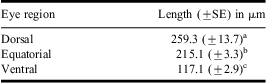
Longitudinal and transverse sections of compound eyes of male and female moths revealed no gender-specific differences in ommatidial microstructure. The convex cornea of each ommatidium has a hexagonal lens (Fig. 4III) and is subtended by a corneal process (Fig. 4I, II). The eucone crystalline cone has four semper cells, and is surrounded by granular pigments (Fig. 4IV). The rhabdom spans the distance from the base of the crystalline cone to the basement membrane (Fig. 4I). Each ommatidium has up to eight retinular cells, six large and two small cells (Fig. 4V). Basal pigment cells occur above the basement membrane. There was no obvious tracheal tapetum. Ommatidia varied significantly in length (F 2,6 = 25.9, P = 0.0011), with the longest ommatidia observed in the dorsal region of the eye and the shortest ommatidia found in the ventral region of the eye (Table 1).
Light micrographs of (I) a longitudinal section through a Synanthedon myopaeformis compound eye revealing the cornea (co), crystalline cones (cc), rhabdoms (r), basal pigment cells (bpc), and basement membrane (bm); (II) Close-up of I with transverse sections taken at A, B, and C through: the cornea (III), a crystalline cone revealing four semper cells (IV), and a rhabdom revealing six large and two small retinular cells demarked by arrows (V).
Mean (±SE) length of ommatidia in the dorsal, equatorial, and ventral regions of the compound eyes (n = 4) in Synanthedon myopaeformis.
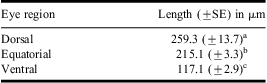
a–cSignificant differences in ommatidia length are denoted by different letters based on the Tukey–Kramer HSD test following a significant ANOVA, α = 0.05.
HSD, honestly significant difference.
Discussion
Eyes of S. myopaeformis were most efficient at detecting UV and green wavelengths, implying their potential for dichromatic vision. The potential for dichromatic vision has been found in diverse insect taxa, including the cockroach Periplana americana (Linnaeus) (Blattodea: Blattidae) (Mote and Goldsmith Reference Mote and Goldsmith1970), the owlfly, Libelloides macaronius (Scopoli) (Neuroptera: Ascalaphidae) (Gogala Reference Gogala1967), several species of ants (Menzel Reference Menzel1973; Mote and Wehner Reference Mote and Wehner1980; Lieke Reference Lieke1981), the moth Synanthedon tipuliformis (Clerk) (Lepidoptera: Sesiidae) (Karalius and Būda Reference Karalius and Būda2007), and the butterfly Parantica sita (Kollar) (Lepidoptera: Nymphalidae) (Eguchi et al. Reference Eguchi, Watanabe, Hariyama and Yamamoto1982). In most other Lepidoptera, however, three photoreceptors (UV, blue, and green) are more common, with some species even possessing a fourth, red receptor (reviewed by Briscoe and Chittka Reference Briscoe and Chittka2001). The broad peak of spectral efficiency to green wavelengths in eyes of S. myopaeformis could be the result of contributions from a blue receptor. However, to unequivocally reveal the specific photoreceptors in the eyes of S. myopaeformis, additional ERGs under adapting light or intracellular single cell recordings should be considered (Goldsmith and Bernard Reference Goldsmith and Bernard1974; Kirchner et al. Reference Kirchner, Doring and Saucke2005).
Eyes of S. myopaeformis were also efficient at absorbing red light of increasing wavelengths from 655 to 700 nm. The potential to detect red wavelengths would make sense because both male and female S. myopaeformis have an orange/red band around their abdomen, which males strike during courtship before females permit copulation (Stüber and Dickler Reference Stüber and Dickler1988). However, red receptors typically are most sensitive or efficient in the 565-nm to 630-nm range (Briscoe and Chittka Reference Briscoe and Chittka2001). An alternative, though less likely, explanation for the increase in efficiency from 655 to 700 nm is that S. myopaeformis eyes have a near-infrared (700–1000 nm) receptor, which we did not test for in this study. To date, infrared receptors have only been found in pits on the abdomen and thorax of some species of Coleoptera (Evans Reference Evans1966; Schmitz et al. Reference Schmitz, Schmitz and Bleckmann2000) and Hemiptera (Takács et al. Reference Takács, Bottomley, Andreller, Zahradnik, Schwarz, Bennett, Strong and Gries2009).
To the human eye, the hoods of A. speciosa inflorescences appear white and the petals pink. Inflorescences that appear white to humans typically do not reflect UV light, and might appear coloured to insects with trichromatic vision (Kevan et al. Reference Kevan, Giurfa and Chittka1996). This is consistent with our measurements showing inflorescences of A. speciosa do not reflect UV light. Bee pollinators typically rely on chromatic rather than achromatic cues to locate floral resources (Giurfa et al. Reference Giurfa, Núñez, Chittka and Menzel1995). “Bee white” inflorescences may be rare because they are difficult to find by pollinators based only on achromatic cues such as contrast against the background or green foliage (Kevan et al. Reference Kevan, Giurfa and Chittka1996). Interestingly, the diurnal hawkmoth, M. stellatarum (Linnaeus) (Lepidoptera: Sphingidae), prefers chromatic over achromatic cues (Kelber Reference Kelber2005), whereas the nocturnal hawkmoth, Manduca sexta (Linnaeus) (Lepidoptera: Sphingidae), uses achromatic cues followed by tactile cues when it probes inflorescences (Goyret Reference Goyret2010). With spectral efficiency for UV and green light, eyes of S. myopaeformis might readily detect plants reflecting UV and green wavelengths using dichromatic colour vision. With the inflorescences and foliage of A. speciosa not reflecting UV, S. myopaeformis could still detect A. speciosa based on contrasting light intensities reflected from inflorescences and foliage or the background using only a green receptor (Dafni and Kevan Reference Dafni and Kevan1997). Alternatively, S. myopaeformis may primarily rely on olfactory cues to locate A. speciosa (Eby et al. Reference Eby, Gardiner, Gries, Judd, Khaskin and Gries2013), and use vision to locate other nectar plants with corresponding visual cues.
Ommatidia in compound eyes of S. myopaeformis contain a rhabdom that is continuous from the crystalline cone to the basement membrane, supporting the conclusion that they have an apposition configuration. The longer rhabdom of ommatidia in the dorsal eye region than in the equatorial and in the ventral eye region likely enhances visual acuity in this region (Rutowski and Warrant Reference Rutowski and Warrant2002). Compound eyes having apposition ommatidia are present in other sesiids (Ehnbom Reference Ehnbom1948; Yagi and Koyama Reference Yagi and Koyama1963) and while this is most common in diurnal Lepidoptera, some possess superposition ommatidia (Horridge et al. Reference Horridge, Giddings and Stange1972, Reference Horridge, McLean, Stange and Lillywhite1977; Warrant et al. Reference Warrant, Bartsch and Günther1999). Superposition eyes may have evolved from apposition eyes via a gradual shift from focal to afocal crystalline cones (Nilsson et al. Reference Nilsson, Land and Howard1988). To date, afocal apposition eyes have only been found in papilionid butterflies (Lepidoptera: Papilionidae) (Warrant et al. Reference Warrant, Kelber and Kristensen2003). Whether S. myopaeformis possess afocal or focal apposition eyes remains unknown, but being phylogenetically more primitive than the papilionids (Tree of Life Web Project 2010), they have likely retained the more ancestral focal apposition eye.
Acknowledgements
The authors thank Tracy Zahradnik, Thomas Cowan, and Dmitri Star for advice; Linda Edwards for providing infested apple wood; and two reviewers for constructive comments. This research was funded by a Natural Sciences and Engineering Research Council of Canada Scholarship (NSERC) to C.E., Agriculture and Agri-food Canada, and by an NSERC-Industrial Research Chair to G.G., with Contech Enterprises Inc. and Global Forest Science as industrial sponsors.