Introduction
Autism spectrum disorder (ASD) is a major neurodevelopmental disorder (APA, 2013). While social communication and interaction deficits, as well as stereotypic behaviour and interests, represent the core problem of ASD, alterations in behavioural and executive control are also evident (Chmielewski & Beste, Reference Chmielewski and Beste2015). An important executive control function is the ability to inhibit responses (Simson et al. Reference Simson, Vaughan and Ritter1977; Pfefferbaum et al. Reference Pfefferbaum, Ford, Weller and Kopell1985; Kok, Reference Kok1986; Jodo & Kayama, Reference Jodo and Kayama1992; Falkenstein et al. Reference Falkenstein, Hoormann and Hohnsbein1999; Bokura et al. Reference Bokura, Yamaguchi and Kobayashi2001; Menon et al. Reference Menon, Adleman, White, Glover and Reiss2001; Bari & Robbins, Reference Bari and Robbins2013). In ASD, findings on response inhibition (RI) are rather ambiguous (for review see Chmielewski & Beste, Reference Chmielewski and Beste2015) with some findings accounting for deficits (Ozonoff et al. Reference Ozonoff, Strayer, McMahon and Filloux1994; Raymaekers et al. Reference Raymaekers, van der Meere and Roeyers2004; Bishop & Norbury, Reference Bishop and Norbury2005; Kana et al. Reference Kana, Keller, Minshew and Just2007), while others account for non-compromised RI functions (Hughes, Reference Hughes1996; Geurts et al. Reference Geurts, Verté, Oosterlaan, Roeyers and Sergeant2004; Schmitz et al. Reference Schmitz, Rubia, Daly, Smith, Williams and Murphy2006; Duerden et al. Reference Duerden, Taylor, Soorya, Wang, Fan and Anagnostou2013; Huster et al. Reference Huster, Enriquez-Geppert, Lavallee, Falkenstein and Herrmann2013). In children with ASD a lack of behavioural deficits in RI is reflected by unchanged connectivity patterns between right inferior frontal and medial frontal areas (Lee et al. Reference Lee, Yerys, Rosa, Foss-Feig, Barnes, James, VanMeter, Vaidya, Gaillard and Kenworthy2009). Other studies accounted for a decreased number of recruited brain regions during RI in ASD (Duerden et al. Reference Duerden, Taylor, Soorya, Wang, Fan and Anagnostou2013), even though behavioural data do not show differences. This suggests that it is important for measures of neuronal activity to deepen the understanding of RI processes in ASD. Apart from functional imaging this can be achieved using neurophysiological (EEG) measures with the advantage that subprocesses during RI can examined. Using event-related potentials (ERPs), two RI subprocesses can be distinguished: One process refers to conflict monitoring and/or pre-motor inhibition processes (NoGo-N2) (Falkenstein et al. Reference Falkenstein, Hoormann and Hohnsbein1999; Nieuwenhuis et al. Reference Nieuwenhuis, Yeung, van den Wildenberg and Ridderinkhof2003, Reference Nieuwenhuis, Yeung and Cohen2004; Beste et al. Reference Beste, Willemssen, Saft and Falkenstein2010; Chmielewski et al. Reference Chmielewski, Mückschel, Roessner and Beste2014, Reference Chmielewski, Roessner and Beste2015c ). The other process, reflected by NoGo-P3, to the motor inhibition process per se and/or evaluation processes of the successful outcome of an inhibition (Bruin et al. Reference Bruin, Wijers and van Staveren2001; Friedman et al. Reference Friedman, Cycowicz and Gaeta2001; Roche et al. Reference Roche, Garavan, Foxe and O'Mara2004; Schmajuk et al. Reference Schmajuk, Liotti, Busse and Woldorff2006; Beste & Saft, Reference Beste and Saft2015; Huster et al. Reference Huster, Enriquez-Geppert, Lavallee, Falkenstein and Herrmann2013; Quetscher et al. Reference Quetscher, Yildiz, Dharmadhikari, Glaubitz, Schmidt-Wilcke, Dydak and Beste2015; Chmielewski et al. Reference Chmielewski, Mückschel, Stock and Beste2015b ; Mückschel et al. Reference Mückschel, Smitka, Hermann, von der Hagen and Beste2015; Wessel & Aron, Reference Wessel and Aron2015). In the current study we explore how far these subprocesses are specifically modulated in ASD.
So far, in healthy subjects it has been shown that RI processes are modulated by the complexity of sensory integration processes (Chmielewski et al. Reference Chmielewski, Mückschel, Dippel and Beste2015a ): when there is a conflict between information from different sensory modalities, RI processes are complicated (Chmielewski et al. Reference Chmielewski, Mückschel, Dippel and Beste2015a ). These cross-modal conflicts during RI processes are of particular relevance for ASD, since deficits in multisensory functions, i.e. in the processing of information across the senses, are increasingly recognized in ASD (Baum et al. Reference Baum, Stevenson and Wallace2015). For example, even though single senses seem not to be explicitly affected in ASD (Baum et al. Reference Baum, Stevenson and Wallace2015), it has been shown that ASD patients have difficulties detecting asynchronies between auditory and visual stimuli, as well as in audio-visual integration processes in general (Kwakye et al. Reference Kwakye, Foss-Feig, Cascio, Stone and Wallace2011; Stevenson et al. Reference Stevenson, Siemann, Woynaroski, Schneider, Eberly, Camarata and Wallace2014). Moreover, it has been shown that ASD patients have difficulties processing complex stimuli (Bertone et al. Reference Bertone, Mottron, Jelenic and Faubert2005; Minshew & Hobson, Reference Minshew and Hobson2008) and do not seem to benefit from redundant information in the same modality (Foxe et al. Reference Foxe, Molholm, Del Bene, Frey, Russo, Blanco, Saint-Amour and Ross2015). Thus, the current study has two questions: (i) Which neurophysiological subprocesses are modulated in ASD during RI?; (ii) How far are RI processes in ASD differentially affected by multisensory integration processes? To answer these questions we use a system neurophysiological approach using high-density EEG recordings and source localization.
Recent findings suggest that RI becomes compromised when conflicting audio-visual information has to be integrated (Chmielewski et al. Reference Chmielewski, Mückschel, Dippel and Beste2015a ). Considering deficits in multisensory integration processes in ASD (Baum et al. Reference Baum, Stevenson and Wallace2015), one hypothesis is that ASD patients may show a superior and impaired RI performance compared to controls, depending on the content of multi-modal stimuli: In case of conflicting multi-modal stimuli, ASD patients might benefit from impairments in multisensory integration processes, since less integrated conflicting information should entail a less compromised RI process. By contrast, in situations where multisensory information usually enhances RI, performance should be decreased in ASD patients compared to controls. However, if multisensory information is properly processed in ASD, then ASD patients may show increased RI deficits under conflicting multisensory information, as this may add up with possible RI deficits reported in ASD.
Regarding neurophysiological processes, we hypothesize the following: Given that multisensory integration processes may be deficient in ASD, it is conceivable that neurophysiological correlates reflecting perceptual or attentional gating and integration processes (i.e. P1 and N1; Herrmann & Knight, Reference Herrmann and Knight2001) are affected. Moreover, as mechanisms of resource allocation, reflected by the P2 ERP (Geisler & Murphy, Reference Geisler and Murphy2000; Campbell & Sharma, Reference Campbell and Sharma2013; Sugimoto & Katayama, Reference Sugimoto and Katayama2013), are modulated by multisensory information during RI (Chmielewski et al. Reference Chmielewski, Mückschel, Dippel and Beste2015a ), it is possible that changes (i.e. a decreased P2) in ASD already occur at the early response selection level. The manipulation of multisensory information should, however, specifically affect conflict monitoring processes and thus the NoGo-N2 amplitude in ASD. That is, depending on the extent of conflict, the strength of the N2 amplitude should systematically vary between ASD patients and controls in conditions with concurrent auditory information. Moreover, the P3 amplitude, i.e. the evaluation of, or the RI process per se (Bruin et al. Reference Bruin, Wijers and van Staveren2001; Friedman et al. Reference Friedman, Cycowicz and Gaeta2001; Roche et al. Reference Roche, Garavan, Foxe and O'Mara2004; Schmajuk et al. Reference Schmajuk, Liotti, Busse and Woldorff2006; Beste & Saft, Reference Beste and Saft2015; Huster et al. Reference Huster, Enriquez-Geppert, Lavallee, Falkenstein and Herrmann2013; Quetscher et al. Reference Quetscher, Yildiz, Dharmadhikari, Glaubitz, Schmidt-Wilcke, Dydak and Beste2015; Chmielewski et al. Reference Chmielewski, Mückschel, Stock and Beste2015b ; Mückschel et al. Reference Mückschel, Smitka, Hermann, von der Hagen and Beste2015; Wessel & Aron, Reference Wessel and Aron2015), should be generally decreased in ASD, given that RI deficits are evident in ASD.
Materials and method
Participants
A group consisting of adolescents with ASD (n = 20, 17 males; mean age 14.8, s.d.= 1.2, age range 13.5–16 years) and a control group (n = 20, 16 males; mean age 15.0, s.d.=1.1, age range 13.5–16 years) was recruited. All ASD patients and controls were right-handed. Both groups did not differ in IQ scores (ASD: 111.31 ± 3.91; controls: 114.06 ± 3.55) (p > 0.5), as obtained using the German version of the Wechsler Intelligence Scale for Children (WISC-IV; Petermann & Petermann, Reference Petermann and Petermann2011). Potential co-morbidities in the ASD group were assessed with the parent questionaire Child Behavior Checklist (CBCL/4–18; Aschenbach, Reference Aschenbach1991) and the MINI-KID (Sheehan et al. Reference Sheehan, Sheehan, Shytle, Janavs, Bannon, Rogers, Milo, Stock and Wilkinson2010). Two ASD patients were diagnosed with co-morbid attention deficit disorder (ADD) and another two with co-morbid ADD. These co-morbidities were either treated with atomoxetine or with Medikinet. All controls reported no history of neurological or psychiatric disorders and were within normal limits in the MINI-KID and were free of medication. Controls and ASD patients had normal or corrected vision and hearing. Participants in the ASD group fulfilled the diagnostic criteria of childhood autism (F84.0, n = 8), Atypical autism (F84.1, n = 3), or Asperger syndrome (F84.5, n = 9) according to ICD-10-GM (Dilling et al. Reference Dilling, Mombour, Schmidt, Schulte-Markwort and Remschmidt2015). To ensure participants in the ASD group fulfilled the diagnostic criteria of ASD the Autism Diagnostic Observation Schedule (ADOS, module 3; Lord & Risi, Reference Lord and Risi1998), an observational instrument based on a series of structured and semi-structured tasks, involving social interaction between the examiner and the subject, was conducted with each participant. ASD participants had an overall score of 14.00 ± 0.99 points (cut-off: 6) in the ADOS. On average 4.26 ± 0.49 points were scored in the communication domain, 7.47 ± 0.49 in the social reciprocity domain, 1.47 ± 0.17 in play skills and imaginative use of objects and 0.89 ± 0.26 in restricted and repetitive behaviours. Moreover on ADOS calibrated severity score (Gotham et al. Reference Gotham, Risi, Pickles and Lord2007, Reference Gotham, Pickles and Lord2009; Shumway et al. Reference Shumway, Farmer, Thurm, Joseph, Black and Golden2012) participants scored on average 7.68 ± 1.70, thus confirming the ASD diagnosis. To complement this, the Autism Diagnostic Interview – Revised (ADI-R; Lord et al. Reference Lord, Rutter and Couteur1994) was conducted with the parents, or caregivers. In this test, an average of 14.47 ± 1.38 points were scored in the reciprocal social interaction domain (cut-off: 10), 11.11 ± 0.99 in the communication and language domain (cut-off: 8) and 3.16 ± 0.48 points in the restricted and repetitive behaviours domain (cut-off: 3). Written informed consent was obtained from all participants (and parents).
Task
We used a task introduced by Chmielewski et al. (Reference Chmielewski, Mückschel, Dippel and Beste2015a ), i.e. a Go/NoGo task, with 70% (672) Go trials, requiring a right-hand response and 30% (288) NoGo trials, requiring withholding of the response. This distribution was chosen to induce a tendency of pre-potent response tendencies (Beste et al. Reference Beste, Dziobek, Hielscher, Willemssen and Falkenstein2009, Reference Beste, Ness, Falkenstein and Saft2011). As stimuli, the German words for ‘stop’ (i.e. stopp) and ‘press’ (drück) were used. Go trials were always presented visually without any second stimulus occurring, to intensify the effects of concurrent stimuli on NoGo trials (Chmielewski et al. Reference Chmielewski, Mückschel, Dippel and Beste2015a ). Out of all NoGo trials, 33% were facilitated (NoGocompatible), and 33% were aggravated (NoGoincompatible) by means of simultaneously presenting either the compatible (same word) or incompatible/conflicting (opposite word) auditory version of the NoGo stimuli verbalized by a female voice. The remaining 33% of NoGo trials were not accompanied by concurrent auditory information (NoGowithout). Auditory stimuli were created using ‘Google translate’ to ensure emotional neutrality. To ensure that no effects of exposure time would occur, the presentation onset and offset of visual and auditory stimuli were equated to 400 ms. Participants were explicitly instructed to only respond to visual stimuli, while ignoring auditory stimuli. Trials were separated by inter-trial intervals jittered between 1700 and 2100 ms. Go trials were coded as misses, when no response was obtained within 1000 ms, while NoGo trials were coded as false alarms (FAs), when a response was given in the same time window. To familiarize subjects with the task, a standardized instruction was given and an exercise with 60 trials was conducted, before the experiment was started.
EEG recording and analysis
EEG data was recorded from 60 Ag/AgCl electrodes mounted in elastic caps (EasyCap Inc., USA) arranged in equidistant positions using BrainAmp amplifiers (Brain Products Inc., USA). The ground electrode was placed at coordinates θ = 58, ϕ = 78 and the reference electrode at θ = 90, ϕ = 90, respectively. A sampling rate of 500 Hz was employed and electrode impedances were kept <5 kΩ. Processing of the data was performed using the BrainVision Analyzer 2 software package (Brain Products). Offline, data was down-sampled to 256 Hz. A band-pass filter from 0.5 to 20 Hz (with a slope of 48 db/oct each) and a notch filter at 50 Hz were applied. After a manual inspection of the data, an automatic independent component analysis (ICA; infomax algorithm; Makeig et al. Reference Makeig, Bell, Jung, Sejnowski and Touretzky1996) was run to remove recurring artifacts. Only ICA components revealing pulse artifacts, blinks and vertical or horizontal eye movements were discarded. Afterwards, the EEG data was segmented for Go trials, NoGo trials without concurrent information (NoGowithout), compatible NoGo trials (NoGocompatible), and incompatible NoGo (NoGoincompatible) trials. Go trials were only included when the correct response was given in a time window until 1000 ms after target onset. Likewise, NoGo trials were only segmented when no response was given in the same time window. The segments were locked to the onset of the target stimulus (Go or NoGo stimulus). After epoching the data, an automated artifact rejection procedure was run for all segments. A difference exceeding 200 μV in a 100 ms interval and an activity <0.5 μV in a 200 ms period, were used as rejection criteria. In order to eliminate reference potential from the data, a current source density (CSD) transformation (Nunez & Pilgreen, Reference Nunez and Pilgreen1991) re-referencing the data was then applied. The resulting CSD values are given in μV/m2. An additional advantage of the CSD transformation is that it serves as a spatial filter (Nunez & Pilgreen, Reference Nunez and Pilgreen1991), which makes it possible to identify electrodes that best reflect activity related to cognitive processes (Cohen, Reference Cohen2014; Mückschel et al. Reference Mückschel, Stock and Beste2014). Afterwards, a baseline correction was applied in the time interval from −500 to −300 ms prior to target onset. For each condition, individual averages were calculated for every participant. The ERP components at the early processing stage were quantified by detecting peaks at electrodes P7 and P8 (P1: 70–150 ms; N1: 150–220 ms). The ERP components at the response selection stage were quantified at electrode Cz (P2: 160–240 ms; N2: 210–350; P3: 340–540 ms). The electrode sites were identified with the help of the scalp topographies. This choice of electrodes was validated using statistical methods (cf. Mückschel et al. Reference Mückschel, Stock and Beste2014). Peak quantification was conducted semi-automatically. For all ERP components, peak-to-baseline amplitudes were computed.
Standardized low-resolution brain electromagnetic tomography (sLORETA) analyses
Source localization was conducted using sLORETA (Pascual-Marqui, Reference Pascual-Marqui2002). sLORETA gives a single linear solution to the inverse problem, based on extra-cranial measurements without a localization bias (Sekihara et al. Reference Sekihara, Sahani and Nagarajan2005). sLORETA reveals high convergence with fMRI data and it has been mathematically proven that sLORETA provides reliable results without localization bias (Sekihara et al. Reference Sekihara, Sahani and Nagarajan2005). There is also evidence of EEG/fMRI and EEG/TMS studies underlining the validity of the sources estimated using sLORETA (e.g. Sekihara et al. Reference Sekihara, Sahani and Nagarajan2005; Dippel & Beste, Reference Dippel and Beste2015). For sLORETA, the intracerebral volume is partitioned into 6239 voxels at 5 mm spatial resolution. The standardized current density at each voxel is calculated in a realistic head model (Fuchs et al. Reference Fuchs, Kastner, Wagner, Hawes and Ebersole2002) using the MNI152 template (Mazziotta et al. Reference Mazziotta, Toga, Evans, Fox, Lancaster, Zilles, Woods, Paus, Simpson, Pike, Holmes, Collins, Thompson, MacDonald, Iacoboni, Schormann, Amunts, Palomero-Gallagher, Geyer, Parsons, Narr, Kabani, Le Goualher, Boomsma, Cannon, Kawashima and Mazoyer2001). In this study, the voxel-based sLORETA images were compared across conditions using the sLORETA built-in voxel-wise randomization tests with 3000 permutations, based on statistical non-parametric mapping. Voxels with significant differences (p < 0.01, corrected for multiple comparisons) between contrasted conditions were located in the MNI brain (www.unizh.ch/keyinst/NewLORETA/sLORETA/sLORETA.htm).
Statistics
For all ANOVAs (i.e. for the behavioural and neurophysiological data), Greenhouse–Geisser correction was applied and the conducted post-hoc tests were Bonferroni-corrected, whenever necessary. All variables included in the analyses were normally distributed, as indicated by Kolmogorov–Smirnov tests (all z < 0.9, p > 0.3). The mean and standard error of the mean (s.e.m.) are given. Statistic analyses were conducted using PASW 20.
Ethical standards
The authors assert that all procedures contributing to this work comply with the ethical standards of the relevant national and institutional committees on human experimentation and with the Helsinki Declaration of 1975, as revised in 2008. The study was approved by the ethics committee of TU Dresden.
Results
Behavioural data
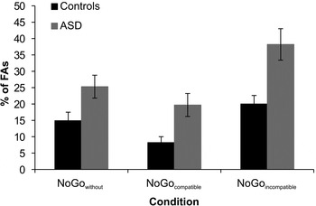
Concerning Go trial reaction times (RTs) and Go trials misses no significant differences could be found between ASD patients (RTs: 474 ± 22 ms; misses: 3.13 ± 0.85%) and the control group (RTs: 511 ± 20 ms; misses: 2.93 ± 0.1.49%) (all t < 1.2, p > 0.4). However, the rate of FAs is the most important behavioural parameter in Go/NoGo tasks. For FAs, repeated-measures ANOVA revealed a main effect of ‘condition’ (F 2,76 = 50.46, p < 0.001, η 2 = 0.570), showing FA rates increasing from the NoGocompatible (14.0 ± 2.0) to the NoGowithout (20.1 ± 2.2) to the NoGoincompatible (29.1 ± 2.7) conditions. Moreover, a main effect of ‘group’ was detected (F 1,38 = 97.15, p < 0.001, η 2 = 0.719), showing the ASD group (27.8 ± 3.0) committed more FAs than controls (14.4 ± 3.0). Most importantly, a ‘condition × group’ interaction was observed (F 2,76 = 3.95, p = 0.023, η 2 = 0.570), which is shown in Fig. 1. Post-hoc paired t tests revealed significant differences between the two groups in the NoGowithout, NoGocompatible and NoGoincompatible conditions (all t ⩾ 2.39, p ⩽ 0.022). We calculated the difference between groups in the FA rate for each condition separately. Post-hoc paired t tests with these group differences (NoGowithout: 10.4 ± 4.4; NoGocompatible: 11.5 ± 4.4; NoGoincompatible: 18.2 ± 5.2) revealed that in the NoGoincompatible condition, group differences were larger than in the NoGowithout and the NoGocompatible conditions (all t ⩾ 2.44, p ⩽ 0.025). The latter conditions did not differ from each other (t 19 = −0.40, p = 0.695).
False alarm (FA) rates (with corresponding s.e.m.s) for both groups in the NoGowithout, NoGocompatible and NoGoincompatible conditions.  , Controls;
 , individuals with autism spectrum disorder (ASD). The bars depict the FA group differences in each NoGo condition.

Regarding the rate of FAs, there was no difference between the first and the second half of trials in each condition in ASD patients and controls (all p > 0.4), suggesting that the length of the experiment did not bias the results obtained for patients relative to the controls.
Neurophysiological data
For all ERP components, there were no latency effects (all F < 1.24, p > 0.189).
Early processing stage
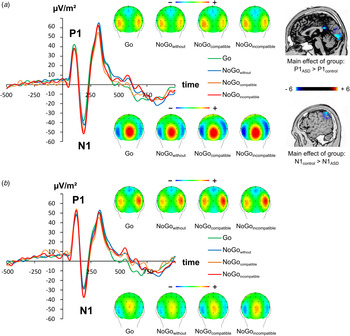
The neurophysiological data for P1 and N1 is shown in Fig. 2. (This data, using no CSD transformation, is shown in Supplementary Fig. S1.)
Event-related potentials (ERPs) on Go and NoGo trials averaged across electrodes P7 and P8. Time-point zero denotes the time-point of Go and NoGo stimulus presentation. The different lines show the NoGowithout condition (blue lines), NoGocompatible condition (orange lines) and the NoGoincompatible condition (red lines). Go trials are coloured in green. The scalp topography plots show the distribution of the scalp electrical potential for the P1 (upper row), and N1 (lower row) on Go and NoGo trials. Panel (a) shows the ERPs and scalp topographies for controls. Additionally sLORETA sources for the (main effect of) group differences between controls and autism spectrum disorder (ASD) patients are displayed for P1 and N1. Panel (b) shows the ERPs and scalp topographies for ASD patients.

Concerning the P1 amplitude, a main effect of ‘group’ was found (F 1,38 = 186.31, p < 0.001, η 2 = 0.831), showing ASD patients (60.5 ± 5.7 μV/m2) exhibiting larger (i.e. more positive) P1 amplitudes than controls (50.4 ± 5.7 μV/m2). sLORETA analysis revealed that this group difference was related to a decreased activation in the cuneus [Brodmann Area (BA) 18] in ASD (Fig. 2). Moreover, a main effect of ‘electrode’ was observed (F 1,38 = 13.84, p = 0.001, η 2 = 0.267), showing the P1 amplitude to be smaller (i.e. less positive) in P7 (47.2 ± 4.5 μV/m2) than in P8 (63.7 ± 7.3 μV/m2). No main effect of ‘condition’ was observed (F 1,38 = 0.56, p = 0.640, η 2 = 0.015). An ‘condition × group’ interaction was observed (F 3,114 = 3.24, p = 0.025, η 2 = 0.079), but post-hoc tests did not withstand Bonferroni-corrected post-hoc testing (all t < 1.65, p > 0.2). No further significant effects were found (all F < 2.03, p > 0.265).
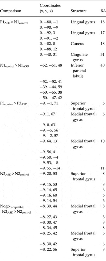
For the N1 amplitudes the repeated-measures ANOVA revealed a main effect of ‘condition’ (F 3,114 = 11.79, p < 0.001, η 2 = 0.237), showing the N1 amplitudes becoming larger (i.e. more negative) from the Go (45.8 ± 4.6 μV/m2) to the NoGowithout (47.5 ± 4.4 μV/m2) to the NoGocompatible (55.6 ± 4.8 μV/m2) to the NoGoincompatible (56.1 ± 5.2 μV/m2) conditions. However, post-hoc paired t tests revealed that all N1 amplitudes differed significantly from each other (all t ⩾ 3.72, p ⩽ 0.001), except for the Go from NoGowithout and the NoGocompatible from NoGoincompatible (all t ⩽ 1.08, p ⩾ 0.2) trials. Moreover, a main effect of ‘group’ was detected (F 1,38 = 126.57, p < 0.001, η 2 = 0.769), showing ASD patients (43.7 ± 7.5 μV/m2) with smaller (i.e. less negative) N1 amplitudes than controls (58.8 ± 5.1 μV/m2). sLORETA analysis revealed that this group difference was related to a decreased activation in the inferior parietal lobe (IPL; BA 40) in ASD (Fig. 2). No further significant effects were found (all F < 1.05, p > 0.374). The results of the sLORETA analysis are given in Table 1.
Results from sLORETA analysis. The Table provides coordinates of obtained activation differences together with the neuroanatomical structure and Brodmann Areas (BAs) for each of the calculated contrasts

Response selection stage
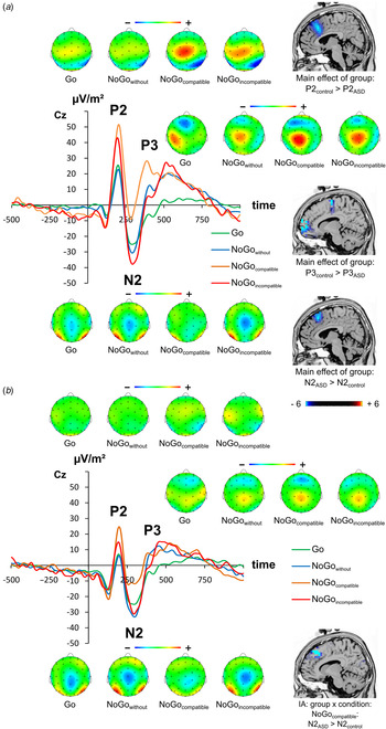
The P2, N2 and P3 ERPs are shown in Fig. 3. (This data, using no CSD transformation, is shown in Supplementary Fig. S2).
Event-related potentials (ERPs) on Go and NoGo trials at the Cz electrode. Time-point zero denotes the time-point of the Go and NoGo stimulus presentation. The different lines show the NoGowithout condition (blue lines), NoGocompatible condition (orange lines) and the NoGoincompatible condition (red lines). Go trials are coloured in green. The scalp topography plots show the distribution of the scalp electrical potential for P2 (upper row), N2 (middle row) and P3 (lower row) on Go and NoGo trials. Panel (a) shows the ERPs and scalp topographies for controls. Additionally sLORETA sources for the P2 main effect of group are displayed in the proximity of the corresponding scalp topographies. Panel (b) shows the ERPs and scalp topographies for autism spectrum disorder (ASD) patients. For N2, the sLORETA source of the group differences in the NoGocompatible condition is displayed.

For the P2 amplitude a main effect of ‘condition’ was observed (F 3,114 = 39.85, p < 0.001, η 2 = 0.512), showing that P2 amplitudes increased from NoGowithout (18.6 ± 2.7 μV/m2) to Go (20.2 ± 2.6 μV/m2) to NoGoincompatible (35.7 ± 3.8 μV/m2) to NoGocompatible (42.3 ± 4.6 μV/m2) trials. Post-hoc paired t tests revealed all conditions to differ significantly from each other (all t ⩾ 2.27, p ⩽ 0.029), except for the Go from NoGowithout trials (t 39 = 1.38, p = 0.175). Moreover, a main effect of ‘group’ was detected (F 1,38 = 85.44, p < 0.001, η 2 = 0.692), showing ASD patients (17.8 ± 4.7 μV/m2) exhibited a smaller (i.e. less positive) P2 than controls (40.6 ± 4.5 μV/m2). sLORETA analysis revealed that this group difference was related to a decreased activation in the middle frontal gyrus (MFG; BA 9) in ASD patients (Fig. 3, Table 1). A ‘condition × group’ interaction was not found (F 3,114 = 1.35, p > 0.15, η 2 = 0.058).
For the N2 amplitude (see Fig. 3), a main effect of ‘condition’ (F 3,114 = 17.80, p < 0.001, η 2 = 0.319) was found: N2 amplitudes increased (in negativity) from NoGocompatible (−22.5 ± 3.0 μV/m2) to Go (−33.9 ± 2.8 μV/m2) to NoGowithout (−38.2 ± 2.9 μV/m2) to NoGoincompatible (−42.1 ± 3.9 μV/m2) trials. All conditions differed significantly from each other (all t ⩾ 2.44, p ⩽ 0.019), except for the NoGowithout from NoGoincompatible (t 39 = 1.33, p = 0.193) trials. Moreover, a main effect of ‘group’ was detected (F 1,38 = 165.23, p < 0.001, η 2 = 0.813), showing ASD patients (−35.9 ± 3.8 μV/m2) exhibited a larger (i.e. more negative) N2 than controls (−32.4 ± 3.8 μV/m2). sLORETA analysis revealed that this group difference was related to an increased activation in the superior frontal gyrus (SFG; BA 8) in ASD (Fig. 3). Most importantly, a ‘condition × group’ interaction was observed (F 3,114 = 3.47, p = 0.019, η 2 = 0.084). Post-hoc tests revealed that this interaction was due to N2 amplitude differences between both groups in NoGocompatible trials (t 38 = 2.10, p = 0.042) (ASD: −28.8 ± 4.6 μV/m2 v. controls: −16.2 ± 3.8 μV/m2). sLORETA analysis revealed that this group difference between compatible trials was related to increased activation in the SFG (BA 8) in ASD patients (Fig. 3 and Table 1). The groups did not differ in the other conditions (all t ⩽ 0.72, p ⩾ 0.4).
For the P3 amplitude, a main effect of ‘condition’ (F 3,114 = 29.13, p < 0.001, η 2 = 0.434) was detected. The P3 amplitude increased from Go (12.3 ± 2.5 μV/m2) to NoGowithout (28.9 ± 3.0 μV/m2) to NoGoincompatible (32.0 ± 2.7 μV/m2) to NoGocompatible (35.8 ± 4.2 μV/m2) trials. Post-hoc paired t tests revealed that all conditions differed significantly from each other (all t ⩾ 2.83, p ⩽ 0.007), except for NoGoincompatible trials, which did not significantly differ from NoGowithout and NoGocompatible trials (all t ⩽ 1.40, p ⩾ 0.16). Moreover, a main effect of ‘group’ was detected (F 1,38 = 106.36, p < 0.001, η 2 = 0.737), showing ASD patients (22.2 ± 3.7 μV/m2) exhibited a smaller (i.e. less positive) P3 amplitude, than controls (32.2 ± 3.7 μV/m2). sLORETA analysis revealed that this group difference was related to a decreased activation in the MFG (BA 6, BA 10, BA 11) and cingulate gyrus (BA 32) in ASD (Fig. 3, Table 1). A ‘condition × group’ interaction was not found (F 3,114 = 1.75, p = 0.161, η 2 = 0.044).
Discussion
In this study we examined the role of conflict in multisensory integration processes during RI in adolescent ASD. The results show that RI performance was compromised in adolescent ASD in all three RI conditions tested (i.e. without concurrent information, with conflicting information and with redundant information), compared to healthy controls. This shows that there is a general RI deficit in adolescent ASD. Interestingly, there was also a ‘group × condition’ interaction effect, showing that multisensory information differentially affected RI processes in ASD and controls. Redundant sensory input did not improve RI performance in ASD more than in controls. By contrast, group differences were evident when the multisensory information was conflicting; i.e. RI processes were more dysfunctional in ASD patients than in controls, when conflicting multisensory information was presented during trials requiring the inhibition of responses. This suggests that auditory information is taken into account along with visual information. Importantly, what seems to be central is the content of the auditory information being processed along with the visual information: conflicting multisensory content leads to differences between ASD patients and controls, while non-conflict multisensory content does not lead to differences between ASD patients and controls when RI function as an instance of executive control functions is concerned. With respect to the field of multisensory processing in ASD (cf. Baum et al. Reference Baum, Stevenson and Wallace2015) this suggests that it is important to consider the content of multisensory information, when cognitive control processes in adolescent ASD are concerned, especially since potential deficits in conflict monitoring processes in ASD have been shown to be related to the involvement of other factors (Chmielewski & Beste, Reference Chmielewski and Beste2015).
The neurophysiological data show which mechanisms in the cognitive processing cascade are modulated between controls and ASD patients, as indexed by increased P1 and decreased N1 amplitudes in ASD deficits already occurring at the perceptual gating and attentional selection level (Herrmann & Knight, Reference Herrmann and Knight2001). These group differences were due to activation differences in the cuneus (BA 18) and IPL (BA 40), which have frequently been shown to be associated with perceptual gating and attentional selection processes (e.g. Salazar et al. Reference Salazar, Kayser and König2004; Schintu et al. Reference Schintu, Hadj-Bouziane, Dal Monte, Knutson, Pardini, Wassermann, Grafman and Krueger2014). It seems that perceptual gating processes (i.e. P1 ERP) are paradoxically enhanced, while attentional selection processes (i.e. N1 ERP) are decreased, which is in line with the assumption that multisensory integration processes might be altered in ASD (Baum et al. Reference Baum, Stevenson and Wallace2015). However, since P1 and N1 are not differentially modulated across NoGo conditions, these modulations cannot explain the observed behavioural effects. Apart from perceptual gating and attentional selection processes, the P2, indexing resource allocation processes (e.g. Geisler & Murphy, Reference Geisler and Murphy2000; Campbell & Sharma, Reference Campbell and Sharma2013; Sugimoto & Katayama, Reference Sugimoto and Katayama2013), was smaller in each of the experimental conditions in ASD patients compared to controls. This suggests that ASD patients are not able to allocate similar large processing resources to the task at hand, which might contribute to the observed behavioural RI deficits. The source localization analyses suggest that modulations seen for P2 are due to functional neuroanatomical structures in MFG (BA 9). These have frequently been shown to be involved in mechanisms of resource allocation (Peelle et al. Reference Peelle, Troiani, Wingfield and Grossman2010). However, since resource allocation mechanisms were not differentially modulated between groups across experimental conditions, this suggests that this process only contributes to RI deficits in ASD non-specifically.
Regarding subsequent neurophysiological correlates of subprocesses during RI, the data show that NoGo-N2 and NoGo-P3 were smaller in ASD patients than in controls, which parallels the finding of a generally increased FA rate in the behavioural data. However, only NoGo-N2, but not NoGo-P3 revealed differential effects across conditions. This suggests that conflict and/or pre-motor inhibition processes (reflected by NoGo-N2) (Falkenstein et al. Reference Falkenstein, Hoormann and Hohnsbein1999; Nieuwenhuis et al. Reference Nieuwenhuis, Yeung, van den Wildenberg and Ridderinkhof2003, Reference Nieuwenhuis, Yeung and Cohen2004; Beste et al. Reference Beste, Willemssen, Saft and Falkenstein2010; Chmielewski et al. Reference Chmielewski, Mückschel, Roessner and Beste2014, Reference Chmielewski, Roessner and Beste2015c ), but not subprocesses related to motor inhibition process per se and/or evaluation processes of the successful outcome of an inhibition (Schmajuk et al. Reference Schmajuk, Liotti, Busse and Woldorff2006; Beste & Saft, Reference Beste and Saft2015; Huster et al. Reference Huster, Enriquez-Geppert, Lavallee, Falkenstein and Herrmann2013; Quetscher et al. Reference Quetscher, Yildiz, Dharmadhikari, Glaubitz, Schmidt-Wilcke, Dydak and Beste2015; Mückschel et al. Reference Mückschel, Smitka, Hermann, von der Hagen and Beste2015) play a role in the modulation of RI by multisensory information in ASD. Such a dissociation of affected RI subprocesses in ASD has until now not been shown, which is due to the methods applied in studying RI in ASD (Chmielewski & Beste, Reference Chmielewski and Beste2015). In the NoGo condition without concurrent information no differences in N2 amplitude could be observed. Regarding the condition with conflicting concurrent information, the NoGo-N2 amplitudes were not different between ASD patients and controls. However, NoGo-N2 was larger in ASD patients in the compatible condition. As NoGo-N2 is increased on compatible trials, but not affected in conditions without concurrent information, this suggests that the processing of multisensory information comes with increased demands in patients in ASD. As this increase in N2 amplitude is not evident in the conflicting condition, this result pattern suggests that conflict monitoring processes cannot sufficiently be triggered in ASD patients when conflicting multisensory information has to be controlled. This may in turn lead to the increased FA rates in the conflicting NoGo condition. The source localization analysis suggests that networks in the SFG (BA 8) are differentially modulated between controls and ASD patients in the time range of NoGo-N2. These areas have frequently been shown to be modulated by RI processes (Chmielewski et al. Reference Chmielewski, Mückschel, Dippel and Beste2015a ). Moreover, they are known to show alterations in ASD (for review see Chmielewski & Beste, Reference Chmielewski and Beste2015), suggesting that the functional changes observed are in line with structural changes found in ASD patients. It may, therefore, be speculated that the observed functional changes are due to structural changes in this brain region. The precise relation, however, remains to be tested in future studies. It can also not be ruled out that the generally deficient resource allocation processes (cf. P2 effects), just prior to the conflict monitoring or pre-motor inhibition subprocess, play a role. That is, due to the decreased allocation of processing resources conflict monitoring processes may not unfold to an extent necessary to resolve the conflict imposed by multisensory information.
Regarding clinical relevance it has been suggested that the assessment of cognitive control functions may serve as useful cognitive biomarkers for pharmacological and behavioural treatment studies in ASD (Chmielewski & Beste, Reference Chmielewski and Beste2015). The experimental paradigm used in this study yields strong effects between ASD patients and controls. Therefore, processes examined in this study may be considered for inclusion in clinical studies and in the assessment of cognitive control abilities in ASD patients in routine clinical care.
A limitation of this study is that sample size of the ASD group is relatively low and that the ASD group included childhood autism, atypical autism and Asperger syndrome. It is therefore unclear whether there are differences in the examined cognitive processes between different forms of ASD. This may be a subject for future studies. However, the effect sizes were strong, suggesting that the effects obtained are reliable. Future studies may also extend the scope to ASD in adults.
In summary, the study shows that RI performance is compromised in adolescent ASD and that multisensory processes modulate RI. The neurophysiological processes related to the changes affect the entire processing cascade from perceptual gating to RI subprocesses. However, conflict monitoring and pre-motor RI processes were specifically modulated between ASD patients and controls depending on multisensory information. The data suggest that conflict monitoring processes are overstrained in adolescent ASD upon conflicting multisensory information, which leads to further declines in RI processes. Multisensory integration processes have very specific effects on cognitive control processes in ASD and constitute an important factor in future research to disentangle cognitive control deficits in ASD.
Supplementary material
For supplementary material accompanying this paper visit http://dx.doi.org/10.1017/S0033291716001008.
Acknowledgements
We thank all participants for their participation. This work was supported by a grant from the Deutsche Forschungsgemeinschaft (DFG) BE4045/10-2.
Declaration of Interest
V.R. has received payment for consulting and writing activities from Lilly, Novartis, and Shire Pharmaceuticals, lecture honoraria from Lilly, Novartis, Shire Pharmaceuticals, and Medice Pharma, and support for research from Shire and Novartis. He has carried out (and is currently carrying out) clinical trials in cooperation with the Novartis, Shire, and Otsuka companies. C.B. has received payment for consulting from GlaxoSmithKline, and Teva. W.X.C., N.W. and M.M. declare no competing or potential conflicts of interest.