Introduction
Termites are a group of eusocial insects that, until recently, have been classified at the taxonomic rank of order Isoptera but are now accepted as the epifamily Termitoidae of the cockroach order Blattaria. Termites are commonly known, especially in Australia, as ‘white ants’, but they are not closely related to true ants (Engel and Krishna, Reference Engel and Krishna2004).
A typical termite nest consists of different castes of individuals: workers, soldiers and reproductives. Workers are the main caste in the colony for the digestion of cellulose in food and are those most likely to be found in infested wood. The winged reproductive has been documented to be a source of food to many people in certain parts of the world (Cornelius and Osbrink, Reference Cornelius and Osbrink2001).
Because of their wood-eating habits, many termite species can cause great damage to unprotected buildings and other wooden structures (Potter, Reference Potter and Moreland1997). Their habit of remaining concealed often results in their presence being undetected until the timbres are severely damaged and exhibit surface changes. Once termites have entered a building, they do not limit themselves to wood; they also damage paper, cloth, carpets and other cellulosic materials. Unfortunately, human activity has also moved many wood-eating species between continents (Cornelius and Osbrink, Reference Cornelius and Osbrink2001)
As a result of the damage caused by termites to wood and wood products, it becomes imperative to reduce and control their population where necessary. However, as the application of chemical agents has been known to have lethal side effects on people and the environment, there is a pressing need to develop much safer biological alternatives with reduced side effects. The aims of this study were to assess the pathogenic efficacy of some entomopathogenic bacteria on termites as a biocontrol measure and to assess the effect of exposure to a magnetic field on their potency.
Materials and methods
Collection of insects and construction of insect cages
The worker termites used (Macrotermes bellicosus (Smeathman) (Isoptera: Termitidae)) were collected from their natural habitats in the environment with the aid of a termite trap, artificially constructed using synthetic materials placed in a dug-out hole and filled with moist cellulosic materials. The trapped termites were taken to the laboratory, placed in cages made from plastic materials, kept in a dark place and supplied with a food source composed of sterilized cellulosic material in the form of moist cardboard.
Observation of the termite population for the onset of disease symptoms
The collected termites were allowed to acclimatize in the cages. Termites exhibiting morbid symptoms in the form of reduced activity, lethargy, colour change, reduced feeding rate and mortality were separated from the population for maceration and subsequent isolation of organisms (Lacey and Brooks, Reference Lacey, Brooks and Lacey1997).
Isolation of bacteria from termite cadavers
Termite cadavers were picked aseptically and macerated in a sterile mortar. Serial dilutions of the macerate were carried out and the appropriate diluents plated on both general and selective bacterial media. Nutrient agar, MacConkey agar, eosin methylene blue agar, deoxycholate citrate agar and mannitol salt agar were used in the experiment. These media were prepared according to the manufacturers' specifications and sterilized in the autoclave at 121 °C for 15 min. They were allowed to cool to a suitable temperature before pour plating. The media were allowed to solidify and the plates incubated at a temperature of 37 °C for 24 h.
Purification and identification of isolated bacterial cells
Each of the distinct and visible colonies obtained on the growth media after the incubation period was purified by repeated streaking of each morphological type on fresh, aseptically prepared media. The identities of colonies were ascertained by means of both cultural and biochemical characterization: Gram's staining reaction (Fawole and Oso, Reference Fawole and Oso2001), spore staining (Barnett et al., Reference Barnett, Payne and Yarrow2000), catalase test, coagulase test and sugar fermentation test (Harrigan, Reference Harrigan1998).
Preparation of bacterial suspensions for the infection of termites
Broth cultures of all the isolated bacteria were prepared and centrifuged to obtain the cells. The cells were washed and reconstituted using sterile water to make the suspension.
Infection of termites with all the isolated bacteria
Termites were infected by immersion for 60 s in microbial suspensions prepared from all the isolated bacteria in accordance with the methods of Samish et al. (Reference Samish, Ginsberg and Glazer2004). The infected termites were observed for the manifestation of disease symptoms for 120 h. Controls were immersed in sterile water.
Selection of entomopathogens from all the isolated bacteria
Pathogenicity tests were carried out on all the isolated bacteria. Entomopathogens were selected by observing the bacteria able to cause infection in the insects in the form of morbidity characterized by reduced activity, lethargy, reduced feeding and death. Reduced feeding was examined by observing the number of perforations made by termites in the moist thick cardboard placed in each of the cages. Cardboard having more perforations indicated a higher feeding rate than that with fewer holes. Bacteria able to exert morbid and mortal effects on termites after pathogenicity tests were selected as entomopathogens.
Determination of the minimum lethal concentration needed for pathogenicity
The minimum bacterial concentration needed for pathogenicity was determined by infecting the termites with various concentrations of the isolated entomopathogens. Infecting concentrations used were 101, 102, 103, 104, 105, 106, 107, 108 and 109colony-forming units (cfu)/ml. The lowest concentration having pathogenic activity was calculated and recorded after 120 h of observation.
Exposure of entomopathogens to varying intensities of magnetic fields
The magnetic fields were generated using a home-made solenoid of 300 turned cylindrical coils (2.5 cm radius and 20 cm length). The minimum lethal concentrations of each entomopathogen were prepared in two sets. A complete set was exposed to varying intensities of static magnetic fields having values of 460, 480 and 500 mT through the variable resistor of 10 KΩ for 30, 60, 90, 120 and 150 min. Trioden for Alternating Current (TRIAC) was used as a high-current switching device controlling the flow of current through the coils. The whole circuit was enclosed in a locally made Perspex case serving as a guide for the field. The equipment was calibrated using a standard electromagnetic generating system. The other set of concentrations served as a positive control and was not exposed to the magnetic fields before being used to infect the insects.
Infection of termites with irradiated cells
The entomopathogens became irradiated after being exposed to varying amounts of magnetic radiation for varying periods of time. These irradiated cells were used to infect a new healthy batch of insects. Non-radiated cells of the same concentrations were also prepared for each of the bacterial entomopathogens and used to infect a different batch of termites to serve as a positive control.
Statistical analysis
In each treatment, 20 termites were used, and all the experiments were carried out in triplicate. All data are expressed as means ± SEM. Statistical analysis was carried out using analysis of variance. Inter-group comparison was achieved by Duncan's multiple range test using SPSS 15.0 for Windows. Differences were judged to be statistically significant when P was less than 0.05.
Results
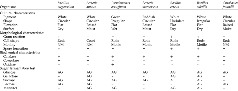
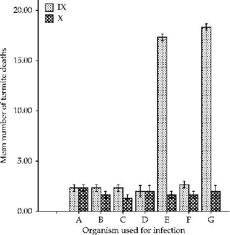
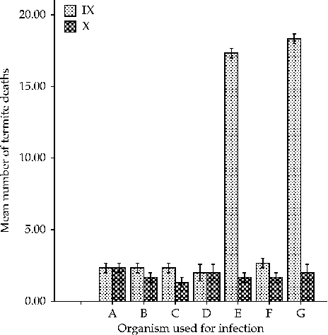
Seven bacterial species were isolated from termites after plating the appropriate macerate dilutions and incubation. These were Staphylococcus aureus, Pseudomonas aeruginosa, Serratia marcescens, Bacillus cereus, Bacillus subtilis, Bacillus megaterium and Citrobacter freundii before and after infection with the entomopathogens (Table 1). After infection with all the isolated bacteria, only B. subtilis and S. marcescens were found to have entomopathogenic effects on termites. This is shown in Fig. 1, where the organisms used for infection are plotted against the mean number of termite deaths.
Characteristics of bacterial isolates obtained from macerated Macrotermes bellicosus worker termite cadavers before and after infection with the entomopathogens

+, positive; − , negative; NM, non-motile; AG, acid and gas production; A, acid production.
Infection of termites with isolated bacteria after 120 h. (A) Bacillus cereus, (B) Citrobacter freundii, (C) Bacillus megaterium, (D) Pseudomonas aeruginosa, (E) Serratia marcescens, (F) Staphylococcus aureus and (G) Bacillus subtilis. IX, infected termites; X, control termites. X-axis represents the organisms used in the infection of termites. Y-axis represents the mean number of termite deaths that occurred after infection. Error bars: ± 1 SE.
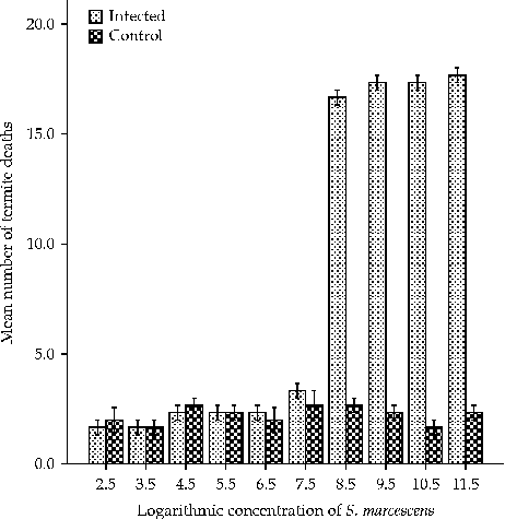
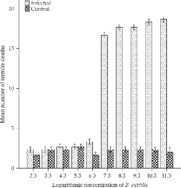
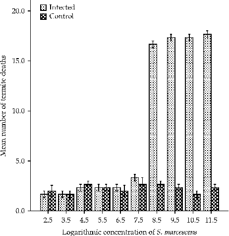
The concentration values of 108cfu/ml for S. marcescens and 107cfu/ml for B. subtilis were the lowest bacterial concentrations that resulted in the symptoms of pathogenicity in the infected termites. These are shown in Figs 2 and 3, where the organism concentration used is plotted against the mean number of termite deaths.
Minimum lethal concentration of Serratia marcescens against termites. X-axis represents the logarithmic values in colony-forming units/ml of S. marcescens concentration used to infect termites. Y-axis represents the mean number of termite deaths that occurred after infection with S. marcescens. Error bars: ± 1 SE.
Minimum lethal concentration of Bacillus subtilis against termites. X-axis represents the logarithmic values in colony-forming units/ml of B. subtilis concentration used to infect termites. Y-axis represents the mean number of termite deaths that occurred after infection with B. subtilis. Error bars: ± 1 SE.
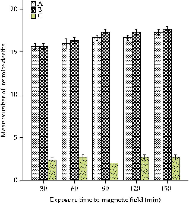
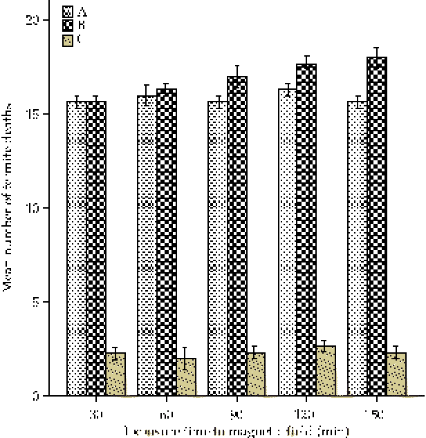
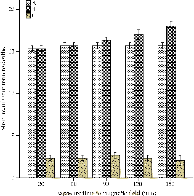
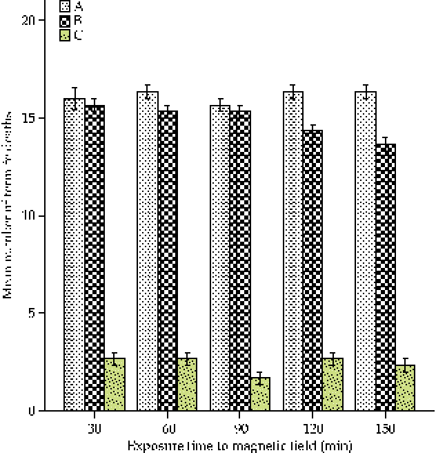
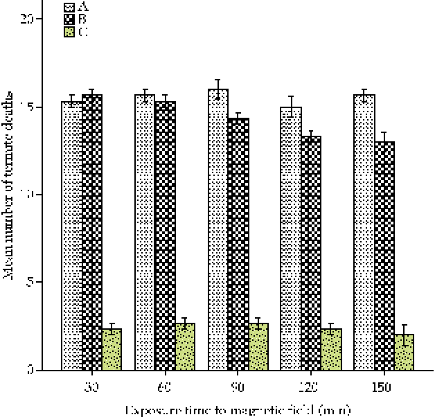
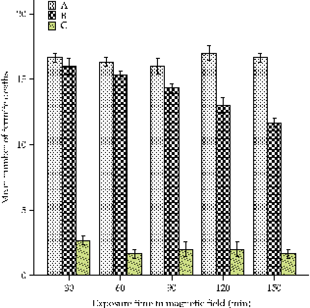
The entomopathogenic potency of S. marcescens was found to be slightly increased with an increase in the time and intensity of exposure at 460 mT (Fig. 4). However, the increase in potency peaked at 480 mT, and no further significant increase in potency was observed at 500 mT (Figs 5 and 6). This is shown in Figs 8 and 9, where the exposure time of B. subtilis suspension to a magnetic field is plotted against the mean number of termite deaths that occurred after infection with the exposed bacteria. Furthermore, Figs 7 and 8 show an initial slight reduction in the entomopathogenic potency of B. subtilis against termites after exposure to 460 and 480 mT magnetic fields. The decrease in potency became more pronounced with an increase in the time and intensity of exposure at 500 mT (Fig. 9).
(colour online) Infection of termites with Serratia marcescens exposed to a 460 mT magnetic field. (A) Termites infected with exposed cells; (B) termites infected with unexposed cells; and (C) uninfected termites. X-axis represents the exposure time (min) of S. marcescens suspension to a magnetic field. Y-axis represents the mean number of termite deaths that occurred after infection with exposed S. marcescens. Error bars: ± 1 SE.
(colour online) Infection of termites with Serratia marcescens exposed to a 480 mT magnetic field. (A) Termites infected with exposed cells; (B) termites infected with unexposed cells; and (C) uninfected termites. X-axis represents the exposure time (min) of S. marcescens suspension to a magnetic field. Y-axis represents the mean number of termite deaths that occurred after infection with exposed S. marcescens. Error bars: ± 1 SE.
(colour online) Infection of termites with Serratia marcescens exposed to a 500 mT magnetic field. (A) Termites infected with exposed cells; (B) termites infected with unexposed cells; and (C) uninfected termites. X-axis represents the exposure time (min) of S. marcescens suspension to a magnetic field. Y-axis represents the mean number of termite deaths that occurred after infection with exposed S. marcescens. Error bars: ± 1 SE.
(colour online) Infection of termites with Bacillus subtilis exposed to a 460 mT magnetic field. (A) Termites infected with exposed cells; (B) termites infected with unexposed cells; and (C) uninfected termites. X-axis represents the exposure time (min) of B. subtilis suspension to a magnetic field. Y-axis represents the mean number of termite deaths that occurred after infection with exposed B. subtilis. Error bars: ± 1 SE.
(colour online) Infection of termites with Bacillus subtilis exposed to a 480 mT magnetic field. (A) Termites infected with exposed cells; (B) termites infected with unexposed cells; and (C) uninfected termites. X-axis represents the exposure time (min) of B. subtilis suspension to a magnetic field. Y-axis represents the mean number of termite deaths that occurred after infection with exposed B. subtilis. Error bars: ± 1 SE.
(colour online) Infection of termites with Bacillus subtilis exposed to a 500 mT magnetic field. (A) Termites infected with exposed cells; (B) termites infected with unexposed cells; and (C) uninfected termites. X-axis represents the exposure time (min) of B. subtilis suspension to a magnetic field. Y-axis represents the mean number of termite deaths that occurred after infection with exposed B. subtilis. Error bars: ± 1 SE.
Discussion
Bacteria isolated from termites have also been recorded in previous research studies and these include S. marcescens (Osbrink et al., Reference Osbrink, Williams, Connick, Wright and Lax2001), Citrobacter spp. (Harazono et al., Reference Harazono, Yamashita, Shinzato, Watanabe, Fukatsu and Kurane2003) and P. aeruginosa (Tsunoda et al., Reference Tsunoda, Ohmura and Yoshimura1993).
This study has demonstrated that B. subtilis is a potential entomopathogen, capable of causing lethargic effects that subsequently result in the death of termites used. This is in accordance with the findings of Lacey and Joel (Reference Lacey and Joel2000), who reported Bacillus spp. to be a potential entomopathogen that has been used extensively for the control of insect and pest populations in domestic animals, crops, forests and the aquatic environment. This has also been established by Mulla (Reference Mulla1991) during his studies on the entomopathogenic potency of Bacillus spp. in controlling populations of mosquitoes in developing countries. Backman et al. (Reference Backman, Wilson, Murphy, Rechcigl and Rechcigl1997) also described some strains of B. subtilis to have innate insecticidal properties that are useful in biocontrol activities.
The entomopathogenic potential of S. marcescens, as demonstrated in this study, is possibly a result of the specific plasmid (pADAP)-encoding gene. The pADAP protein is known to be present in Serratia spp. During the 41st Annual Meeting of the Society for Invertebrate Pathology and the 9th International Conference on Bacillus thuringiensis, 3–7 August 2008 (SIP, 2008), entomopathogens other than Bacillus spp. were researched. The host responds to the release of this bacterial protein by the expulsion of the gut contents to the hindgut and the release of frass pellets. This eventually leads to the weakening of internal tissues, thus leading to bacterial invasion of the haemocoel, causing septicaemia and death.
According to Pothakamury et al. (Reference Pothakamury, Babosa-Canovas and Swanson1993), B. subtilis can be inhibited in terms of potency after being subjected to treatment with an electromagnetic field. This can be attributed to the loosening of the bond between the vital cell protein and its ions, which are responsible for the normal physiological functioning of the cells, thus leading to malfunction in the cells and a subsequent loss of entomopathogenicity. It can also be attributed to the breaking down of the covalent bonds in the DNA molecule and subsequent inhibition of the growth and attributes of the microorganism.
In addition, it has been revealed through this study that S. marcescens is slightly stimulated with regard to its entomopathogenic ability after being exposed to an electromagnetic field and used in the infection of termites. This can be attributed to the low positive response or reaction of the organism to the magnetic field. This finding is in agreement with the submissions of Gerencer et al. (Reference Gerencer, Barnothy and Barnothy1962) that S. marcescens exhibits an initial small or nil response to both static and heterogeneous magnetic fields but upon further exposure may be slightly stimulated in terms of growth or other properties.
Conclusion
Bacillus subtilis and S. marcescens were shown to have insecticidal effects on termites. They can, therefore, be employed in biocontrol activities against termites, especially on wood and wood products. In addition, the exposure of S. marcescens to a magnetic field was shown to have mild stimulatory effects on its potency; this can be used subsequently for increasing the entomopathogenic potential of the organism against termites. By contrast, exposure of B. subtilis to an electromagnetic field resulted in a reduction in its insecticidal activity. Therefore, further research is needed into other ways of stimulating the entomopathogenic potency of the organism against termites.