Implications
In this study, expression of adipocyte-specific genes was analyzed. As some of these genes are regulated by overfeeding, they represent good candidate genes and their expression could be taken into account the select ducks with a greater ability to produce fatty liver. Ducks also appear as promising models to answer questions according to non-alcoholic fatty liver disease and relevant of human health.
Introduction
Associated with the obesity and being recognized as the hepatic manifestation of metabolic syndrome, non-alcoholic fatty liver disease (NAFLD) has become the most common cause of human liver disease in the industrialized world. Non-alcoholic fatty liver disease is the result of hepatic steatosis, that is fat accumulation in the liver. Huge efforts are made to elucidate the mechanisms underlying hepatic steatosis which at least include hepatic fatty acid uptake, de novo lipogenesis, fatty acid oxidation and fatty acid export (Musso et al., Reference Musso, Gambino and Cassader2009; Fabbrini et al., Reference Fabbrini, Sullivan and Klein2010). According to de novo lipogenesis, several studies have established that hepatic steatosis in mammals is associated to overexpression of lipogenic genes (Tamura and Shimomura, Reference Tamura and Shimomura2005; Lavoie and Gauthier, Reference Lavoie and Gauthier2006; Parekh and Anania, Reference Parekh and Anania2007; Ferré and Foufelle, Reference Ferré and Foufelle2010). It has also been shown that adipokines produced by the adipose tissue are involved in hepatic steatosis (Stojsavljevic et al., Reference Stojsavljevic, Gomercic Palcic, Virovic Jukic, Smircic Duvnjak and Duvnjak2014; Panera et al., Reference Panera, Della Corte, Crudele, Stronati, Nobili and Alisi2016; Polyzos et al., Reference Polyzos, Kountouras and Mantzoros2016). Interestingly, some studies have shown that expression of peroxisome proliferator-activated receptor gamma (PPARG), a key regulator of adipogenesis, and of some adipocyte-specific genes or adipokines are expressed in hepatic steatosis (Gavrilova et al., Reference Gavrilova, Haluzik, Matsusue, Cutson, Johnson, Dietz, Nicol, Vinson, Gonzalez and Reitman2003; Matsusue et al., Reference Matsusue, Haluzik, Lambert, Yim, Gavrilova, Ward, Brewer, Reitman and Gonzalez2003; Yu et al., Reference Yu, Matsusue, Kashireddy, Cao, Yeldandi, Yeldandi, Rao, Gonzalez and Reddy2003; Schadinger et al., Reference Schadinger, Bucher, Schreiber and Farmer2005), leading to the concept of ‘adipogenic hepatic steatosis’ or ‘hepatic adiposis’ (Yu et al., Reference Yu, Matsusue, Kashireddy, Cao, Yeldandi, Yeldandi, Rao, Gonzalez and Reddy2003). Most of these data relatives to ‘adipogenic hepatic steatosis’ were generated in obese mouse models or after manipulation of gene expression and the relevance of this concept to other species and more physiological models should be better addressed.
Fatty liver production in waterfowls appears very similar to human NAFLD (Ray, Reference Ray2013; Osman et al., Reference Osman, Shao, Liu, Xia, Sun, Zheng, Wang, Zhang, Zhang, Zhang, Gong and Geng2016) making these animals very interesting models for a better understanding of liver steatosis. Indeed, liver steatosis can occur spontaneously in waterfowls as a result of energy storage before migration and can be induced by overfeeding in a short period of time to produce fatty liver (foie gras). Liver steatosis in these birds exists in isolation, a degenerative event rarely occurs, and is reversible when overfeeding is stopped (Hermier et al., Reference Hermier, Guy, Guillaumin, Davail, André and Hoo-Paris2003). We and others have demonstrated that hepatic steatosis in waterfowls also involves de novo lipogenesis and overexpression of related genes, triacylglycerol (TG) synthesis, uptake of circulating lipids and finally accumulation of lipids in the liver (Davail et al., Reference Davail, Rideau, Guy, Andre, Hermier and Hoo-Paris2003; Hermier et al., Reference Hermier, Guy, Guillaumin, Davail, André and Hoo-Paris2003; Hérault et al., Reference Hérault, Saez, Robert, Al Mohammad, Davail, Chartrin, Baéza and Diot2010). However, few data were produced on adipocyte-specific gene expression in waterfowls fatty liver.
The present study was thus conducted to analyze the expression of PPARG and other ‘adipocyte-specific genes’ in the liver of ducks fed ad libitum or overfed and compared with those observed in adipose tissue. Adipocyte-specific genes analyzed were adiponectin (ADIPOQ), thrombospondin receptor (CD36), complement factor D also known as adipsin (CFD), adipocyte fatty acid-binding protein 4 (FABP4) also known as adipocyte protein 2 (aP2) and leptin receptor (LEPR). Expressions of adipogenic transcription factor CCAAT/enhancer-binding protein beta (CEBPB) and of hepato-specific albumin gene (ALB) were also analyzed. These analyses were conducted in the liver of two duck species, Pekin (Anas platyrhynchos) and Muscovy ducks (Cairina moschata), as metabolic response to overfeeding differs according to these species, Muscovy ducks having a greater ability to synthesize and store lipids in the liver than Pekin ducks (Hermier et al., Reference Hermier, Guy, Guillaumin, Davail, André and Hoo-Paris2003; Chartrin et al., Reference Chartrin, Bernadet, Guy, Mourot, Hocquette, Rideau, Duclos and Baéza2006; Saez et al., Reference Saez, Baéza, Davail, Durand, Bauchart and Gruffat2009; Hérault et al., Reference Hérault, Saez, Robert, Al Mohammad, Davail, Chartrin, Baéza and Diot2010). These ducks were those previously analyzed for expression of genes involved in lipid and carbohydrate metabolisms (Hérault et al., Reference Hérault, Saez, Robert, Al Mohammad, Davail, Chartrin, Baéza and Diot2010) in order to provide additional information on genes that are regulated by overfeeding in the same experimental design.
Material and methods
Experimental design, animals and housing
Experimental design, animals and housing have been described previously (Chartrin et al., Reference Chartrin, Bernadet, Guy, Mourot, Hocquette, Rideau, Duclos and Baéza2006). At 12 weeks of age, 10 male ducks per species were overfed with corn and corn meal and 10 male ducks per species were fed ad libitum with the growing diet for 14 days. Immediately after bleeding, liver and abdominal fat samples were frozen in liquid nitrogen and stored at −70°C until RNA was prepared.
Ribonucleic acid isolation and reverse transcription
Total RNA was extracted from liver and adipose tissue samples using NucleoSpin® RNA L (Macherey-Nagel SARL, Hoerdt, France) according to the manufacturer’s instructions. This kit involves guanidinium thiocyanate, silica membrane and on-column RNase-free DNase digestion. RNA concentration was determined using a ND-1000 Spectrophotometer (NanoDrop Technologies, Wilmington, DE, USA). Integrity of RNA was checked with Lab-on-a-Chip Eukaryote Total RNA Nano and Bionalyzer 2100 (Agilent Technologies, Les Ullis, France). RNA with absorbance ratio λ260 nm/λ280 nm and λ260 nm/λ230 nm >1.8 and RNA integrity number (Schroeder et al., Reference Schroeder, Mueller, Stocker, Salowsky, Leiber, Gassmann, Lightfoot, Menzel, Granzow and Ragg2006) or RIN >7.4 were selected.
Quantitative real-time polymerase chain reaction
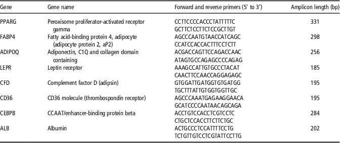
Expression of genes in Pekin and Muscovy duck livers and adipose tissues was analyzed by real-time quantitative PCR (Q-PCR) amplifications with SYBR-Green® as described previously (Bourneuf et al., Reference Bourneuf, Hérault, Chicault, Carré, Assaf, Monnier, Mottier, Lagarrigue, Douaire, Mosser and Diot2006; Hérault et al., Reference Hérault, Saez, Robert, Al Mohammad, Davail, Chartrin, Baéza and Diot2010). First strand cDNA synthesis was performed by reverse transcription of 10 μg of total RNA with the High Capacity cDNA Archive Kit (Applied Biosystems, Foster City, CA, USA) including random primers, in a total volume of 100 µl and according to the manufacturer’s instructions. Gene-specific primer pairs (Table 1) were designed according to sequences in GenBank. Each primer pair was tested for PCR amplification efficiency with cDNA dilutions from 1/2 to 1/2048. The efficiencies obtained were in a range of 99.8±0.3%. Q-PCR was then performed in triplicate in an IQ-cycler (BioRad, Hercules, CA, USA) in the presence of 1× of IQTM SYBR-Green® Supermix (BioRad), 300 nm specific primers, and 5 µl of 1/10 diluted cDNA in a 25-µl total volume, and using a two-step PCR protocol (95°C for 2 min, followed by 45 cycles of 95°C for 15 s and 59°C for 45 s). To ascertain the specificity of the reactions, the quality of PCR amplicons was monitored using post-PCR melt curve analysis of amplification reactions (in triplicate from all samples) and by sequencing amplification products.
Duck primer design and PCR amplification in ducks

Results are expressed as means±standard errors of the means (SEM) of
 $$2^{{{\minus}\Delta \Delta _{{Ct}} }} $$
values (Livak and Schmittgen, Reference Livak and Schmittgen2001) with ΔΔ
Ct
=(Ct
ij
−Ct
18Sj
)−(Ct
i1−Ct
18S1), where Ct
ij
and Ct
18Sj
are the Ct for gene i and for 18S in a sample (named j) and where Ct
i1 and Ct
18S1 are the Ct in the liver of Pekin ducks fed ad libitum, expressed as the standard.
Statistical analyses
Statistical analyses were performed using the R software environment (http://www.r-project.org/). Feeding, genotype and interaction effects were determined by ANOVA on ΔΔ Ct values. They were considered significant when P-value was 0.05 or less.
Results
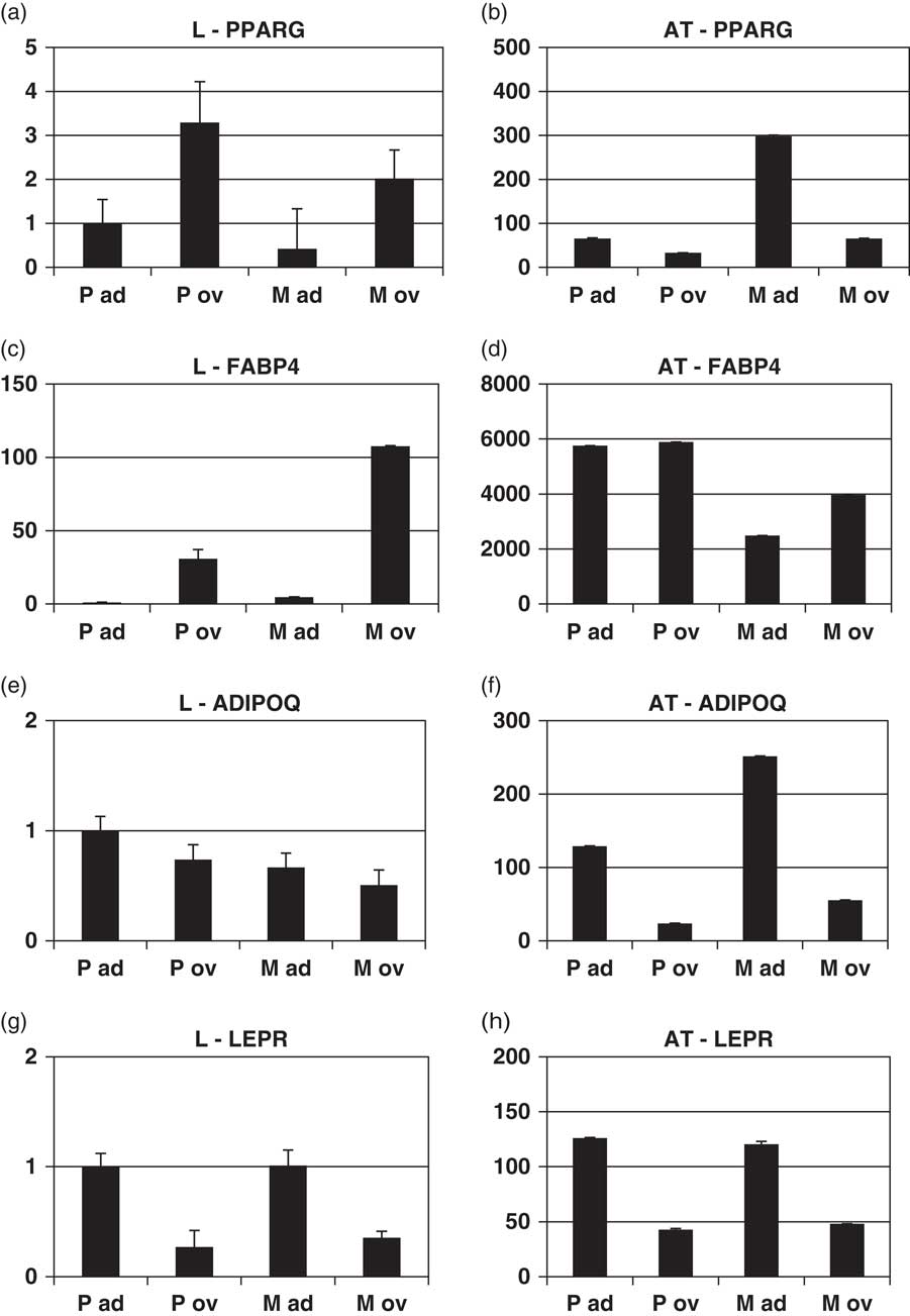
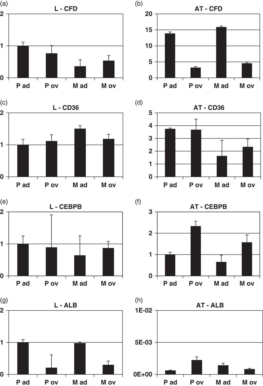
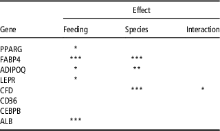
Specific primers were designed (Table 1) in order to analyze RNA levels of ‘adipocyte-specific’ genes in the liver and adipose tissue of Pekin and Muscovy ducks fed ad libitum and overfed. These genes encode PPARG, FABP4, ADIPOQ, LEPR, CFD and CD36. Due to the important role of LEP (leptin) in mammals, design of primers was also conducted according to A. platyrhynchos leptin sequence in Genbank (accession no. AY555727.1). However, PCR amplifications were unsuccessful and LEP expression could not be analyzed. As shown in Figure 1, PPARG, FABP4, ADIPOQ and LEPR were expressed in the liver of Pekin and Muscovy ducks (Figure 1a, c, e and g) although these expression levels were rather low when compared with those observed in adipose tissues (Figure 1b, d, f and h). PPARG and FABP4 hepatic expressions were induced by overfeeding (Figure 1a and c; Table 2) whatever the species. In adipose tissue, PPARG expression was reduced by overfeeding (Figure 1b; Table 2) whereas FABP4 expression was not affected by overfeeding (Figure 1d). A negative effect of overfeeding was observed on ADIPOQ and LEPR expressions in the liver (Figure 1e and g and Table 2) and in adipose tissue (Figure 1f and h). A negative effect of overfeeding was also observed on ADIPOQ and LEPR expressions in adipose tissues (Figure 1f and h). CFD and CD36 expressions in the liver were also rather low when compared with those observed in adipose tissue and were not affected by overfeeding (Figure 2a and c). A negative effect of overfeeding was observed in adipose tissue on CFD expression (Figure 2b) whereas CD36 expression in adipose tissue was not affected (Figure 2d).
Quantitative real-time PCR analysis of PPARG, FABP4, ADIPOQ and LEPR gene expression in Pekin and Muscovy ducks. Analyses were conducted in the liver (L) and adipose tissue (AT) of Pekin (P) and Muscovy (M) ducks fed ad libitum (ad) or overfed (ov). Results are expressed as means and standard errors (n=10) and according to expression level observed in the liver of Pekin ducks fed ad libitum (P ad). (a) PPARG expression in the liver, (b) PPARG expression in adipose tissue, (c) FABP4 expression in the liver, (d) FABP4 expression in adipose tissue, (e) ADIPOQ expression in the liver, (f) ADIPOQ expression in adipose tissue, (g) LEPR expression in the liver, (h) LEPR expression in adipose tissue. Complete gene names are indicated in Table 1.

Quantitative real-time PCR analysis of CFD, CD36, CEBPB and ALB gene expression in Pekin and Muscovy ducks. Analyses were conducted in the liver (L) and adipose tissue (AT) of Pekin (P) and Muscovy (M) ducks fed ad libitum (ad) or overfed (ov). Results are expressed as means and standard errors (n=10) and according to expression level observed in the liver of Pekin ducks fed ad libitum (P ad). (a) CFD expression in the liver, (b) CFD expression in adipose tissue, (c) CD36 expression in the liver, (d) CD36 expression in adipose tissue, (e) CEBPB expression in the liver, (f) CEBPB expression in adipose tissue, (g) ALB expression in the liver, (h) ALB expression in adipose tissue. Complete gene names are indicated in Table 1.

Feeding and species effects and interaction on hepatic gene expressions

Significant feeding effects (ad libitum v. overfed) and duck species effects (Anas platyrhynchos Pekin ducks v. Cairina moschata Muscovy ducks) on hepatic gene expressions and effect interactions were determined using ANOVA and are indicated as follows: ***P<0.001, **P<0.01, *P<0.05. Complete gene names are indicated in Table 1.
As expected, CEBPB expression in adipose tissue was induced by overfeeding (Figure 2f) but no difference was observed in the liver (Figure 2e). ALB was found to be expressed to high levels in the liver with a negative effect of feeding (Figure 2g and Table 2) and nearly not expressed in adipose tissue. Species effects on hepatic gene expressions were observed for FABP4, ADIPOQ and CFD (Table 2).
Discussion
Expression of PPARG and of other ‘adipocyte-specific genes’ were analyzed in the liver of ducks fed ad libitum or overfed and compared with those observed in adipose tissue in order to better characterize differences between normal and fatty liver, especially with regard to adipokines and adipocyte-specific gene expression.
Our results clearly indicate that PPARG, FABP4, ADIPOQ, LEPR, CFD and CD36 are expressed in fatty livers. PPARG and FABP4 expressions are increased by overfeeding, as also observed in another study (Tavernier et al., Reference Tavernier, Davail, Ricaud, Bernadet and Gontier2017). Many studies in rodents have established a link between PPARG expression and obesity and/or hepatic steatosis (Tontonoz and Spiegelman, Reference Tontonoz and Spiegelman2008; Lefterova et al., Reference Lefterova, Haakonsson, Lazar and Mandrup2014). PPARG was demonstrated to be overexpressed in the liver of obese mouse models compared with lean littermates (Vidal-Puig et al., Reference Vidal-Puig, Jimenez-Linan, Lowell, Hamann, Hu, Spiegelman, Flier and Moller1996; Memon et al., Reference Memon, Tecott, Nonogaki, Beigneux, Moser, Grunfeld and Feingold2000; Bedoucha et al., Reference Bedoucha, Atzpodien and Boelsterli2001; Rahimian et al., Reference Rahimian, Masih-Khan, Lo, van Breemen, McManus and Dube2001; Colombo et al., Reference Colombo, Haluzik, Cutson, Dietz, Marcus-Samuels, Vinson, Gavrilova and Reitman2003). Liver-specific PPARG knockout reduced hepatic steatosis in obese mice (Gavrilova et al., Reference Gavrilova, Haluzik, Matsusue, Cutson, Johnson, Dietz, Nicol, Vinson, Gonzalez and Reitman2003; Matsusue et al., Reference Matsusue, Haluzik, Lambert, Yim, Gavrilova, Ward, Brewer, Reitman and Gonzalez2003) and overexpression of PPARG in the liver of PPARG −/− mice induced hepatic steatosis and increased lipogenesis-related genes together with adipocyte-specific genes (Yu et al., Reference Yu, Matsusue, Kashireddy, Cao, Yeldandi, Yeldandi, Rao, Gonzalez and Reddy2003). Expression of FABP4 is highly induced during adipocyte differentiation and transcriptionally controlled by PPARG (Cook et al., Reference Cook, Lucas, Sibley, Bolanowski, Christy, Kelly and Lane1988; Amri et al., Reference Amri, Bertrand, Ailhaud and Grimaldi1991; Distel et al., Reference Distel, Robinson and Spiegelman1992; Kletzien et al., Reference Kletzien, Foellmi, Harris, Wyse and Clarke1992; Melki and Abumrad, Reference Melki and Abumrad1993; Tan et al., Reference Tan, Shaw, Vinckenbosch, Liu, Yasmin, Desvergne, Wahli and Noy2002). Our findings confirm the adipogenic role of these two genes in hepatic steatosis in duck species also. Conversely, ADIPOQ and LEPR expression are decreased in fatty liver of overfed animals when compared with those observed in normal liver of Pekin and Muscovy ducks fed ad libitum. These results are also similar to those observed in mammals. Adiponectin, the protein encoded by ADIPOQ, was first isolated from differentiated 3T3-L1 adipocytes as an abundant and adipose tissue-specific adipokine (Scherer et al., Reference Scherer, Williams, Fogliano, Baldini and Lodish1995) but was also shown to be expressed in hepatocytes as a response to hepatic injury (Yoda-Murakami et al., Reference Yoda-Murakami, Taniguchi, Takahashi, Kawamata, Saito, Choi-Miura and Tomita2001). Contrary to other adipokines, adiponectin is decreased when adipose mass increases (Arita et al., Reference Arita, Kihara, Ouchi, Takahashi, Maeda, Miyagawa, Hotta, Shimomura, Nakamura, Miyaoka, Kuriyama, Nishida, Yamashita, Okubo, Matsubara, Muraguchi, Ohmoto, Funahashi and Matsuzawa1999; Polyzos et al., Reference Polyzos, Kountouras, Zavos and Tsiaousi2010). These results on adipokines expression in overfed ducks agree with the concept of ‘adipogenic transformation of hepatocytes’ developed by Yu et al. (Reference Yu, Matsusue, Kashireddy, Cao, Yeldandi, Yeldandi, Rao, Gonzalez and Reddy2003). However, the situation is more complex as no effect was observed on CD36 and CFD expression. Furthermore, adipokine expression in overfed ducks was shown to be much lower in fatty liver than in adipose tissue suggesting that adipokine effects could be more endocrine than autocrine and paracrine. It is also important to note that albumin expression was decreased in duck fatty liver when compared with normal ones. Another study has also reported a decrease of albumin and some other hepato-specific genes expressions in overfed goose fatty livers (Zhu et al., Reference Zhu, Meng, Duan, Xu, Zhang and Gong2011). Altogether, these results suggest that hepatic steatosis partly affects hepato-specific functions and is linked to adipokine expression, although fatty liver is not fully transformed to adipose tissue.
In a recent publication, Seroussi et al. (Reference Seroussi, Cinnamon, Yosefi, Genin, Smith, Rafati, Bornelov, Andersson and Friedman-Einat2016) reported the identification of the duck leptin gene. The authors indicated that duck leptin gene was difficult to sequence due to an extreme GC content (~70%) and that they failed to obtain this sequence by RT-PCR ‘in spite of applying a variety of specialized protocols such as that used for PCR amplification of GC-rich dove leptin.’ We also failed to analyze duck leptin expression by RT-PCR. Surprisingly, Seroussi et al. (2016) also reported low expression level (analyzed by RNA sequencing) of leptin and its receptor in chicken and duck liver and adipose tissues, and no difference in expression level of LEP and LEPR between lean and fat chicken lines. This contrasts with our results showing that LEPR is expressed to a high level in duck adipose tissue and to a lower extent in the liver, and that LEPR expression is strongly decreased in fatty liver when compared with normal liver. The reasons of this discrepancy are not clear but can in part be attributed to differences in species and experimental conditions. To our knowledge, differences in expression levels between lean and fat chickens are much lower than those observed between ducks fed ad libitum or overfed (Bourneuf et al., Reference Bourneuf, Hérault, Chicault, Carré, Assaf, Monnier, Mottier, Lagarrigue, Douaire, Mosser and Diot2006; Hérault et al., Reference Hérault, Saez, Robert, Al Mohammad, Davail, Chartrin, Baéza and Diot2010). Nevertheless, LEPR expression in duck liver and adipose tissues and its negative correlation to hepatic steatosis raises the debate about the meaning of LEP signaling in avian species. More investigations are now needed to address this point.
In conclusion, PPARG and some adipokine expressing genes were found to be expressed in the liver of ducks, PPARG and FABP4 being upregulated and ADIPOQ and LEPR downregulated by overfeeding. However, these expression levels are much lower than those observed in adipose tissue suggesting that fatty liver cells are not transformed to adipocytes, although some hepato-specific functions are decreased in fatty liver when compared with normal liver. More study are now needed at least to analyze in more details the role of LEP/LEPR pathway and more broadly to better understand the mechanisms involved in hepatic steatosis. Ducks appear excellent models to answer these questions, relevant of both livestock species production and human health.
Acknowledgments
This research was supported by Institut National de la Recherche Agronomique (INRA). The authors greatly acknowledge Grimaud Frères Selection for producing and providing the animals, the technical staff of Unité Expérimentale Palmipèdes à Foie Gras UE0089 for rearing and overfeeding ducks, Capucine Le Mare and Guillaume Ayral for technical assistance and Gregory Guernec for advices in statistics.
Declaration of interest
We declare no conflict of interest.
Ethics statement
Experimental work has been conducted in accordance with French national legislation on the use of animals for research.
Software and data repository resources
Not applicable.