INTRODUCTION
A number of common, medically significant parasitic diseases are, ironically, neglected diseases (Van der Pol, Reference Van der Pol2007). Within this group are diseases caused by protozoan flagellates that reside extracellularly and in oxygen-deprived environments (anaerobic) within their hosts. These include enteric human parasites such as Giardia duodenalis, Entamoeba histolytica, Cryptosporidium and the urogenital track parasite, Trichomonas vaginalis. Pathogenesis of enteric parasites results from physical parasite damage to the intestinal mucosal membrane causing malabsobtion, and mild-to-severe chronic gastroenteritis associated with increased secretion that induce diarrhoea and reduced growth in children (Buret, Reference Buret2008; Raza et al. Reference Raza, Iqbal, Muhammad, Khan and Hanif2013; Checkley et al. Reference Checkley, White, Jaganath, Arrowood, Chalmers, Chen, Fayer, Griffiths, Guerrant, Hedstrom, Huston, Kotloff, Kang, Mead, Miller, Petri, Priest, Roos, Striepen, Thompson, Ward, Van Voorhis, Xiao, Zhu and Houpt2015). In addition, Entamoeba infections can advance to more serious manifestations including acute necrotizing colitis that mask other underlying pathogenesis (Petri and Singh, Reference Petri and Singh1999; Bercu et al. Reference Bercu, Petri and Behm2007). On the other hand, T. vaginalis causes physical damage to the genital mucosa of female hosts resulting in severe vaginitis and facilitates the spread of the HIV virus (Wegayehu et al. Reference Wegayehu, Adamu and Petros2013; Fletcher et al. Reference Fletcher, Caprarelli, Merif, Andresen, Van Hal, Stark and Ellis2014). The epidemiology of neglected protozoan parasitic diseases predominantly correlates with socio-economic attributes of developing countries with poor sanitary conditions (Raza et al. Reference Raza, Iqbal, Muhammad, Khan and Hanif2013; Betancourt et al. Reference Betancourt, Duarte, Vasquez and Gurian2014; Fletcher et al. Reference Fletcher, Caprarelli, Merif, Andresen, Van Hal, Stark and Ellis2014; Checkley et al. Reference Checkley, White, Jaganath, Arrowood, Chalmers, Chen, Fayer, Griffiths, Guerrant, Hedstrom, Huston, Kotloff, Kang, Mead, Miller, Petri, Priest, Roos, Striepen, Thompson, Ward, Van Voorhis, Xiao, Zhu and Houpt2015). A 2014 World Health Organisation report estimated that the global disability-adjusted life year (DALY) for diarrhoeal disease in low and middle income countries, expressed as the number of years lost due to illness or death, was 33 793 DALYs due to inadequate water supply (WHO, 2014). Cryptosporidium, a significant cause of moderate-to-severe diarrhoea, was ranked as the second most significant agent of diarrhoeal disease in sub-Saharan African and south Asia by the Global Enteric Multicentre Study (GEMS) (Kotloff et al. Reference Kotloff, Nataro, Blackwelder, Nasrin, Farag, Panchalingam, Wu, Sow, Sur, Breiman, Faruque, Zaidi, Saha, Alonso, Tamboura, Sanogo, Onwuchekwa, Manna, Ramamurthy, Kanungo, Ochieng, Omore, Oundo, Hossain, Das, Ahmed, Qureshi, Quadri, Adegbola and Antonio2013). While GEMS failed to associate Giardia as a significant cause of severe diarrhoea (Kotloff et al. Reference Kotloff, Nataro, Blackwelder, Nasrin, Farag, Panchalingam, Wu, Sow, Sur, Breiman, Faruque, Zaidi, Saha, Alonso, Tamboura, Sanogo, Onwuchekwa, Manna, Ramamurthy, Kanungo, Ochieng, Omore, Oundo, Hossain, Das, Ahmed, Qureshi, Quadri, Adegbola and Antonio2013), parasite burden is clearly prominent in countries of lower socio-economic status. For instance, a recent study conducted in Ethiopia revealed 13·8% of clinical samples were Giardia-positive, while in the more developed state of New South Wales in Australia, 1% of clinical samples examined were Giardia-positive (Betancourt et al. Reference Betancourt, Duarte, Vasquez and Gurian2014; Fletcher et al. Reference Fletcher, Caprarelli, Merif, Andresen, Van Hal, Stark and Ellis2014). As the most common, non-virally sexually transmitted pathogen, the urogenital flagellate T. vaginalis has a prevalence of up to 46·9% in developed countries (Petrin et al. Reference Petrin, Delgaty, Bhatt and Garber1998; Rendon-Maldonado et al. Reference Rendon-Maldonado, Espinosa-Cantellano, Soler, Torres and Martinez-Palomo2003). The recommended drug for anaerobic parasite infections has persistently been the 5-nitroimidazole derivatives such as metronidazole, despite growing concerns of parasite resistance and host-toxicity (Gookin et al. Reference Gookin, Copple, Papich, Poore, Stauffer, Birkenheuer, Twedt and Levy2006; Dunn et al. Reference Dunn, Burgess, Krauer, Eckmann, Vanelle, Crozet, Gillin, Upcroft and Upcroft2010; LeVine et al. Reference LeVine, Gookin, Papich and Davidson2014; Meltzer et al. Reference Meltzer, Lachish and Schwartz2014). Discovery of novel drug options has, potentially, been hindered by leaden research progress due to the unique and complex biology of anaerobic parasites.
Knowledge of prokaryotic and higher eukaryotic cell biology has encountered major headway over the past three decades as a result of advancements in and integration of various scientific tools. The merger of molecular methods, imaging technologies and increasingly affordable high-throughput sequencing technology has been particularly sweeping. Emanating from this combination is the use of fluorescent protein tags (FPs), engineered to label proteins at the genetic level within living cells. The essence of the approach is at the DNA level, where the FP tag is fused to the DNA sequence of a known protein-of-interest (POI) in an expression vector and subsequently expressed as a fusion-protein in the parasite. Fluorescent microscopy is used to track the journey of the focus protein through the cell enabling visualisation of secretory pathways, localization, protein dynamics and interaction within the parasite (Fernandez-Suarez and Ting, Reference Fernandez-Suarez and Ting2008). The physical association of the FP with the expressed protein of interest and the built-in ability of FPs to generate fluorescence upon excitation by a specific wavelength of light facilitates an enhanced level of specificity in protein tracking (Wang and Hazelrigg, Reference Wang and Hazelrigg1994; Dhandayuthapani et al. Reference Dhandayuthapani, Via, Thomas, Horowitz, Deretic and Deretic1995; Waller et al. Reference Waller, Keeling, Donald, Striepen, Handman, Lang-Unnasch, Cowman, Besra, Roos and McFadden1998, Reference Waller, Reed, Cowman and McFadden2000). As such, the contribution of specific proteins within the dynamic live-cell environment can be visualized, pin-pointing protein–protein interactions or interactions with organelles.
The burgeoning research activity that followed endogenous tagging technology boosted our understanding of cellular events; however, only islands of success have been achieved in parasitology. In parasitic protozoans, exploitation of FPs to their full potential is restricted to oxygen-dwelling protozoans (aerobic protozoans), or oxygenated stages of the parasite lifecycle and has contributed to ground-breaking research in the field. On the other hand, there has been limited application for FPs as a live-parasite protein tag in anaerobic protozoans due to clashes between the physio-chemical properties of common FPs and the anaerobic nature of bio-chemical pathways in anaerobic parasites. Despite well-established molecular parasitology methods for introducing novel genes into anaerobic parasites (Delgadillo et al. Reference Delgadillo, Liston, Niazi and Johnson1997; Singer et al. Reference Singer, Yee and Nash1998; Jerlstrom-Hultqvist et al. Reference Jerlstrom-Hultqvist, Stadelmann, Birkestedt, Hellman and Svard2012; Manna et al. Reference Manna, Ehrenkaufer and Singh2014; Vinayak et al. Reference Vinayak, Pawlowic, Sateriale, Brooks, Studstill, Bar-Peled, Cipriano and Striepen2015), the long-standing obstacle for parasitologist working with these parasites has been choosing the correct FP to avoid physiological artefact, while simultaneously maximizing FP signal. This has proven difficult as the commonly used FPs do not tolerate the anaerobe intracellular environment and as such, fail to capture cellular events in their true dynamic state. In this review, we aim to explore the anaerobic biology of common oxygen-sensitive protozoans and the physio-chemical aspects of routinely used FP tags. We will examine aspects of anaerobic protozoa biology that clash with aspects of FPs that have hindered their use as live-parasite protein tags. We further delve into untapped FP-tagging opportunities and discuss their potential for application as a protein-tag in this group of parasites.
PROTEIN TAGGING
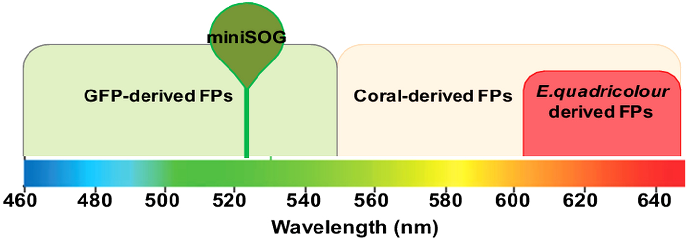
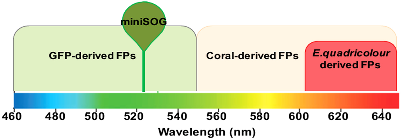
FPs emitting a rainbow of colours have emanated from three decades of molecular engineering verve (Fig. 1). Genetically encoded FPs cloned from the jelly fish Aequorea victoria or the mushroom coral, Discosoma are the patriarch of the most routinely used genetic-level protein tags (Prasher et al. Reference Prasher, Eckenrode, Ward, Prendergast and Cormier1992; Chalfie et al. Reference Chalfie, Tu, Euskirchen, Ward and Prasher1994). As tags, they are powerful tools for tracking proteins of interest in a particular organism in vivo to characterize protein function, mechanism of action and intermediary protein–protein interactions. Intracellular localization of proteins, however, is not limited to FPs and various well-established techniques can be employed. They include direct and indirect immuno-localization of the protein using an antibody raised to an epitope of the POI or antibody localization of affinity tags or FPs that are genetically fused to the protein of interest. Antibody localization to FP-fused parasite proteins has, to date, been the highly successful for protein localization in anaerobic protozoans. The technique, however, is labour intensive, time-consuming and expensive. In addition, all antibody-based techniques require manipulation of the sample through fixation and permeabilization, and the exogenous addition of synthetic dyes or enzyme substrate for signal production. Unless monoclonal antibodies are available, the use of polyclonal antibodies for sub-cellular protein localization can be highly unspecific (Lipman et al. Reference Lipman, Jackson, Trudel and Weis-Garcia2005). In contrast, FP localizations are less likely to contain experimental artefacts that arise from the use of exogenous reagents, hence the protein of interest can be observed in their natural dynamic state within the parasite. There are considerations that have to be taken into account prior to adopting FPs for live-cell protein tracking. The protein tag should be non-toxic to the organism that expresses it, and in return, the cell has to provide the precise physio-chemical environment to guarantee chromophore maturation and photostability which are key in determining its applicability as a protein tag. An exact chromophore architecture is required for fluorescence emission which is dictated by correct folding of the FP (Waldo et al. Reference Waldo, Standish, Berendzen and Terwilliger1999; Pedelacq et al. Reference Pedelacq, Piltch, Liong, Berendzen, Kim, Rho, Park, Terwilliger and Waldo2002). Intracellular conditions (i.e. pH) and interference from the fused POI are factors that may induce misfolding or aggregation of a protein, particularly as a result of over-expression (King et al. Reference King, HaasePettingell, Robinson, Speed and Mitraki1996; Makrides, Reference Makrides1996; Pedelacq et al. Reference Pedelacq, Piltch, Liong, Berendzen, Kim, Rho, Park, Terwilliger and Waldo2002; Costantini and Snapp, Reference Costantini and Snapp2013). Despite development of FPs with a lower propensity to aggregate, clumping of expressed FPs can occur as a result of the increased interaction between fusion proteins within small sub-organelle spaces (Costantini and Snapp, Reference Costantini and Snapp2013). Over-expression of FP-fusions should be modulated by careful selection of flanking promotor sequences. Protein misfolding and aggregation not only affects the effectiveness of the FP as a fluorescent marker, but also the ability of the fusion-protein to translocate across membranes, resulting in aberrant protein localization (Waldo et al. Reference Waldo, Standish, Berendzen and Terwilliger1999). Therefore, in choosing an FP for localization in an anaerobic protozoan, there is a need to understand various aspect of FPs such as the excitation and emission spectra, brightness and photostability. In addition, the chromophore maturation time, the tendency for the FP to aggregate, pH stability and the requirement for oxygen will affect the suitability of an FP in a particular parasite.
Emission spectrum range of FPs. FPs emit at different wavelengths depending on the FP family. Abbreviation: FP, fluorescent protein.
Common FPs
Green FPs (GFPs) and its derivatives
Since its discovery and isolation from the jelly fish Aequorea victoria and first use as a genetically-encodable tag (Prasher et al. Reference Prasher, Eckenrode, Ward, Prendergast and Cormier1992; Chalfie et al. Reference Chalfie, Tu, Euskirchen, Ward and Prasher1994), the GFP and its derivatives has endured as the most commonly used live-cell protein tag in the microbiologist's toolkit. The GFP is made up of 238 amino acids residues (27 kDa) held in a β-barrel by 11 β-strands that contain an α-helix at its core (Ormo et al. Reference Ormo, Cubitt, Kallio, Gross, Tsien and Remington1996). The GFP chromophore is ultimately responsible for fluorescence and develops from the amino acid triad Ser65-Tyr66-Gly67 buried deep within the central α-helix. In the presence of oxygen, a three-stage spontaneous, self-catalysed post-translational modification of the triad occurs to form the mature chromophore (Yang et al. Reference Yang, Moss and Phillips1996). The final step of chromophore maturation is a rate-limiting oxidation reaction that creates the fluorescence-emitting chromophore, a five-membered conjugated imidazolone ring system with a p-hydroxybenzylidene substituent (Barondeau et al. Reference Barondeau, Putnam, Kassmann, Tainer and Getzoff2003). Emission of fluorescence results from a protonation-deprotonation equilibrium of chromophore species with absorbances at ~395 and ~470 nm (Heim et al. Reference Heim, Prasher and Tsien1994). Fluorescence emitted from GFP peaks at 509 nm and can be visualized using conventional fluorescein isothiocyanate (FITC) excitation-emission filter cubes.
Although unhampered by exogenous co-factor requirements, GFP fluorescence emission is highly dependent on expression, correct folding within the cellular milieu and an absolute presence of high oxygen concentrations (Heim et al. Reference Heim, Prasher and Tsien1994; Cormack et al. Reference Cormack, Valdivia and Falkow1996; Reid and Flynn, Reference Reid and Flynn1997). In the wild-type GFP (WT-GFP), this occurs optimally at low temperatures and a minimum exposure to 4 h of oxygen (Heim et al. Reference Heim, Prasher and Tsien1994). Changes to the spectral emission can be achieved by manipulation of the residues within, and surrounding the chromophore and has resulted in a wide collection of GFP derivatives now readily available to microbiologists as protein markers (Heim et al. Reference Heim, Cubitt and Tsien1995; Cormack et al. Reference Cormack, Valdivia and Falkow1996; Heim and Tsien, Reference Heim and Tsien1996). For instance, the introduction of two mutations (S65T, F64L) in the chromophore region of WT-GFP improved the emission by 21-fold, decreased the time required for oxygen exposure to 8 min and increased the excitation spectra (Cormack et al. Reference Cormack, Valdivia and Falkow1996). Following years of subsequent mutagenesis, a multitude of GFP variants emitting an assortment of fluorescent colours ranging the spectrum from blue to yellow have been developed; each having varied protein- and photo-stability (Crameri et al. Reference Crameri, Whitehorn, Tate and Stemmer1996; Siemering et al. Reference Siemering, Golbik, Sever and Haseloff1996; Kimata et al. Reference Kimata, Iwaki, Lim and Kohno1997; Patterson et al. Reference Patterson, Knobel, Sharif, Kain and Piston1997). The emission wavelength limit of GFP derivatives, however, does not surpass the yellow FPs (YFPs), of which Citrins and Venus are the most useful derivatives (Griesbeck et al. Reference Griesbeck, Baird, Campbell, Zacharias and Tsien2001; Nagai et al. Reference Nagai, Ibata, Park, Kubota, Mikoshiba and Miyawaki2002). The true value of YFPs has been in pairings with other GFP variants of distinct wavelengths as acceptors for fluorescence resonance energy transfer (FRET) to greatly improve the photostability of signals (Miyawaki and Tsien, Reference Miyawaki and Tsien2000; Griesbeck et al. Reference Griesbeck, Baird, Campbell, Zacharias and Tsien2001). A different method to ensure bright GFP signals have been developed as the SunTag system, where the POI is fused with up to 24 peptide epitopes instead of GFP and is then co-expressed within the cell with GFP-tagged single-chain antibodies that host the epitope binding site, recruiting up to 24 GFP molecules to the POI (Tanenbaum et al. Reference Tanenbaum, Gilbert, Qi, Weissman and Vale2014). While SunTag enhances the brightness of GFP fluorescent signals, the system results in a cluster of proteins of greater size than obtained when using single GFP tagging (up to 1400 kDa) (Tanenbaum et al. Reference Tanenbaum, Gilbert, Qi, Weissman and Vale2014). Despite these advances, all GFP derivatives are limited to emit at wavelengths below 600 nm, discounting the red spectral range (>600 nm) and limiting their use in cells that exhibit green autofluorescence (Niswender et al. Reference Niswender, Blackman, Rohde, Magnuson and Piston1995).
The development of GFP derivatives led to the successful expression of GFP in bacteria, plant cells, the yeast model, Saccharomyces cerevisiae and the fruit fly Drosophila melanogaster within the first decade (Wang and Hazelrigg, Reference Wang and Hazelrigg1994; Dhandayuthapani et al. Reference Dhandayuthapani, Via, Thomas, Horowitz, Deretic and Deretic1995; Niedenthal et al. Reference Niedenthal, Riles, Johnston and Hegemann1996; Harper et al. Reference Harper, Mabon, Leffel, Halfhill, Richards, Moyer and Stewart1999). Among these reports was also the first report describing the expression of the GFP in a protozoan aerobe, Plasmodium falciparum, the malaria causing parasite (Chalfie et al. Reference Chalfie, Tu, Euskirchen, Ward and Prasher1994; VanWye and Haldar, Reference VanWye and Haldar1997). Later, GFP was used to elucidate function and targeting signals of the apicoplast, a newly discovered chloroplast-derived organelle at the time, by fusing GFP to a P. falciparum nuclear gene encoding fatty acid biosynthesis (Waller et al. Reference Waller, Keeling, Donald, Striepen, Handman, Lang-Unnasch, Cowman, Besra, Roos and McFadden1998). Further research using various GFP-tagged proteins led to an understanding of the critical role of the apicoplast on parasite survival, redirecting drug-target discovery efforts to the organelle (Waller et al. Reference Waller, Keeling, Donald, Striepen, Handman, Lang-Unnasch, Cowman, Besra, Roos and McFadden1998; He et al. Reference He, Shaw, Pletcher, Striepen, Tilney and Roos2001; Goodman and McFadden, Reference Goodman and McFadden2013). In Trypanosoma brucei, the human aerobic parasite responsible for African sleeping sickness, GFP was used to resolve the long standing conundrum of whether the parasite undergoes meiotic recombination or exists as a clonal population (Bingle et al. Reference Bingle, Eastlake, Bailey and Gibson2001; Gibson et al. Reference Gibson, Peacock, Ferris, Williams and Bailey2006, Reference Gibson, Peacock, Ferris, Williams and Bailey2008; Peacock et al. Reference Peacock, Bailey, Carrington and Gibson2014). The mode of genetic exchange in parasites relates to parasite epidemiology, host adaptations and drug resistance. By mating GFP-tagged parasites, the hybrid colours of T. brucei obtained not only established that co-infections with different T. brucei strains are more successful than single-strain infections, but also proved that only a proportion of flies are responsible for distributing trypanosome infections to humans (Peacock et al. Reference Peacock, Ferris, Bailey and Gibson2007). While these instances involve aerobic protozoa, the studies exemplify the potential of live-cell tagging if employed in anaerobic protozoa.
Red FPs (RFPs)
The motivation to expand the colour palette of FPs from the limited blue-yellow (emission beyond 550 nm) range of the GFPs led to the first red FPs from the non-bioluminescent Indo-Pacific anthozoa species of coral (Matz et al. Reference Matz, Fradkov, Labas, Savitsky, Zaraisky, Markelov and Lukyanov1999). Through homology prediction at predicted conserved sites within GFP, six red-fluorescing coral proteins were isolated (Matz et al. Reference Matz, Fradkov, Labas, Savitsky, Zaraisky, Markelov and Lukyanov1999). These proteins exhibit remarkable sequence and folding conservation with GFP in the chromophore region, although, on the whole, only 26–30% is shared amino-acid similarity between the GFP and coral RFPs (Matz et al. Reference Matz, Fradkov, Labas, Savitsky, Zaraisky, Markelov and Lukyanov1999; Yarbrough et al. Reference Yarbrough, Wachter, Kallio, Matz and Remington2001). The first red-shifted FP that was made commercially available, DsRed, is similar in size and structure to the GFP consisting of 225 amino acids (26 kDa) arranged into an 11-stranded β-barrel with a central helix and α-helical caps on the barrel ends (Matz et al. Reference Matz, Fradkov, Labas, Savitsky, Zaraisky, Markelov and Lukyanov1999; Gross et al. Reference Gross, Baird, Hoffman, Baldridge and Tsien2000; Yarbrough et al. Reference Yarbrough, Wachter, Kallio, Matz and Remington2001). The chromophore of DsRed, as in GFP, is a p-hydroxybenzylideneimidazolidinone, formed as a result of a final oxidation step in the presence of oxygen (Gross et al. Reference Gross, Baird, Hoffman, Baldridge and Tsien2000). In contrast to GFP, the DsRed chromphore hosts more complex chromophore maturation events that consists of a GFP-like stage (excitation: 475 nm; emission: 500 nm), prior to red-shifting to the excitation and emission spectra of 558 and 583 nm, respectively, of mature DsRed (Matz et al. Reference Matz, Fradkov, Labas, Savitsky, Zaraisky, Markelov and Lukyanov1999; Gross et al. Reference Gross, Baird, Hoffman, Baldridge and Tsien2000; Yarbrough et al. Reference Yarbrough, Wachter, Kallio, Matz and Remington2001). Conversion between the green and red spectral species is the rate-limiting step, taking days to occur, after which, at least 50% of the green species remains (Baird et al. Reference Baird, Zacharias and Tsien2000). Unlike the GFPs, DsRed is more tolerant to surrounding pH, whereby emission is unaffected between pH 4 and 12, and structurally exists as a closely packed obligate tetramer (Baird et al. Reference Baird, Zacharias and Tsien2000; Gross et al. Reference Gross, Baird, Hoffman, Baldridge and Tsien2000; Yarbrough et al. Reference Yarbrough, Wachter, Kallio, Matz and Remington2001). While attributes such as the red emission wavelength, photostability and environmental resistance of DsRed makes it valuable as an FP tag, it has several disadvantages that render it unsuitable for use in anaerobic protozoa. Slow and incomplete maturation ensues that real-time collection of biological information cannot be entertained, and its tendency to aggregate as a result of its tetrameric arrangement results in aberrant localization and difficulty for the fusion-tag to cross some organelle membranes (Baird et al. Reference Baird, Zacharias and Tsien2000; Lauf et al. Reference Lauf, Lopez and Falk2001; Campbell et al. Reference Campbell, Tour, Palmer, Steinbach, Baird, Zacharias and Tsien2002).
Currently, the most commonly used RFP are monomeric variants of DsRed, known collectively as mFruits since FP members are named according to their emission spectra. Marketed by Clontech, a variety of colours exists in the mFruits collection with each member possessing a unique compilation of physical properties, excitation and emission spectra and fluorescence quantum yield (Shaner et al. Reference Shaner, Campbell, Steinbach, Giepmans, Palmer and Tsien2004). Compared to DsRed, mFruits boasts reduced chromophore maturation times (<1 h) and are less likely to aggregate upon expression (Shaner et al. Reference Shaner, Campbell, Steinbach, Giepmans, Palmer and Tsien2004). These benefits, however, are undermined by compromised photostability and emission quantum yield of monomeric RFPs owing to lower protection of the chromophore from the external environment surrounding the protein (Campbell et al. Reference Campbell, Tour, Palmer, Steinbach, Baird, Zacharias and Tsien2002). Even the brightest member of mFruits, mOrange, suffer from remarkably fast photobleaching (Shaner et al. Reference Shaner, Campbell, Steinbach, Giepmans, Palmer and Tsien2004). Photobleaching in these proteins arises as a result of the reduced structural stability of the monomeric mFruits compared to the self-supported tetrameric structure of DsRed. Furthermore, alterations introduced in the structure of the β-barrels resulted in gap openings leading to the chromophore and more structural fluctuation in the protein (Chapagain et al. Reference Chapagain, Regmi and Castillo2011; Laurent et al. Reference Laurent, Mironov, Chapagain, Nemukhin and Krylov2012). This allows extended oxygen contact with the mature chromophore at core within the barrel, which acts to significantly reduce photostability (Chapagain et al. Reference Chapagain, Regmi and Castillo2011; Regmi et al. Reference Regmi, Bhandari, Gerstman and Chapagain2013). Phenotypically selecting for increased resistance to photobleaching in mFruits is attainable, but comes at a cost of decreasing FP brightness (Shaner et al. Reference Shaner, Lin, McKeown, Steinbach, Hazelwood, Davidson and Tsien2008). As improving one aspect of an mFruit member has, so far, resulted in depressing another, the most commonly used member of mFruits is mCherry which exhibits intermediary attributes. Despite not being the brightest in the mFruits collection, mCherry displays superior qualities of having the lowest maturation time (~30 min), high pH resistance, a very similar excitation and emission spectrum to parent RFPs (587 and 610 nm, respectively) and enhanced resistance to photobleaching (Shaner et al. Reference Shaner, Campbell, Steinbach, Giepmans, Palmer and Tsien2004). The enhanced photostability has rendered mCherry on par with synthetic secondary labelling dyes for single-molecule localization and has an attainable resolution of as low as 23 nm (Winterflood and Ewers, Reference Winterflood and Ewers2014).
Far-red FPs
In the search for RFPs with a lower predisposition to aggregate, FPs from the sea anemone Entacmaea quadricolor has been isolated using a similar homology-to-GFP method used to obtain coral RFPs (Wiedenmann et al. Reference Wiedenmann, Schenk, Rocker, Girod, Spindler and Nienhaus2002; Merzlyak et al. Reference Merzlyak, Goedhart, Shcherbo, Bulina, Shcheglov, Fradkov, Gaintzeva, Lukyanov, Lukyanov, Gadella and Chudakov2007). These FPs, however, represent a different class of FPs to the original GFP and coral RFPs by possession of a unique chromophore-forming triad; Met63-Tyr64-Gly65 (Wiedenmann et al. Reference Wiedenmann, Schenk, Rocker, Girod, Spindler and Nienhaus2002; Merzlyak et al. Reference Merzlyak, Goedhart, Shcherbo, Bulina, Shcheglov, Fradkov, Gaintzeva, Lukyanov, Lukyanov, Gadella and Chudakov2007). As for the GFPs and RFPs discussed so far, the autocatalytic formation of the E. quadricolor RFP chromophore require only the presence of molecular oxygen (Regmi et al. Reference Regmi, Bhandari, Gerstman and Chapagain2013). In their natural state, these proteins are dimeric, offer bright fluorescence, fast chromophore maturation with an excitation and emission spectra peaking at 552 and 578 nm, respectively (Merzlyak et al. Reference Merzlyak, Goedhart, Shcherbo, Bulina, Shcheglov, Fradkov, Gaintzeva, Lukyanov, Lukyanov, Gadella and Chudakov2007). Probably the most advantageous characteristic of the E. quadricolor FP, eqFP578, over DsRed is that it exists in a dimeric arrangement with little propensity to aggregate. As with the coral-derived RFPs, the E. quadricolor FP chromophore undergoes a two-step maturation process that progresses from a green species to a red species, however, unlike the coral-derived FPs, the E. quadricolor FP chromophore undergoes complete maturation (Wiedenmann et al. Reference Wiedenmann, Schenk, Rocker, Girod, Spindler and Nienhaus2002; Merzlyak et al. Reference Merzlyak, Goedhart, Shcherbo, Bulina, Shcheglov, Fradkov, Gaintzeva, Lukyanov, Lukyanov, Gadella and Chudakov2007; Shcherbo et al. Reference Shcherbo, Merzlyak, Chepurnykh, Fradkov, Ermakova, Solovieva, Lukyanov, Bogdanova, Zaraisky, Lukyanov and Chudakov2007). After 12 h, 90% of eqFP611 presents as the red species, with less than 1% of the green species remaining after complete maturation (Wiedenmann et al. Reference Wiedenmann, Schenk, Rocker, Girod, Spindler and Nienhaus2002). An enhanced version of the FP, TurboRFP, produced as the next generation eqFP578, displays a further decrease maturation time, high pH stability, increased brightness (approx. 1·7-fold higher than DsRed2) and can be expressed in mammalian cell lines for up to a week without aggregation (Merzlyak et al. Reference Merzlyak, Goedhart, Shcherbo, Bulina, Shcheglov, Fradkov, Gaintzeva, Lukyanov, Lukyanov, Gadella and Chudakov2007). As monomeric FPs are favourable for in vivo protein tagging applications, successful attempts to monomerize TurboRFP resulted in TagRFP. When compared with a similar monomeric RFP, mCherry, TagRFP boasted a 2·2-fold increase in fluorescence and a longer average fluorescence half-life (2·2 ns compared with 1·4 ns in mCherry), however it has a less red-shifted fluorescent emission maxima of 584 nm (Merzlyak et al. Reference Merzlyak, Goedhart, Shcherbo, Bulina, Shcheglov, Fradkov, Gaintzeva, Lukyanov, Lukyanov, Gadella and Chudakov2007). Alternatively, subsequent generations of eqFP611 produced mRuby which has an excitation maxima of 558 nm and an emission maxima of 605 nm and is 2·7 times brighter than mCherry (Kredel et al. Reference Kredel, Oswald, Nienhaus, Deuschle, Roecker, Wolff, Heilker, Nienhaus and Wiedenmann2009).
The success of TurboRFP and TagRFP as non-aggregating FPs prompt further development to increase their excitation maxima (far-red) and photostability (Shcherbo et al. Reference Shcherbo, Merzlyak, Chepurnykh, Fradkov, Ermakova, Solovieva, Lukyanov, Bogdanova, Zaraisky, Lukyanov and Chudakov2007). The resultant FPs are the dimeric Katushka (evolving from TurboRFP) and the monomeric mKate (evolving from TagRFP). These FPs exhibit fast chromophore maturation with half-times of 20 min for Katushka and 75 min of mKate (Shcherbo et al. Reference Shcherbo, Merzlyak, Chepurnykh, Fradkov, Ermakova, Solovieva, Lukyanov, Bogdanova, Zaraisky, Lukyanov and Chudakov2007). Perhaps the most interesting property of Katushka and mKate is their superiority as bright far-red shifted alternatives to their parent FPs and RFPs of other families. In vitro, Katushka and mKate abide by an almost identical excitation and emission spectra of 588 and 635 nm, respectively (Shcherbo et al. Reference Shcherbo, Merzlyak, Chepurnykh, Fradkov, Ermakova, Solovieva, Lukyanov, Bogdanova, Zaraisky, Lukyanov and Chudakov2007). Structural analysis reveals that mKate folds into a 11-stranded β-barrel protein sealed on either end by loop caps, within which the chromophore, once matured from the Met63-Tyr64-Gly65 triad, sits at the core of an internal α-helix (Pletnev et al. Reference Pletnev, Shcherbo, Chudakov, Pletneva, Merzlyak, Wlodawer, Dauter and Pletnev2008). Chromophore formation occurs in a similar fashion to GFP whereby in the presence of oxygen a two ring-conjugated core is formed which contains a five-membered imidazolinone heterocycle containing a p-hydroxybenzylidene (Pletnev et al. Reference Pletnev, Shcherbo, Chudakov, Pletneva, Merzlyak, Wlodawer, Dauter and Pletnev2008). Nonetheless, they exhibit other superior qualities compared with previously available RFPs such as improved photostability at 37 °C, a high pH tolerance, and the highly advantageous quality to not aggregate at high concentrations (Shcherbo et al. Reference Shcherbo, Merzlyak, Chepurnykh, Fradkov, Ermakova, Solovieva, Lukyanov, Bogdanova, Zaraisky, Lukyanov and Chudakov2007). In most of the far-red FPs, chemical interactions with the first amino acid Met63 are responsible for the longer wavelength of the emission spectrum.
CONSTRAINTS OF ANAEROBIC PARASITE PHYSIOLOGY TO FP EMISSION
The anaerobic lifestyle and its limitations to FP tagging
Despite widespread use of GFP-like protein tags (GFPs and RFPs), precise environmental conditions need to be met to overcome the physio-chemical constraints of the chromophore for fluorescent signal induction. Two factors that are most likely to influence the emission and intensity of fluorescence emission in anaerobic protozoans as a direct result of the anaerobic cellular milieu are the presence of oxygen and pH.
Oxygen
The indispensable requirement for molecular oxygen within the vicinity of the expressed fusion-protein for chromophore maturation is most cited as the reason for the unavailing use of FPs in anaerobes. In these parasites, the metabolic role of oxygen is repudiated and, as in the case of the T. vaginalis, the oxygen-utilizing mitochondria is replaced by a reduced version of the organelle known as hydrogenosomes which generate adenosine triphosphate (ATP) by substrate-level phosphorylation (Lindmark and Muller, Reference Lindmark and Muller1973; Leipe et al. Reference Leipe, Gunderson, Nerad and Sogin1993; Tovar et al. Reference Tovar, Fischer and Clark1999). Instead of oxygen, hydrogenosomes rely on protons as terminal acceptors to assimilate free electrons (Muller and Lindmark, Reference Muller and Lindmark1978; Lindmark et al. Reference Lindmark, Eckenrode, Halberg and Dinbergs1989). Within the organelle, electron transfer is facilitated by two particularly oxygen-sensitive enzymes, the iron-sulphur containing pyruvate:ferredoxin oxidoreductase and iron-containing (Fe:Fe) hydrogenase (Lloyd and Kristensen, Reference Lloyd and Kristensen1985; PageSharp et al. Reference PageSharp, Behm and Smith1996). This raises the question:
-
• How does the presence of molecular oxygen required for chromophore maturation of routinely used FPs affect anaerobic parasite physiology?
The oxygen-sensitive iron-sulphur proteins that participate in core-energy metabolism of anaerobes induce diverse oxygen-stress responses between the different anaerobic flagellate parasites. The flagellates can, in fact, tolerate a small amount of oxygen (Mack and Muller, Reference Mack and Muller1978a ; Weinbach et al. Reference Weinbach, Claggett, Keister, Diamond and Kon1980; Cedeno and Krogstad, Reference Cedeno and Krogstad1983). Exposure to oxygen, however, has an effect on carbohydrate metabolism in the flagellates and the nature of metabolic end-products. For instance, under low oxygen tension, the end-products of Giardia metabolism are mainly ethanol, alanine, CO2 and acetate, but when the oxygen concentration is increased to 46 µ m, acetate and CO2 become more important, with these shifts being irreversible at higher oxygen tensions (Paget et al. Reference Paget, Raynor, Shipp and Lloyd1990, Reference Paget, Kelly, Jarroll, Lindmark and Lloyd1993). Even trace levels of oxygen, as low as below 0·25 µ m is enough to induce modifications in the carbon flux of Giardia (Paget et al. Reference Paget, Kelly, Jarroll, Lindmark and Lloyd1993). Oxygen-driven shifts in the transcription of Giardia metabolic enzymes has recently been confirmed, revealing that cell-wide changes in response to oxidative stress extends to regulatory components that include proteins used for post-transcriptional regulation (Raj et al. Reference Raj, Ghosh, Mukherjee, Nozaki and Ganguly2014). In T. vaginalis, the presence of oxygen results in a switch from hydrogenosomal metabolism to cytoplasmic fermentative metabolism causing marked changes in carbon flux. Under anaerobic conditions, T. vaginalis accumulates an equal ratio of lactate to acetate and glycerol. While there are no significant changes to this ratio upon the addition of trace levels of oxygen (<0·25 mm), supplementing oxygen with carbon dioxide diminishes hydrogenosomal metabolism (decreased acetate), and promotes glycolytic pathways that produce lactate and glycerol (Paget and Lloyd, Reference Paget and Lloyd1990). To some extent, these adaptations allow the trichomonads to divide successfully in the presence of low-level oxygen (1% (v/v)) in culture (Mack and Muller, Reference Mack and Muller1978b ; Ellis et al. Reference Ellis, Yarlett, Cole, Humphreys and Lloyd1994). Only above the concentrations of 10% (v/v) and 20% (v/v) is growth completely abolished in Tritrichomonas foetus and T. vaginalis, respectively (Mack and Muller, Reference Mack and Muller1978a ).
The low oxygen availability for FP maturation in micro-anaerobic systems has been solved, in part, by the development of new FP derivatives with lowered oxygen requirements (Cormack et al. Reference Cormack, Valdivia and Falkow1996; Heim and Tsien, Reference Heim and Tsien1996; Baird et al. Reference Baird, Zacharias and Tsien2000). For instance, the minimal concentration of dissolved oxygen required for chromophore maturation of the GFP mutant Gfpmut3* (S2R, S65G, S72A) (Cormack et al. Reference Cormack, Valdivia and Falkow1996) to fluoresce is 0·1 p.p.m (parts per million) (Hansen et al. Reference Hansen, Palmer, Udsen, White and Molin2001). This is 1000-fold less than the maximal oxygen concentration that will enable satisfactory growth of trichomonads in culture (Mack and Muller, Reference Mack and Muller1978a ). However, although parasite viability is not affected under micro-aerobic conditions, slight changes in environmental oxygen results in altered anaerobic protozoan physiology (Gould et al. Reference Gould, Woehle, Kusdian, Landan, Tachezy, Zimorski and Martin2013; Raj et al. Reference Raj, Ghosh, Mukherjee, Nozaki and Ganguly2014), running the risk of producing artefactual results in anaerobic parasites. Reducing the parasite exposure to oxygen to decrease these effects will result in reduced fluorescent signals and result in inefficient tagging signals. This effect may be more pronounced when the FP chromophore does not fully mature, as in the case of the coral-derived RFPs, where chromophore maturation only reaches to 50% completion (Baird et al. Reference Baird, Zacharias and Tsien2000; Gross et al. Reference Gross, Baird, Hoffman, Baldridge and Tsien2000). Over-expression of the FP-fusion protein within the parasite is needed to achieve minimal fluorescence, resulting in the likely spill-over of expressed protein in unintended cellular compartments causing aberrant fluorescent signals (Heim et al. Reference Heim, Prasher and Tsien1994).
pH
The benefits gained by the lower oxygen requirement of GFP derivatives are counteracted if the surrounding intracellular environment does not provide an adequate pH environment. Contrary to WT-GFP, new generations of GFP are sensitive to changes in pH causing a shift in fluorescence brightness (Ward et al. Reference Ward, Prentice, Roth, Cody and Reeves1982; Kneen et al. Reference Kneen, Farinas, Li and Verkman1998; Elsliger et al. Reference Elsliger, Wachter, Hanson, Kallio and Remington1999). The optimal fluorescence emission of GFP occurs between pH 5·5 and 10 where the chromophore exists in one of the two excitation conformations (protonation state and excited at 395 nm or deprotonation state and excited at 475 nm) (Miesenbock et al. Reference Miesenbock, De Angelis and Rothman1998). Outside of the optimal pH range, the GFP chromophore is committed to only one conformation with evidence suggesting that fluorescence intensity of the two absorbance spectra of GFP shifts in opposing directions with acidification (i.e. acidification causes an increase in absorbance at 395 nm, but a decrease in absorbance at 475 nm) (Kneen et al. Reference Kneen, Farinas, Li and Verkman1998). A 50% decrease in emission occurs in the popular GFP mutant S65T with every change of 1 pH unit around its apparent pKa, causing protonation of residues that result in decreased absorbance at 480 nm with decreasing pH (Kneen et al. Reference Kneen, Farinas, Li and Verkman1998). The sensitivity of GFP derivatives to acidic pH is thought to be the result of the protonation state of a phenolic group within the chromophore and high-resolution crystal structures have shown structural transformations due to breaking of hydrogen bonds in S65T, depending on intracellular pH (Elsliger et al. Reference Elsliger, Wachter, Hanson, Kallio and Remington1999). The instability of the GFP chromophore in acidified environment, however, has been circumvented in the pH-sensitive GFP class of proteins (pHluorins) through targeted mutagenesis (Miesenbock et al. Reference Miesenbock, De Angelis and Rothman1998), although, this has yet to be utilized in acidic organelles of anaerobic protozoans.
The optimal pH range required by GFPs for chromophore maturation falls within the neutral intracellular and extracellular pH range of the majority of cells and parasites (Elsliger et al. Reference Elsliger, Wachter, Hanson, Kallio and Remington1999; Morin-Adeline et al. Reference Morin-Adeline, Fraser, Stack and Slapeta2015a ). In anaerobic protozoa, maturation of the GFP chromophore has been achieved in cytoplasmic and cytoskeletal structures (Poxleitner et al. Reference Poxleitner, Carpenter, Mancuso, Wang, Dawson and Cande2008; House et al. Reference House, Richter, Pham and Dawson2011), however, not in vesicles and double-membrane organelles (Regoes et al. Reference Regoes, Zourmpanou, Leon-Avila, van der Giezen, Tovar and Hehl2005). Although successful fluorescence emission in the parasite cytoplasm suggests that pH may not be a factor for chromophore maturation in these parasites, studies reporting intrinsic GPF-derived fluorescence are scarce. It is plausible that GFP signal brightness is low due to the fermentative or substrate-level phosphorylation-type metabolism which occurs, for example, within hydrogenosome of trichomonads (Lindmark and Muller, Reference Lindmark and Muller1973). The end-products of these processes include lactate, acetate and carbon dioxide, all of which are derived from weak acids and their production fluctuates with nutrient and oxygen availability (Lindmark and Muller, Reference Lindmark and Muller1974, Reference Lindmark, Eckenrode, Halberg and Dinbergs1989; Paget et al. Reference Paget, Raynor, Shipp and Lloyd1990, Reference Paget, Kelly, Jarroll, Lindmark and Lloyd1993). Sudden metabolic shifts, as observed in the presence of oxygen (Gould et al. Reference Gould, Woehle, Kusdian, Landan, Tachezy, Zimorski and Martin2013; Raj et al. Reference Raj, Ghosh, Mukherjee, Nozaki and Ganguly2014), has the capacity to temporarily alter intracellular pH, leading to intermittent spikes of acidification which would disrupt the GFP chromophore maturation and stability. This would affect fluorescence emission resulting in unreliable signals, even for cytoplasmic FP localizations in these protozoans. The negative effects of increased fermentative end-products on GFP signal brightness has been confirmed in batch cultures of the bacteria Streptococcus gordonii, where fluorescence emission from GFP-transformed bacteria showed significantly lower intensity with increasing lactic acid (Hansen et al. Reference Hansen, Palmer, Udsen, White and Molin2001). As of yet, localization of protein in double membrane organelles such as the hydrogenosome of amitochondrial protozoans have been visualized by fluorescence immunolocalization of expressed GFP or via immunolocalization to genetic epitope tags such as the human influenza hemagglutinin (HA)-epitope (Regoes et al. Reference Regoes, Zourmpanou, Leon-Avila, van der Giezen, Tovar and Hehl2005; Wexler-Cohen et al. Reference Wexler-Cohen, Stevens, Barnoy, van der Bliek and Johnson2014). It is plausible that the supplementary use of immunolocalization to visualize FPs in double-membraned organelles is required due to the restrictive effect of intra-organelle pH. Therefore, even with the use of oxygen-sensitive GFP derivatives, localizations with GFP-like FPs in anaerobic protozoa are limited.
THE CURRENT STATE OF ANAEROBIC PROTOZOA RESEARCH
FP tagging in anaerobic protozoans
To date, very little success has been achieved with FP tagging in anaerobic protozoans. Efforts to obtain fluorescence from anaerobic parasites expressing GFP-fusion proteins have been contradictory as some groups have reported intrinsic fluorescence emission from GFP-fused proteins in anaerobic parasites. Microscopic imaging of the anaerobic parasite Giardia post-transfection with GFP as a marker showed that although the GFP fluorescence was attainable, significant heterogeneity is seen between parasites (Singer et al. Reference Singer, Yee and Nash1998). This manifested as inconsistencies in the brightness of expressed GFP by individual Giardia parasites. A positive correlation between the vector copy number and the brightest parasites was observed, which was also higher in parasites that stably integrated GFP within its DNA compared with parasites that held the vector as autonomously replicating plasmids within the cytoplasm (episome) (Singer et al. Reference Singer, Yee and Nash1998). Even so, the authors acknowledged the potential for additional inhibitory effects of oxygen-deprivation for the inconsistencies in fluorescence that may have had an effect on the GFP chromophore maturation. GFP-derived fluorescence in transfected Giardia has also been used for the localization of proteins in several structural components of the parasite such as the cyst wall (Hehl et al. Reference Hehl, Marti and Kohler2000), microtubule structures and the flagella (Dawson et al. Reference Dawson, Sagolla, Mancuso, Woessner, House, Fritz-Laylin and Cande2007). Furthermore, GFP and mCherry have been used in Giardia to investigate the practicality of the Cre-Lox recombination system, a site-specific recombination technique (Wampfler et al. Reference Wampfler, Faso and Hehl2014). The vector consisted of the coding region for mCherry, flanked on either end by the N-terminus or the C-terminus of the GFP coding region. mCherry fluorescence of transgenic parasites emitted a 2·4-fold greater red fluorescence than non-transgenic parasites, however, expressed re-constituted GFP used as a split-GFP system was only visualized by immunofluorescence (Wampfler et al. Reference Wampfler, Faso and Hehl2014). In the majority of cases that report intrinsic GFP fluorescence in FP transgenic anaerobic parasites in the absence of immunolocalization, the parasites were exposed to a period of between 30 min and 4 h of oxygen interaction. This creates an artificial environment that the parasites are unlikely to encounter within their oxygen-deprived niche within their hosts. Therefore, caution must be executed to ensure that in such cases, the targeted POI is not a component of an oxygen-sensitive physiological pathway. In the last instance, defining the functionality of the Cre-system in Giardia using GFP and mCherry as markers (Wampfler et al. Reference Wampfler, Faso and Hehl2014) is unlikely to be influenced by oxygen exposure. Another application of GFP tagging in anaerobic parasites has been to elucidate sub-cellular organelle structure. By continuous expression of endoplasmic reticulum (ER)-targeted GFP, Teixeira and Huston (Reference Teixeira and Huston2008) refuted previous suggestions that the Entamoeba ER is compartmentalized. The application of this technique for elucidating internal organelle structure can be conflicting as it is known that anaerobic parasites sustain damage to internal compartments after extended exposure to oxygen (Lloyd et al. Reference Lloyd, Harris, Maroulis, Biagini, Wadley, Turner and Edwards2000; Regoes and Hehl, Reference Regoes and Hehl2005). Furthermore, the presence of FP-fusions within tight organelle spaces can induce aggregation which can be extreme resulting in complete ruin of internal organelle architecture (Costantini and Snapp, Reference Costantini and Snapp2013). While it is possible that GFP tagging has revealed the true internal structure of Entamoeba ER, additional techniques, such as transmission electron microscopy (TEM) should be considered to support structural findings under these circumstances. Nevertheless, these studies demonstrate that the GFP protein undergoes correct folding within the cellular milieu of anaerobic parasites and factors including oxygen availability are responsible for the fluctuating success rate of GFP expressed in anaerobic parasites.
Where are we now?
Adopting the repository of traditional DNA and proteomics tools commonly used for anaerobic protozoan research has been straight forward; but has offered only a non-dynamic snapshot of anaerobic parasite biology. The advent of cheap high-throughput sequencing platforms has resulted in sequencing projects of several parasite genomes and transcriptomes with the hope of obtaining a more complete representation of cellular processes. To date, four of the major human anaerobic parasites, T. vaginalis, Giardia, Entamoeba and Cryptosporidium have sequenced genomes accompanied by public database resources (Adam, Reference Adam2000; Puiu et al. Reference Puiu, Enomoto, Buck, Abrahamsen and Kissinger2004; Xu et al. Reference Xu, Widmer, Wang, Ozaki, Alves, Serrano, Puiu, Manque, Akiyoshi, Mackey, Pearson, Dear, Bankier, Peterson, Abrahamsen, Kapur, Tzipori and Buck2004; Loftus et al. Reference Loftus, Anderson, Davies, Alsmark, Samuelson, Amedeo, Roncaglia, Berriman, Hirt, Mann, Nozaki, Suh, Pop, Duchene, Ackers, Tannich, Leippe, Hofer, Bruchhaus, Willhoeft, Bhattacharya, Chillingworth, Churcher, Hance, Harris, Harris, Jagels, Moule, Mungall and Ormond2005; Carlton et al. Reference Carlton, Hirt, Silva, Delcher, Schatz, Zhao, Wortman, Bidwell, Alsmark, Besteiro, Sicheritz-Ponten, Noel, Dacks, Foster, Simillion, Van de Peer, Miranda-Saavedra, Barton, Westrop, Mueller, Dessi, Fiori, Ren, Paulsen, Zhang, Bastida-Corcuera, Simoes-Barbosa, Brown, Hayes, Mukherjee, Okumura, Schneider, Smith, Vanacova and Villalvazo2007; Aurrecoechea et al. Reference Aurrecoechea, Brestelli, Brunk, Carlton, Dommer, Fischer, Gajria, Gao, Gingle, Grant, Harb, Heiges, Innamorato, Iodice, Kissinger, Kraemer, Li, Miller, Morrison, Nayak, Pennington, Pinney, Roos, Ross, Stoeckert, Sullivan, Treatman and Wang2009). In addition, transcriptomic approaches detailing the expressed blueprints or whole-cell environmental responses have added to our appreciation of host-preferences and parasitic lifestyle (Gilchrist et al. Reference Gilchrist, Houpt, Trapaidze, Fei, Crasta, Asgharpour, Evans, Martino-Catt, Baba, Stroup, Hamano, Ehrenkaufer, Okada, Singh, Nozaki, Mann and Petri2006; Gould et al. Reference Gould, Woehle, Kusdian, Landan, Tachezy, Zimorski and Martin2013; Morin-Adeline et al. Reference Morin-Adeline, Lomas, O'Meally, Stack, Conesa and Slapeta2014, Reference Morin-Adeline, Mueller, Conesa and Slapeta2015b ). While the integration of parasite genome sequencing with their transcriptomes is beginning to address the infective process and disease outcomes as a whole, there is a need for more focused research on specific protein components of parasites and their interaction within the system.
The lack of reliable tools for protein tagging in anaerobic protozoan parasites has, without doubt, impeded advancement in cell biology research in oxygen-sensitive parasites. Biomolecular pathways derived from sequencing data alone is not definitive and requires validation using alternative approaches. Genetic tagging technologies are currently the best mechanism to elucidate protein–protein interactions and visualize components of the cellular system in its state of dynamism. The techniques and resources employed to achieve tagging at the genetic level, such as building gene carrying vectors and transfection protocols are well-established in various anaerobic parasites (Delgadillo et al. Reference Delgadillo, Liston, Niazi and Johnson1997; Singer et al. Reference Singer, Yee and Nash1998; Liu et al. Reference Liu, Li, Zhang, Liu, Liu, Gong, Zhang, Yao and Zhang2008; Jerlstrom-Hultqvist et al. Reference Jerlstrom-Hultqvist, Stadelmann, Birkestedt, Hellman and Svard2012). Furthermore, the recently developed genomic engineering technology, the clustered regularly interspaced short palindromic repeat (CRISPR)/Cas9 system, which involves a double-stranded DNA (dsDNA) endonuclease guided to specific DNA targets by small guide (sg) RNA has been used to specifically label genomic sequences structures with FPs (Chen et al. Reference Chen, Gilbert, Cimini, Schnitzbauer, Zhang, Li, Park, Blackburn, Weissman, Qi and Huang2013). The successful application of the CRISPR/Cas9 system for genomic modifications in Cryptosporidium (Vinayak et al. Reference Vinayak, Pawlowic, Sateriale, Brooks, Studstill, Bar-Peled, Cipriano and Striepen2015) reveals a novel platform for using FPs to understand aspects of the DNA cell cycle in anaerobic parasites. Full advantage of these labelling techniques cannot be realised in anaerobic parasites with routinely used GFP-like FPs and new labelling alternatives have to be considered. The application of chemical tags such as the SNAP-tag and the Halo-tag in Giardia and E. histolytica has been successful in an attempt to dodge the problems associated with the use of GFP-like FPs in these parasites (Regoes and Hehl, Reference Regoes and Hehl2005; Martincova et al. Reference Martincova, Voleman, Najdrova, De Napoli, Eshar, Gualdron, Hopp, Sanin, Tembo, Van Tyne, Walker, Marcincikova, Tachezy and Dolezal2012; Sateriale et al. Reference Sateriale, Roy and Huston2013). Although these tags circumvent the need for oxygen, they require the addition of synthetic fluorophores for the generation of fluorescent signals (Keppler et al. Reference Keppler, Gendreizig, Gronemeyer, Pick, Vogel and Johnsson2003). This creates a similar conundrum to the one faced with GFP as parasites are exposed to artificial environments and reagents.
LIGHT, OXYGEN AND VOLTAGE (LOV)-INSPIRED PROTEIN TAGGING – A NEW ERA OF FPS
In recent years, the latest family of genetically encodable fluorescent tags, flavin-based FP (FbFP) has been developed to fulfil the demand for oxygen-independent protein tags for low oxygenated or anoxic systems (Drepper et al. Reference Drepper, Eggert, Circolone, Heck, Krauss, Guterl, Wendorff, Losi, Gaertner and Jaeger2007; Chapman et al. Reference Chapman, Faulkner, Kaiserli, Garcia-Mata, Savenkov, Roberts, Oparka and Christie2008; Shu et al. Reference Shu, Lev-Ram, Deerinck, Qi, Ramko, Davidson, Jin, Ellisman and Tsien2011). All FbFP tags, whether of bacterial or plant origin, are derived from the Light, Oxygen and Voltage (LOV) domain of phototropin which contain a molecule of flavin mononucleotide (FMN), a ubiquitous co-factor within the cellular milieu (Kasahara et al. Reference Kasahara, Swartz, Olney, Onodera, Mochizuki, Fukuzawa, Asamizu, Tabata, Kanegae, Takano, Christie, Nagatani and Briggs2002; Baier et al. Reference Baier, Maisch, Maier, Engel, Landthaler and Baeumler2006). The FMN within the LOV2 domain of Arabidopsis, with an excitation and emission maxima of 450 and 535 nm, respectively, acts as the tag chromophore, curbing the requirement for oxygen in FbFPs-tagged systems.
In nature, the LOV domains responds to environmental blue light (450 nm) to regulate a range of physical activities, such as stress response in bacteria or for the optimization of photosynthesizing power in plants (Kasahara et al. Reference Kasahara, Swartz, Olney, Onodera, Mochizuki, Fukuzawa, Asamizu, Tabata, Kanegae, Takano, Christie, Nagatani and Briggs2002; Herrou and Crosson, Reference Herrou and Crosson2011). Illumination by blue light induces a photocycle creating a covalent adduct between the flavin chromophore and a conserved cysteine residue, resulting in the conformational changes needed to carry out biological functions (Salomon et al. Reference Salomon, Christie, Knieb, Lempert and Briggs2000; Krauss et al. Reference Krauss, Losi, Gartner, Jaeger and Eggert2005; Herrou and Crosson, Reference Herrou and Crosson2011). Disruption of the natural photocycle through the replacement of the conserved cysteine residue within the FMN binding pocket in FbFPs has facilitated their use as genetically encodable fluorescent tags (Swartz et al. Reference Swartz, Corchnoy, Christie, Lewis, Szundi, Briggs and Bogomolni2001). In initial versions of bacteria-derived FbFPs, the conserved cysteine residue was replaced with an alanine (Cys→Ala). In this way, the N-terminal LOV domain of the Bacillum subtilis stress protein, YtvA, was altered to produce the first FbFP known as BsFbFP, which was subsequently codon adjusted for Escherichia coli to the variant EcFbFP (Drepper et al. Reference Drepper, Eggert, Circolone, Heck, Krauss, Guterl, Wendorff, Losi, Gaertner and Jaeger2007). In the same work, the authors produced a second FbFP derived from the Pseudomonas putida SB2 protein (Drepper et al. Reference Drepper, Eggert, Circolone, Heck, Krauss, Guterl, Wendorff, Losi, Gaertner and Jaeger2007). As tags, FbFPs are smaller (~12–16 kDa) compared with members of the GFP (~27 kDa) and are ideal for investigations where the size of the protein tag impedes microscopic resolution or restricts transfer across membranes (Chapman et al. Reference Chapman, Faulkner, Kaiserli, Garcia-Mata, Savenkov, Roberts, Oparka and Christie2008; Shu et al. Reference Shu, Lev-Ram, Deerinck, Qi, Ramko, Davidson, Jin, Ellisman and Tsien2011). Unlike the YFP, a GFP variant, FbFPs fluoresces when expressed in both, aerobic or anaerobic environments (Drepper et al. Reference Drepper, Eggert, Circolone, Heck, Krauss, Guterl, Wendorff, Losi, Gaertner and Jaeger2007), showcasing the potential of this protein family as an anaerobic FP tag.
Following the development of FbFPs from bacteria blue-light sensors, several other FbFPs derived from the plant based LOV domains of phototropin have been engineered. The blue-light sensor plant phototropin contains two LOV domains, LOV1 and LOV2. While the LOV1 domain polypeptides of phototropin 1 and 2 form dimers, the LOV2 domain of phototropin 2 exists as a monomer (Nakasako et al. Reference Nakasako, Iwata, Matsuoka and Tokutomi2004). This property has made the LOV2 domain of phototropin2 an ideal candidate for further improvements of FbFP and indeed, several FbFP tags have been developed from LOV2. iLOV was developed by the intergration of four Arabidopsis LOV2-coding sequences through DNA shuffling, all containing the Cys462→Ala462 mutation previously used for development of the bacterial FbFP (Chapman et al. Reference Chapman, Faulkner, Kaiserli, Garcia-Mata, Savenkov, Roberts, Oparka and Christie2008). These changes significantly improve fluorescent output and reduce photobleaching of iLOV (Chapman et al. Reference Chapman, Faulkner, Kaiserli, Garcia-Mata, Savenkov, Roberts, Oparka and Christie2008). In addition, photobleaching of iLOV was shown to be reversible due to FMN core properties, an attribute thought to be dependent on unknown environmental conditions (Chapman et al. Reference Chapman, Faulkner, Kaiserli, Garcia-Mata, Savenkov, Roberts, Oparka and Christie2008; Wingen et al. Reference Wingen, Potzkei, Endres, Casini, Rupprecht, Fahlke, Krauss, Jaeger, Drepper and Gensch2014). The use of iLOV for successfully tracing the movement of viral particles in plant cells showcased its superiority over GFP as in addition to bright fluorescent signals; the small iLOV tag outperformed the bulky GFP tag which physically interfered with the cellular environment (Chapman et al. Reference Chapman, Faulkner, Kaiserli, Garcia-Mata, Savenkov, Roberts, Oparka and Christie2008).
miniSOG
In more recent FbFP development, the replacement of the conserved cysteine (Cys462) with a glycine in the LOV2 domain of Arabidopsis thaliana phototropin2 resulted in a unique variant known as miniSOG (Shu et al. Reference Shu, Lev-Ram, Deerinck, Qi, Ramko, Davidson, Jin, Ellisman and Tsien2011). Like all FbFPs, the monomeric miniSOG tag fluoresces independently of oxygen; a property that increases its superiority over GFP-like FPs for anaerobic systems. Blue light illumination of miniSOG (a peak at 448 nm and a second peak at 473 nm) results in the emission of fluorescence from the FMN core with two emission peaks at 500 nm and 528 nm. Comparisons with currently available FbFPs has revealed that miniSOG is the second brightest FbFP (5820 m −1 cm−1) with an equally ranked quantum yield (ɸF = 0·41 ± 0·01), only after the bacteria-orginated dimeric FbFP, EcFbFP (6380 m −1 cm−1, ɸF = 0·44 ± 0·01) (Wingen et al. Reference Wingen, Potzkei, Endres, Casini, Rupprecht, Fahlke, Krauss, Jaeger, Drepper and Gensch2014). miniSOG, however, is relatively faint compared with WT-GFP (brightness; 21 000 m −1 cm−1, ɸF = 0·77 (Heim and Tsien, Reference Heim and Tsien1996)).
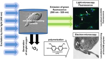
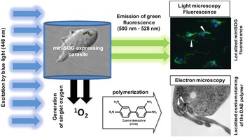
As in previous FbFPs, the removal of the conserved cysteine results in prohibition of the natural photocycle, but in miniSOG, replacement with a glycine causes redirection of the dissipated FMN energy (Shu et al. Reference Shu, Lev-Ram, Deerinck, Qi, Ramko, Davidson, Jin, Ellisman and Tsien2011). As a result, illuminating miniSOG with blue light results in an additional route of energy dissipation other than fluorescence emission, namely, the generation of singlet oxygen (Shu et al. Reference Shu, Lev-Ram, Deerinck, Qi, Ramko, Davidson, Jin, Ellisman and Tsien2011) (Fig. 2). Spin-orbital interactions permit excited electrons to change spin direction resulting in an intersystem conversion of the first-excited-singlet-state (S1) to the first-excited-triplet-state (T1) (i.e. S1→T1) (Climent et al. Reference Climent, Gonzalez-Luque, Merchan and Serrano-Andres2006). The energy transfer from the FMN in the T1 state at the core of miniSOG induces a type-2 photooxidation mechanism causing the localized conversion of molecular oxygen (3O2) into a reactive singlet oxygen (1O2) species (Shu et al. Reference Shu, Lev-Ram, Deerinck, Qi, Ramko, Davidson, Jin, Ellisman and Tsien2011). As reactive oxygen species, such as singlet oxygen, are damaging in protozoa (Fernandes and Assreuy, Reference Fernandes and Assreuy1997; Mariante et al. Reference Mariante, Guimaraes, Linden and Benchimol2003; Apel and Hirt, Reference Apel and Hirt2004), miniSOG disobeys the desired non-toxic nature of FPs. Even so, useful applications for this characteristic have been developed. Singlet oyxgen production by miniSOG has been exploited for ablation of cellular structures in a light-dosage dependent manner that result in regional damage use to explore the function of whole organelles or specific organelle compartments (Ruiz-Gonzalez et al. Reference Ruiz-Gonzalez, Cortajarena, Mejias, Agut, Nonell and Flors2013). The specificity of genetically encoded miniSOG tagging, the controllability of the reaction and contained nature of the singlet oxygen in the vicinity of production allows for highly targeted ablation. In the nematode Caenorhabditis elegans for instance, mitochondrial-targeted miniSOG was successfully used to investigate the role of various neurons in movement by selective miniSOG ablation (Qi et al. Reference Qi, Garren, Shu, Tsien and Jin2012). An alternative use of the miniSOG-induced photoconverted oxygen has been in the field of correlative microscopy (Shu et al. Reference Shu, Lev-Ram, Deerinck, Qi, Ramko, Davidson, Jin, Ellisman and Tsien2011; Asakawa et al. Reference Asakawa, Hiraoka and Haraguchi2014). Singlet oxygen causes the polymerization of diaminobenzidine (DAB) in vicinity of the target protein, which can be subsequently stained and visualized by TEM. Consequently, fluorescent miniSOG signals can be directly translated to the ultrastructural level enabling correlation of microscopy techniques.
Energy dissipation of miniSOG upon blue light illumination and its uses. Upon blue light illumination of organisms expressing the miniSOG-fusion protein, energy is released as green fluorescence which can be used for live-parasite localization of the protein of interest using fluorescent microscopy. For correlative TEM, cells are fixed and additional blue light illumination results in another route of energy dissipation which allows the conversion of molecular oxygen into the singlet oxygen. The addition of DAB causes the formation of DAB deposits which can be strained by osmium for TEM. Abbreviations: DAB, diaminobenzidine; TEM, transmission electron microscopy.
Other than oxygen-independence, miniSOG hosts several other advantages over variants of GFPs, YFPs and RFPs. Physically, miniSOG is a smaller protein (15 kDa) than common FPs (~28 kDa) and possess a less complex quaternary structure (Shu et al. Reference Shu, Lev-Ram, Deerinck, Qi, Ramko, Davidson, Jin, Ellisman and Tsien2011). miniSOG exists as a non-aggregating monomeric protein in solution and does not seem to interfere with protein movement across organelle membranes (Shu et al. Reference Shu, Lev-Ram, Deerinck, Qi, Ramko, Davidson, Jin, Ellisman and Tsien2011). As a photosensitizer, however, it has been revealed that the high singlet oxygen quantum yield (ɸ∆) claimed in the primary literature (ɸ∆ = 0·47 ± 0·05) (Shu et al. Reference Shu, Lev-Ram, Deerinck, Qi, Ramko, Davidson, Jin, Ellisman and Tsien2011), is approximately 15-fold lower than originally claimed (ɸ∆ = 0·03 ± 0·01), rendering miniSOG a poorer photosensitizer than previously thought (Pimenta et al. Reference Pimenta, Jensen, Breitenbach, Etzerodt and Ogilby2013; Ruiz-Gonzalez et al. Reference Ruiz-Gonzalez, Cortajarena, Mejias, Agut, Nonell and Flors2013). The reduced efficiency of photosensitization is a direct result of caging of the FMN molecule, which is a highly efficient photosensitizer in aqueous solutions (Pimenta et al. Reference Pimenta, Jensen, Breitenbach, Etzerodt and Ogilby2013). The protein environment significantly affects singlet oxygen quantum yield because it contains an abundance of amino acids that act as singlet oxygen quenchers (Ruiz-Gonzalez et al. Reference Ruiz-Gonzalez, Cortajarena, Mejias, Agut, Nonell and Flors2013). In particular, an irradiation-dependent increase in absorbance at ~350 nm is consistent with the oxidation of a tryptophan residue in the protein cage (Pimenta et al. Reference Pimenta, Jensen, Breitenbach, Etzerodt and Ogilby2013). Furthermore, in addition to type-2 photooxidation whereby energy is transferred solely to oxygen, miniSOG is capable of undergoing type-1 photooxidation (Pimenta et al. Reference Pimenta, Jensen, Breitenbach, Etzerodt and Ogilby2013; Ruiz-Gonzalez et al. Reference Ruiz-Gonzalez, Cortajarena, Mejias, Agut, Nonell and Flors2013). Therefore, in the presence of suitable acceptor substrates other than oxygen, the FMN core is capable of donating electrons directly to the electron acceptor resulting in reduced 1O2 production (Ruiz-Gonzalez et al. Reference Ruiz-Gonzalez, Cortajarena, Mejias, Agut, Nonell and Flors2013). The occurrence of a type-1 reaction with the substrate anthracene-9, 10-dipropionic acid as a fluorescent trap used originally by Shu et al. (Reference Shu, Lev-Ram, Deerinck, Qi, Ramko, Davidson, Jin, Ellisman and Tsien2011) is anticipated as the reason for the claimed higher singlet oxygen quantum yield (Pimenta et al. Reference Pimenta, Jensen, Breitenbach, Etzerodt and Ogilby2013).
Is miniSOG the future of protein tagging in anaerobic parasitology?
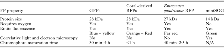
Although miniSOG has yet to be trailed in anaerobic parasites, miniSOG offers several advantages for protein localization in anaerobes over the conventional GFP derivatives and homologues Table 1. The most obvious benefit is the FMN chromophore of miniSOG, which does not undergo oxygen-dependent maturation, unlike the GFP chromophore which undergoes dynamic conformational maturation processes fluorescence emission (Chalfie et al. Reference Chalfie, Tu, Euskirchen, Ward and Prasher1994; Reid and Flynn, Reference Reid and Flynn1997; Shu et al. Reference Shu, Lev-Ram, Deerinck, Qi, Ramko, Davidson, Jin, Ellisman and Tsien2011). This provides opportunities to characterize dynamic molecular events in oxygen-sensitive parasites under true anaerobic conditions, reducing the likelihood of artefactual physiological changes as a result of exposure to oxygen. It is likely that miniSOG will fluoresce in anaerobic protozoa as the required FMN chromophore of miniSOG is the major flavin found in these parasites, being a critical component of oxygen detoxifying flavodiiron proteins (Lo and Reeves, Reference Lo and Reeves1979; Zhao et al. Reference Zhao, Cruz and Ferry2001; Smutna et al. Reference Smutna, Goncalves, Saraiva, Tachezy, Teixeira and Hrdy2009, Reference Smutna, Pilarova, Tarabek, Tachezy and Hrdy2014; Vicente et al. Reference Vicente, Vy, Pinto, Teixeira and Singh2012). Although, the fainter fluorescence emission of miniSOG compared with GFP is likely to restrict its usage as a protein tag. The incorporation of miniSOG instead of GFP into the SunTag system (Tanenbaum et al. Reference Tanenbaum, Gilbert, Qi, Weissman and Vale2014) may enhance signals from miniSOG, however this is yet to be tested. If executed, the miniSOG-SunTag combination will be smaller than the GFP-SunTag combination, due to the smaller size of the miniSOG tag. Being smaller in size than GFP-like FPs, miniSOG may be more successful for elucidating secretory pathways without interfering as space-occupying factor and hence not intrude on the cell's natural process (Shu et al. Reference Shu, Lev-Ram, Deerinck, Qi, Ramko, Davidson, Jin, Ellisman and Tsien2011; Pietra, Reference Pietra2014).
Comparison of commonly used genetically encodable GFP and GFP-like tags with the novel miniSOG
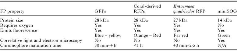
FP, fluorescent protein; GFP, green fluorescent protein; RFP, red fluorescent proteins; N/A, not applicable.
As it stands, miniSOG is theoretically an ideal protein tag for anaerobic protozoans due to its small protein size, independence of oxygen, protein stability and the use of a ubiquitous co-factor found within the anaerobic cellular milieu as a chromophore. In addition, the ability for miniSOG to function as a tag for electron microscopy increases its usefulness, allowing the direct translation of dynamic live-cell fluorescent signals into high-resolution ultrastructural localization. The use of FP tags, however, is complex and requires correct post-translational folding for signal production, as well as non-interference with the host protein to which is it fused. As miniSOG has yet to be used in anaerobic protozoans, it is difficult to conclude the true applicability and stability of this tag in these parasites. The restricted colour palette of miniSOG to green fluorescence is also restrictive because hydrogenosomes of anaerobic protozoans emit green autofluoresence as a result of reduced nicotinamide nucleotides and oxidized flavins (Lloyd et al. Reference Lloyd, Harris, Biagini, Hughes, Maroulis, Bernard, Wadley and Edwards2004; Millet et al. Reference Millet, Williams, Hayes, Hann, Cable and Lloyd2013). Furthermore, reactive oxygen species, such as the singlet oxygen generated by miniSOG is detrimental to anaerobic protozoa (Lloyd et al. Reference Lloyd, Harris, Maroulis, Biagini, Wadley, Turner and Edwards2000; Husain et al. Reference Husain, Sato, Jeelani, Soga and Nozaki2012), which could limit temporal-based studies. These studies should, instead, be conducted under absolute anaerobic environment to avoid oxygen which will be photooxidised upon blue-light illumination of miniSOG.
In conclusion, the common trait shared by anaerobic protozoans is their tropism for niche environments within their hosts that are deprived of oxygen (Lindmark and Muller, Reference Lindmark and Muller1973; Mack and Muller, Reference Mack and Muller1978a ; Cedeno and Krogstad, Reference Cedeno and Krogstad1983). Tagging proteins of interest with FPs is currently the best method of visualizing dynamic events occurring in live parasites. The adoption of FP tagging in anaerobic parasitology has been slow, even if well-established protocols for introducing the tags into the parasites have been developed for most medically significant protozoans (Purdy et al. Reference Purdy, Mann, Pho and Petri1994; Delgadillo et al. Reference Delgadillo, Liston, Niazi and Johnson1997; Liu et al. Reference Liu, Li, Zhang, Liu, Liu, Gong, Zhang, Yao and Zhang2008; Jerlstrom-Hultqvist et al. Reference Jerlstrom-Hultqvist, Stadelmann, Birkestedt, Hellman and Svard2012; Sateriale et al. Reference Sateriale, Roy and Huston2013; Vinayak et al. Reference Vinayak, Pawlowic, Sateriale, Brooks, Studstill, Bar-Peled, Cipriano and Striepen2015). Slow progress is attributed to the unreliability of current, widely-used GFPs and its homologues as live-parasite FP tags. Since these protozoans are anaerobic, the prolonged oxygen contact required for chromophore maturation is not only likely to alter parasite behaviour and physiology, but induce physical membrane and organelle damage which could lead to death (Lloyd et al. Reference Lloyd, Harris, Maroulis, Biagini, Wadley, Turner and Edwards2000; Gould et al. Reference Gould, Woehle, Kusdian, Landan, Tachezy, Zimorski and Martin2013; Raj et al. Reference Raj, Ghosh, Mukherjee, Nozaki and Ganguly2014). As a result, there is a lag in anaerobic protozoa research and an urgent update to the parasitologist toolbox is needed to advance the field.
Novel genetically encodable FPs, such as miniSOG, has potential as a live-protein tag for anaerobic parasitology because they emit fluorescence independent of oxygen (Shu et al. Reference Shu, Lev-Ram, Deerinck, Qi, Ramko, Davidson, Jin, Ellisman and Tsien2011). As a tag, miniSOG is smaller than the GFPs which facilitates transmembrane localization studies. In addition, the ability to use miniSOG as a correlative microscopy tag will augment understanding of how proteins localize and interact (Shu et al. Reference Shu, Lev-Ram, Deerinck, Qi, Ramko, Davidson, Jin, Ellisman and Tsien2011). Therefore, miniSOG has the potential to re-ignite primary research in these medically important parasites. Although met with the challenges of their unusual biology, the novel approaches for incorporation of FPs and novel types of FPs discussed in this review are promising for live-cell imaging of anaerobic protozoa to obtain dynamic understanding of these protozoans. Even so, traditional techniques should not be abandoned, but instead used to compliment and validate tagging results. The power of combining traditional and contemporary techniques such as genomics, transcriptomics, FP tagging, animal experiments and proteomics will be instrumental. With the increase in accessibility of sequenced genomes and transcriptomes, researchers should take advantage of these tools and datasets which can be integrated with tagging technology for a more focused approach to understand anaerobic protozoans.
FINANCIAL SUPPORT
V. M. A. is supported by the International Postgraduate Award (IPRS) and the Australian Postgraduate Award (APA) tenable at the University of Sydney.