Iron oxides belong to the most abundant mineral family and occur with a variety of stoichiometries, structures and properties. The most important are FeO (wustite), λ-Fe2O3 (maghaemite), α-Fe2O3 (haematite) and Fe3O4 (magnetite) with rock-salt, inverse-spinel, corundum and inverse-spinel structures, respectively. Of these iron oxides, FeO and λ-Fe2O3 are thermodynamically less favourable and α-Fe2O3 is the most oxidized. Red iron oxide, α-Fe2O3 (haematite), is one of the main constituents of Earth and other similar planets such as Mars. Depending on the particle size, haematite has been categorized as red (grain diameter less than 10 μm) and grey (grain diameter over 10 μm). Using Earth as a guide to haematite formation, researchers believe that haematite might have formed on Mars by thermal oxidation of iron-rich volcanic eruptive products during eruption or by chemical precipitation when iron-rich water circulated through the pre-existing layers of volcanic material.
The presence of adsorbed liquid water has been emphasized to have a remarkable influence on the physical transport and material properties of soil, such as electrical conductivity and permittivity, optical and spectral properties at the surface, internal friction, heat capacity and thermal conductivity (Mohlmann Reference Mohlmann2005). The recent discoveries through Mars exploration have proved the existence of abundant water in the form of seas and lakes on Mars. Also, the presence of key organic molecules in Murchisons meteorite has compelled us to think about the occurrence of a period of chemical evolution on ancient Mars. Here we present the results of studies on the interaction of ribose nucleotides with haematite prepared by aqueous hydrolysis of Fe3+ followed by calcinations at different temperatures. The calcination of haematite from a lower to a higher temperature could be used to monitor the water content in the lattice. The crystallinity of metal oxides has been found to increase with temperature. It is found that haematite with water in its lattice has a higher adsorption affinity towards ribose nucleotides compared with the haematite with no water in its lattice.
The powder, FeOOH, was prepared by drop-wise addition of aqueous ammonia into about 500 ml of 0.05 M solution of Fe (NO3)3 until the solution reached a pH of 10. Iron hydroxide thus precipitated was filtered and washed thoroughly with double-distilled water. The precipitates were dried and calcined at different temperatures (70–700°C) in a muffle furnace for about five hours. The metal oxide thus synthesized was ground and sieved through a 200 mesh sieve and then subjected to various measurements such as thermal gravimetric analysis/differential thermal analysis (TGA/DTA), surface area measurement, magnetic susceptibility, infrared, X-ray diffraction (XRD) and adsorption studies.
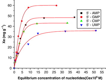
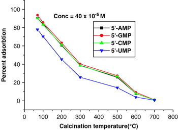
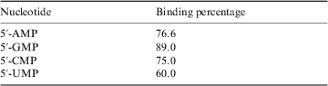
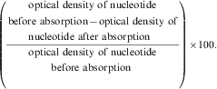
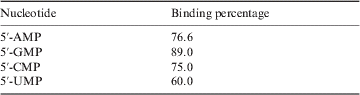
The preliminary studies on the interaction of ribose nucleotides such as 5′-AMP, 5′-GMP, 5′-CMP and 5′-UMP with iron oxide were carried out over a wide pH range (4.0–9.0) at room temperature (~25°C) and the maximum adsorption was found to occur at neutral pH (~7.0). Therefore, the subsequent adsorption studies were carried out at neutral pH only. A plot of the equilibrium concentration (Ce) of nucleotides versus the amount of adsorbed nucleotide (Xe) was sigmoidal in nature (Fig. 1). The initial portion of the isotherms represents a linear relationship between the amount adsorbed and the equilibrium concentration of the ribonucleotides. At a higher concentration range, the isotherms showed saturation phenomena indicating no further adsorption. The binding percentage was calculated from the data of the curve at saturation and is shown in Fig. 2. It is evident that the binding percentage of all four ribose nucleotides decreases drastically with increasing crystallinity of the haematite. The very high binding percentage of the ribose nucleotides with haematite precursor obtained at 70°C is also shown separately in Table 1. The binding percentages in Table 1 were calculated as
Adsorption isotherms of nucleotides on FeOOH (the precursor of iron oxide). Equilibrium time ~24 h, temperature ~25°C, amount of adsorbent ~25 mg.

Binding percentage of nucleotides on iron oxide with increasing calcination temperature.

Binding percentage of nucleotides on the precursor of haematite, FeOOH, obtained through aqueous hydrolysis of Fe3+ by aqueous ammonia and dried at 70°C
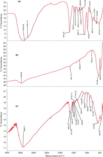
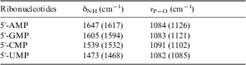
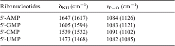
The nature of interaction between ribonucleotides and iron oxide has also been studied by infrared spectral studies. The infrared spectra of 5′-AMP, iron oxide and iron oxide–nucleotide adduct are depicted in Figs 3(a), (b) and (c), respectively. A shift in wavelengths of the characteristic frequencies of ribonucleotides in the nucleotide–iron oxide adduct indicates an interaction between the ribose nucleotides and iron oxide (Table 2). It seems that the binding sites of nucleotides are N-1 and N-7 for 5′-AMP, N-1 and N-7 for 5′-GMP, N-3 for 5′-CMP and N-3 for 5′-UMP. The adsorption is presumably related to the involvement of N-1, N-3 and N-7 sites of the base residues, the lone pair on the nitrogen atom as well as to the dissociation of two available protons of the phosphate group. A shift towards a lower wavelength of N—H bending from 1647 to 1617 cm−1 shows the involvement of the −NH2 group in the interaction of ribonucleotide with iron oxide. A strong band at 1084 cm−1 in 5′-AMP corresponding to the C—O—P vibration has been shifted to 1126 cm−1 indicating an interaction between the amino and phosphate group of ribonucleotides with iron oxide. The fact that there was no significant change in the typical infrared frequencies of metal oxide suggests that the ribonucleotide molecules do not affect the lattice of the metal oxide. Similar observations were also found in our studies on the interaction of ribose nucleotides with ZnO (Arora & Kamaluddin Reference Arora and Kamaluddin2006).
Infrared spectra of (a) AMP, (b) FeOOH and (c) AMP–FeOOH adduct.

Typical infrared spectral frequencies of ribonucleotides before (and after) adsorption on the precursor of haematite, FeOOH, obtained through aqueous hydrolysis of Fe3+ by aqueous ammonia and dried at 70°C
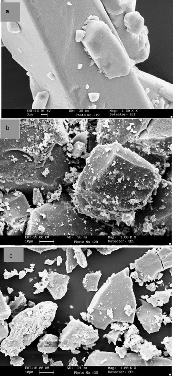
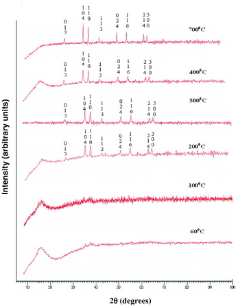
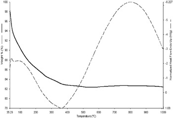
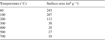
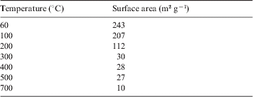
Scanning electron microscopy (SEM) studies of the AMP, iron oxide and AMP–iron oxide adduct were also performed and are shown in Fig. 4. It can be seen from the SEM images that after adsorption the surface of iron oxide becomes smooth and there was a noticeable change in colour of the surface to that of AMP. CHN analysis of the AMP–iron adduct showed 0.742% of N, 0.159% H and 1.672% of C on the surface of iron oxide. This confirms the adsorption of ribose nucleotide on iron oxide as the C:N ratio found above matches that in 5′-AMP; the calculated percentages of C, H and N on the iron oxide adduct were found to be 0.799% N, 0.1714% of H and 1.7136% of C, which are very close to the values obtained from the CHN analysis. The surface area of iron oxide after calcinations at different temperatures was determined by the Brunauer–Emmett–Teller (BET) method and was found to decrease with increased calcination temperature (Table 3). Powder XRD patterns show (Fig. 5) that the relative intensities and interplanar spacing (d) values are in good agreement with α-Fe2O3 (JCPDS 1971). The XRD data reveals that calcinations at higher temperature increased the crystallinity of iron oxide as the diffraction peaks became sharper. The TGA/DTA study of the sample shows an endothermic peak at 364°C (see Fig. 6). This simply indicates that when FeOOH is heated, it absorbs energy and water molecules are removed. So, for the formation of iron oxide, temperatures above 364°C are required. The iron oxides prepared with higher calcination temperatures showed less adsorption of ribose nucleotides.
SEM photographs of (a) AMP, (b) FeOOH and (c) AMP–FeOOH adduct.

XRD pattern of the precursor (FeOOH) heat treated at different temperatures in air.

TGA/DTA curve for the precursor to iron oxide.

Surface area of precursor (FeOOH) heated at various temperatures (the heat treatment leads to the formation of Fe2O3 at temperatures above 364°C)
The present observations reveal the importance of water, the presence of which aided the adsorption of ribose nucleotides on iron oxide. This also supports the theory proposed by Bibring et al. (Reference Bibring2006) about the role of water in helping the processes of chemical evolution in the Phyllosean era on Mars. The present observation is also in accordance with the proposed hypothesis by Mohlmann (Reference Mohlmann2005) on the enhanced physical properties of water-adsorbed Martian soil.
Acknowledgement
The project was funded by ISRO under the RESPOND Programme.