Introduction
Various inborn leukoencephalopathies have been identified, including disorders of lysosomes, mitochondria, peroxisomes, amino and organic acids, and myelin protein defects.Reference Henneke, Preuss, Engelbrecht, Aksu, Bertini and Bibat1 However, these disorders, including cystic leukoencephalopathy or anterior temporal cysts, have not been detected with any underlying genetic or biochemical defects.
Cystic leukoencephalopathy without megalencephaly presents clinically with early-onset severe psychomotor impairment, non-progressive encephalopathy, and either a normal head circumference or microcephaly.Reference Soni, Biswas and Kumar2 It was previously thought to result from congenital cytomegalovirus infection. Generally, surgeons are reluctant to attempt cochlear implantation in children with neuroradiological abnormalities and psychomotor retardation because they might have a delay in speech perception and aural rehabilitation post-implantation.
Case report
A four-year-old female child born out of consanguineous marriage (third degree) at 36 weeks of gestation presented to the out-patient clinic with a history of inability to achieve age-appropriate speech and language milestones. She was unable to respond to sounds or pronounce any words. There was no history of trauma, prolonged hospitalisation, fever or seizures. There was no family history of neurological disorders. She weighed 1.56 kg at birth, with length and head circumference between the 50th and 75th centile. She had standard otoacoustic emissions results during the neonatal screening test. She was kept in the neonatal intensive care unit because of low birth weight and was discharged after one week. She had no signs of hypotonicity or movement disorder or any red flag signs of developmental delay until one year of age.
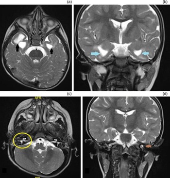
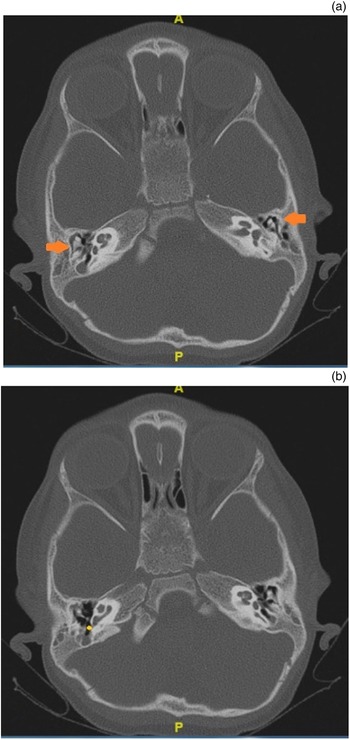
The child had achieved age-appropriate gross motor, fine motor and social developmental milestones, and her development quotient was 80. Her head circumference was measured at 45 cm. Ophthalmology and cardiology consultations showed no abnormalities. Brainstem evoked response audiometry detected absent wave V at 90 dB HL (i.e. bilateral severe sensorineural hearing loss). She was evaluated for cochlear implantation, and radiological investigations were performed as part of the routine investigation for cochlear implantation. Magnetic resonance imaging (MRI) of the brain showed bilateral anterior temporal lobe cysts with multiple asymmetric T2-weighted-fluid-attenuated inversion recovery white matter hyperintensities (Figure 1a and b) and normal-appearing internal ear structures on both sides (Figure 1c and d). The signal intensity of cyst content was similar to cerebrospinal fluid. High-resolution computed tomography of the temporal bone showed the same findings with normal middle-ear structures (Figure 2a and b). However, the child did not have the symptoms of psychomotor retardation or intellectual impairment shown by a non-progressive variant of cystic leukoencephalopathy without megalencephaly.
Magnetic resonance imagining of the brain showing: (a) bilateral anterior temporal cysts (black arrows) in axial view; (b) bilateral occipital cysts (blue arrows) in coronal view; (c) right normal cochlea and lateral semicircular canal (yellow circle) in axial view and (d) left normal cochlea in coronal view (brown arrow).

High-resolution computed tomography of the temporal bone showing: (a) bilateral normal middle-ear structures (i.e. malleus and incus complex showing ice cream (malleus head) and cone (body of incus) appearance (orange arrow) and (b) right round window niche (yellow dot) in axial view. A = anterior; P = posterior.

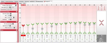
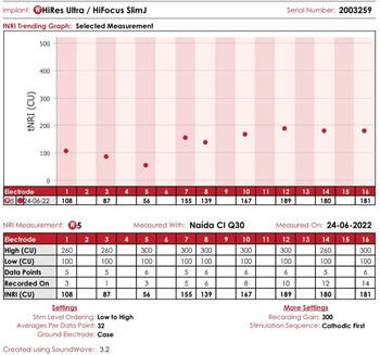
Haematological and biochemical studies were normal, including liver function tests and metabolic screening for the very-long-chain fatty acids pyruvate and lactate. The possibility of congenital infections like cytomegalovirus and toxoplasma was ruled out using viral screening tests. The child underwent right ear cochlear implantation. Intra-operative neural response telemetry was found to be normal (Figure 3). The post-operative stay in the hospital was uneventful. The device was switched on, and mapping was performed on the 21st post-operative day. A threshold level of 12–17 charge units and a comfortable level of 123–173 charge units was found to be optimal for auditory stimulation. After six months of implantation, neural response telemetry showed comfortable and threshold levels for evoked compound action potential, with evoked compound action potential being within the normal range for all electrodes (Figure 4). On follow up, the child showed signs of speech recognition and started uttering monosyllable words, such as ‘amma’ and ‘appa’.
Intra-operative neural response telemetry showing responses (evoked compound action potential) in all electrodes with T-level of 12–17 CU and M-level of 123–173 CU. APW = automatic pulse width; IDR = input dynamic range; AGC = automatic gain control; RF = radio frequency; M = most comfortable level; T = threshold level; tNRI = threshold neural response imaging; Imped = impedance; Ctr = centre.

Neural response telemetry at six months post implantation showing threshold neural response imaging and evoked compound action potential within normal limits. NRI = neural response imaging; CU = charge units.

Discussion
In children, white matter abnormalities can be found in various medical conditions, such as Aicardi–Goutières syndrome, megalencephalic leukoencephalopathy (Van der Knaap disease), vanishing white matter syndrome and congenital cytomegalovirus infection. Clinical findings in the present case were suggestive of cystic leukoencephalopathy without megalencephaly (first described by Olivier et al. in 1998).Reference Olivier, Lenard, Aksu and Gärtner3 This study identified three patients of Turkish origin who had similar MRI and clinical findings comprising delayed motor development and impaired intellectual function. There was an autosomal recessive mode of inheritance identical to the child in the present study, who was born out of a consanguineous marriage. This disease is characterised by non-progressive, severe psychomotor impairment, including tone and reflex abnormalities, normocephaly or microcephaly, and speech delay. Most patients can walk with aid, but some can barely sit or crawl.Reference Henneke, Preuss, Engelbrecht, Aksu, Bertini and Bibat1,Reference Soni, Biswas and Kumar2 Genetic testing in these children has shown a homozygous or compound heterozygous loss of function of RNAase T2 gene on chromosome 6q27, which interferes with myelination and brain development.Reference Henneke, Diekmann, Ohlenbusch, Kaiser, Engelbrecht and Kohlschütter4 Van der Knaap et al. described a similar disease in eight children, with a mild clinical course, but all patients had megalencephaly, unlike the child in the present study.Reference van der Knaap, Barth, Stroink, van Nieuwenhuizen, Arts and Hoogenraad5
Henneke et al.Reference Henneke, Preuss, Engelbrecht, Aksu, Bertini and Bibat1 analysed clinical history, biochemical and genetic analysis, and radiological imaging of 15 children with the same entity and found that the disease had a non-progressive course with autosomal recessive inheritance with characteristic MRI findings of bilateral anterior temporal lobe cysts with white matter abnormalities and pericystic abnormal myelination.
In a study on congenital cytomegalovirus infection by Barkovich and Lindan, MRI findings were identical to the present case except for microcephaly.Reference Barkovich and Lindan6 This study showed that congenital cytomegalovirus infection should have cortical dysplasia or diffuse lissencephaly (migration delay) and cerebellar hypoplasia. Congenital cytomegalovirus infection also shows a similar diagnostic pattern of neuroradiological features, but hearing loss occurs during the second year of life and not usually at birth.Reference Faria, Arita, Peruchi, Lin, Masruha and Vilanova7 All these cases describe children with white matter abnormalities and significant psychomotor retardation. In contrast, the child in the present study did not have any motor disability or intellectual impairment despite having almost similar neuroradiological abnormalities.
There have been studies regarding the outcome of cochlear implantation in children with congenital cytomegalovirus infection with multiple white matter abnormalities; there was no significant difference in the linguistic outcome.Reference Lee, Lustig, Sampson, Chinnici and Niparko8
Radiology, especially MRI, plays a significant role in diagnosing cystic leukoencephalopathy. Magnetic resonance imaging findings include extensive subcortical anterior temporal lobe cysts, white matter abnormalities, inferior horn dilatation and pericystic abnormal myelination.Reference Henneke, Preuss, Engelbrecht, Aksu, Bertini and Bibat1,Reference Olivier, Lenard, Aksu and Gärtner3 Magnetic resonance imaging spectroscopy in these patients shows white matter with increased myo-inositol and decreased N-acetylaspartate.Reference Bodensteiner, Kerrigan and Johnsen9 Bilateral temporal lobe cysts can also occur following viral infections like rubella (congenital rubella syndrome), cytomegalovirus and measles (subacute sclerosing pan encephalitis).Reference Zaatreh, Bates, Hooper, Palmer, Elmenshawi and Courvoisie10–Reference Stadlan and Sung12 These viral infection manifestations are related to the time of disease and are more severe when acquired during early pregnancy. Migration abnormalities are more likely to occur in infections during early pregnancy. Temporal lobe involvement made us sceptical regarding the outcomes of cochlear implantation, but the post-implantation auditory responses by the child were satisfactory.
Conclusion
This case report described a child with cystic leukoencephalopathy without megalencephaly and bilateral sensorineural hearing loss. This is the first such child in reported literature who underwent successful cochlear implantation.
• Cystic leukoencephalopathy without megalencephaly is otherwise called leukoencephalopathy with bilateral anterior temporal cysts
• It has an autosomal recessive type of inheritance
• Genetic analysis has shown it occurs because of mutation of the RNAase 2 gene involved in myelination and brain development
• Hearing loss associated with this entity is very rare but cochlear implantation can be considered provided the child has normal psychomotor development
• Radiology plays a major role in diagnosing this neurological disorder
Cochlear implantation can be performed with a guarded prognosis in children with white matter abnormalities or temporal lobe involvement as long as the co-existing developmental delays do not hinder speech perception.
Competing interests
None declared