I. INTRODUCTION
Over the past years, semiconductor photocatalysis has received intense attention as a potential solution to the worldwide energy shortage and environmental purification. Reference Tong, Ouyang, Bi, Umezawa, Oshikiri and Ye1–Reference Lü, Mao, Wei, Zhang, Xie and Wei5 Among the various semiconductor photocatalytic materials, TiO2 is considered as one of the best photocatalysts because of its high activity, long-term stability, low price, and availability, but it can absorb only the UV portion of the solar spectrum owing to its wide band gap of 3.2 eV. Reference Asahi, Morikawa, Ohwaki, Aoki and Taga6–Reference Yu, Wei, Li, Chen, Fan and Yu10 Therefore, it is an urgent issue to search for efficient sunlight or visible-light-driven photocatalysts with appropriate band gap, strong oxidative/reductive ability, and high stability in water solution system.
Recently, polymer-like semiconductor material graphitic C3N4 (g-C3N4), which can be used as a metal-free photocatalyst for its nontoxicity, abundance, and medium band gap (2.7 eV), has attracted much attention in the field of photocatalysis, such as solar energy conversion, hydrogen production, and environment purification. Reference Wang, Maeda, Thomas, Takanabe, Xin and Carlsson11–Reference Chen, Shen, Guo, Wang, Su, Zhao and Guo17 Nevertheless, there are many drawbacks of the g-C3N4 material in the field of photocatalysis, such as high recombination rate of photogenerated electron–hole pairs, low specific surface area, and low visible-light utilization efficiency. To solve these problems, many methods have been proposed, especially in the field of improving the photocatalytic activity of g-C3N4 under visible light irradiation. One of the most promising methods is to design metal nanoparticles combining with semiconductor composites to facilitate charge separation, among which metals with plasmonic property are attractive. Reference Jin and Shiraishi18,Reference Yu, Li, Kumar, Kawasaki and Jin19
Silver, due to its specific optical, electrical, magnetic, catalytic, antibacterial features, and particularly cheap compared with platinum or gold, was often combined with semiconductor as an efficient photocatalytic material. Ag-CdMoO4, Reference Adhikari, Malla, Gyawali, Sekino and Lee20 Ag@TiO2, Reference Guan, Wang, Guo, Shao, Zhang and Liu21 and Ag/AgBr Reference Dai, Lu, Dong, Ji, Zhu and Liu22 have been demonstrated to be efficient photocatalysts under visible light irradiation. The possible reason is that the noble metal Ag particles can strongly absorb visible light owing to their localized surface plasmon resonance (SPR) and the fast generation of the photogenerated carriers on semiconductor and then rapid separation and transportation of these photogenerated carriers at the interface of substrate and Ag. Reference El-Sayed23–Reference Yu, Cao, Shu, Bao, Xie, Yu and Yang30 When the frequency of the photon is consistent with the plasma oscillation frequency of metal, it will produce resonance and strong absorption of incident light, which is called local SPR phenomenon. Yang and Ge et al. deposited Ag onto the g-C3N4 surface and improved the photocatalytic activity through photodeposition and precipitation–calcination process, respectively. Reference Yang, Guo, Liu, Yuan, Guo and Zhang31,Reference Ge, Han, Liu and Li32 However, the noble metal Ag particles with larger particle size distribution upon the substrate are not uniform, and tend to aggregate on some area owing to the instability in the process of irradiation or heat treatment.
Herein, we prepared AgNP decorated g-C3N4 (Ag/g-C3N4) sheets through a simple wet-chemical pathway, where DMF is used as an efficient reducing agent. The reaction was conducted under mild condition with easy control of homogeneous distribution and particle growth of AgNPs. To test the influence of AgNPs, different amounts of silver precursor were added to prepare various composites. The visible light photocatalytic tests show that the prepared Ag/g-C3N4 composite possesses enhanced photocatalytic activity for the decolorization of methyl orange (MO), much higher than pure g-C3N4. The possible photodecolorization mechanism was also discussed based on the experimental results and PL spectra.
II. EXPERIMENTAL
A. Materials
All the reagents used were of AR grade and used as received without further purification. Melamine, polyvinylpyrrolidone (K-30), and ethanol were purchased from Aladdin Ltd. (Shanghai, China). Silver nitrate, methyl orange, and N,N-dimethylformamide (DMF) were purchased from Merck, India. Double distilled water was used throughout the course of the investigation.
B. Preparation of g-C3N4
Melamine (10 g) was put into an alumina crucible with a cover under ambient pressure in air, and then the crucible was heated in a tube furnace to 500 °C for 2 h, and a final temperature 520 °C for 2 h at a heating rate of 3 °C min−1. The yielded yellow powder was washed with nitric acid (0.1 mol L−1) and distilled water to remove any residual alkaline species (e.g., ammonia) adsorbed on the surface of the product, and then the product was dried at 80 °C for 12 h.
C. Preparation of Ag/g-C3N4
First, dried g-C3N4 powder (0.27 g) was sonicated in 30 mL of DMF for 1 h in a beaker. In another beaker, 0.25 mmol of AgNO3 and 0.05 g of PVP were stirred in 10 mL of DMF for 30 min. Then two mixtures were taken in a screw-capped test tube and heated on a water bath for 16 h. This treatment resulted in the formation of the product which was centrifuged repeatedly at 4500 rpm with distilled water to remove the adsorbed Ag+ ions and the unbound AgNPs. Finally, the product (Ag(10)/g-C3N4) was washed with ethanol and dried at 50 °C under vacuum for 12 h and used for further characterization and photocatalytic study. The products are denoted as Ag(x)/g-C3N4, where x represents Ag deposited level (wt%) in the products. The other Ag deposited g-C3N4 materials with various Ag loadings (0.5, 1, 2, and 5 wt%) were obtained through the same process by adjusting the amount of g-C3N4 and AgNO3.
D. Characterization
The crystal structure and phase purity of the Ag/g-C3N4 were analyzed by x-ray diffraction (XRD) using D8 Advance XRD (Bruker AXS company, Germany) equipped with Cu Kα radiation (λ = 1.5406 Å), using a scanning rate of 7° min−1 in the 2θ range from 15 to 80°. The samples for XRD analysis were obtained with dried samples. Field emission scanning electron microscopy (FESEM) and energy-dispersive x-ray spectroscopy (EDS) were performed on a scanning electron microscope (Hitachi S-4800, Japan) operated at an acceleration voltage of 10 kV to characterize the morphologies and the compositions of the prepared Ag/g-C3N4. Furthermore, the morphology and size of Ag nanoparticles and g-C3N4 were also examined by transmission electron microscopy (TEM), which was recorded on a JEOL-JEM-2010 (JEOL Ltd., Japan) operating at 200 kV. X-ray photoelectron spectroscopy (XPS) was performed on a VG-ADES400 instrument (VG Ltd., UK) with Mg K-ADES source. UV-vis diffuse reflectance spectra (UV-vis/DRS) were recorded on a Shimadzu UV2450 spectrophotometer (Shimadzu Ltd., Japan). PL of the samples was obtained using a Varian Cary Eclipse spectrometer (Varian Ltd., Palo Alto, CA). The Brunauer-Emmett-Teller (BET) specific surface area was obtained from N2 adsorption/desorption analyses at 77 K (NOVA 2000).
E. Photocatalytic tests
As a stable dye, MO was selected and dissolved into water as the simulated dye wastewater, and its photodecolorization has been widely used as a representative reaction for examining the performance of many visible light active photocatalysts. Reference Yang, Jiang, Shi, Xiao and Yan33,Reference Lin, Huang, Huang, Wang and Shi34 Huo et al. reported a photocatalytic membrane reactor combining a visible-light-driven BiOBr photocatalyst and direct contact membrane distillation for the removal of methyl orange from an aqueous solution. Reference Huo, Xie, Wang, Li, Hoang and Caruso35 Huang et al. have prepared Pt modified TiO2 loaded on natural zeolites by sol–gel technique and photoreductive deposition method. Pt doping is helpful to the enhancement of photocatalytic decolorization and this could be attributed to the Pt particles action as electron traps to inhibit the recombination of photogenerated electron and hole pair. Reference Huang, Xu, Wu, Huang, Lin and Wu36
The photocatalytic activities of Ag/g-C3N4 heterojunction were also evaluated by the decolorization of MO in aqueous solution. All experiments were performed in a self-made quartz photoreactor fitted with a circulation water system to maintain a constant temperature and under visible light irradiation of a 300 W xenon lamp (PLS-SXE300, Trusttech Co. Ltd., Beijing) with a 400 nm cutoff filter to remove the UV irradiation. In each experiment, 50 mg of catalyst was suspended in an aqueous solution (100 mL) of MO in a quartz glass reactor, which was cooled by recycled water to prevent the thermal catalytic effect. The initial concentration of MO solutions was 10 mg L−1. Prior to irradiation, the suspension was stirred in the dark for 0.5 h to ensure the establishment of adsorption–desorption equilibrium. At given irradiation time intervals, 3 mL of the suspension was collected and subsequently centrifuged to remove the catalyst particles. The concentration was analyzed by measuring the maximum absorbance at 464 nm for MO using a Shimadzu UV-2450 spectrophotometer. The decolorization rate of methyl orange was calculated by the following equation:
 $$\eta = {{{C_0} - C} \over {{C_0}}} = {{{A_0} - A} \over {{A_0}}} \times 100\% \quad ,$$
where C 0 and A 0 are the initial concentration and absorbance of methyl orange solution at 464 nm corresponding to maximum absorption wave length; C and A are the concentration and absorbance of methyl orange solution at 464 nm after UV light irradiation at any time. Reference Yang, Guo, Liu, Yuan, Guo and Zhang31
III. RESULTS AND DISCUSSION
A simple wet-chemical approach has been used for the fabrication of silver nanoparticle decorated g-C3N4 nanosheets in DMF medium where DMF plays the dual role of solvent and reducing agent. Reference Dutta, Ray, Sarkar, Pradhan, Negishi and Pal37 The proposed chemical reaction is as follows:
 $${\rm{HCONM}}{{\rm{e}}_2} + 2{\rm{A}}{{\rm{g}}^ + } + {{\rm{H}}_2}{\rm{O}} \to 2{\rm{A}}{{\rm{g}}^0} + {\rm{M}}{{\rm{e}}_2}{\rm{NCOOH}} + 2{{\rm{H}}^ + }\quad {\rm{.}}$$
A keypoint of this reaction is that it proceeds at a meaningful rate even at room temperature. A yellow color was readily observed at room temperature, which deepened as time or temperature increasing. What’s more, the reduction ability in mild conditions points toward a larger tendency of DMF for the reduction of Ag+ than that shown by ethanol or other organic solvents. Herein, the optimal temperature adopted for AgNO3 reduction by DMF is approximately 85 °C in a water bath where PVP acting as a growth controlling agent. It is also important to note that the reaction is performed without degassing the solutions or taking any special care with regard to the presence of oxygen.
A. Structural and physicochemical characterization
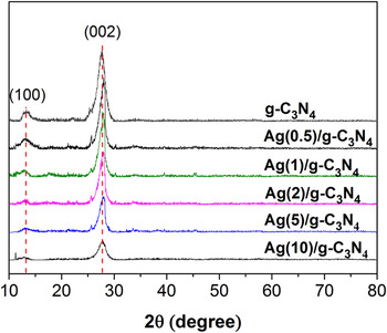
XRD was used to characterize the crystal structure of the as-prepared Ag/g-C3N4 composites, as well as pure C3N4 (Fig. 1). Two distinct peaks of 13.0° and 27.4° in the pure g-C3N4 sample can be indexed as the (100) and (002) diffractions for graphitic materials, corresponding to the in-plane structural packing motif and interlayer stacking of aromatic segments, respectively. Reference Meng, Shen, Chen and Xin38,Reference Cheng, Tian, Liu, Ge, Qusti Abdullah and Asiri Abdullah39 No significant diffraction peaks of any other phases or impurities can be detected in the composite samples, which indicate that the introduction of Ag species does not affect the crystal structure of the g-C3N4 photocatalyst. However, the diffraction intensities of the peak at 27.4° become weaker with increasing Ag contents, which indicate that the introduction of Ag species restrains the growth of crystal structure of g-C3N4. No diffraction peaks of Ag species are detected, which may be explained by the small size and small amounts of Ag species introduced (maximal 10 wt%) in the composite samples.
XRD patterns of pure g-C3N4 and Ag/g-C3N4 composite photocatalysts with different Ag contents.

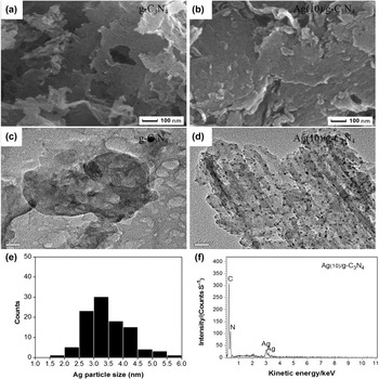
The morphology of the as-synthesized sample Ag(10.0)/g-C3N4 was examined by FESEM. Figures 2(a) and 2(b) show typical FESEM image of the sample. It can be clearly seen that the substrate of g-C3N4 nanocrystals exhibits nanoplate-like morphology with size of hundreds of nanometers and the surface of the composites is rougher than pure g-C3N4. Figure 2(c) presents typical TEM images of g-C3N4 substrate. As shown in Fig. 2(d), a large amount of nanoparticles in spherical shape are observed uniformly on the surface of the g-C3N4. Adding PVP affects the molecular motion of reduced silver and subsequently limits the aggregation of colloids. In addition, it is important to note that the diameter of nanoparticles is in the range of 1.5–6.0 nm with an appropriate distribution width by histogram analysis [Fig. 2(e)]. Figure 2(f) shows the energy-dispersive x-ray spectroscopy (EDS) of the composite, which confirms that the sample contains only the elements of C, N, and Ag.
SEM, TEM images and EDS spectrum of the as-prepared samples. (a) SEM micrograph of pure g-C3N4; (b) SEM micrograph of Ag(10.0)/g-C3N4; (c) TEM micrograph of pure g-C3N4; (d) TEM micrograph of Ag(10.0)/g-C3N4; (e) size distribution of the Ag nanoparticles obtained by counting 100 particles in the TEM image; (f) EDS spectrum of Ag(10.0)/g-C3N4.

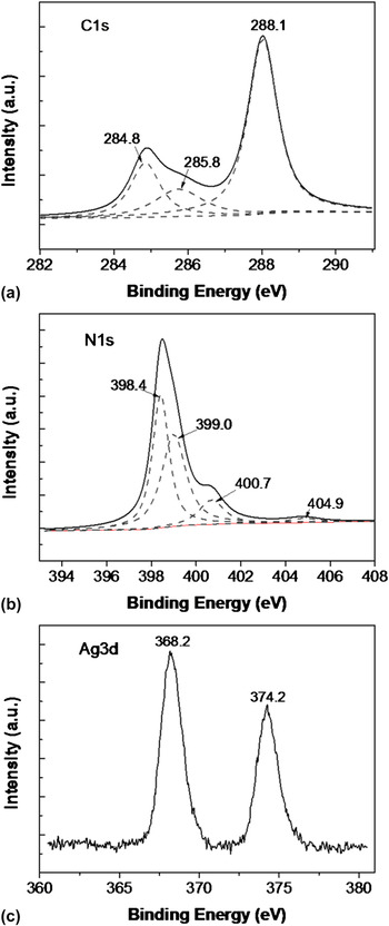
The x-ray photoelectron spectroscopy (XPS) was carried out to determine the chemical composition of the Ag(5.0)/g-C3N4 sample and the valence states of various species present therein. Figure 3(a) shows the high-resolution XPS spectra of C 1s. Two peaks can be distinguished to be centered at 284.8, 285.8, and 288.1 eV. The major peak at 284.8 eV is exclusively assigned to carbon atoms (C–C bonding) in a carbon-containing contamination. Reference Dementjev, de Graaf, van de Sanden, Maslakov, Naumkin and Serov40 The peak at 285.8 and 288.1 eV is identified as originating from sp3-hybrid C atoms (C–(N)3) and sp2-hybrid carbon in the aromatic ring (N–C=N), respectively. Reference Cui, Zhang, Zhang, Huang, Liu and Antonietti41–Reference Ge and Han43 The N 1s spectrum in Fig. 3(b) was deconvoluted into four peaks. The peak centered at 398.4 eV is attributable to sp2 N involved in triazine rings, whereas the contribution at 399.0 eV corresponds to bridged nitrogen atoms N–(C)3. The weak peak at the position of 400.7 eV may be attributed to the –NH2 or =NH groups. Reference Guo, Xie, Wang, Lv, Hou and Liu44 Charging effects or positive charge localization in heterocycles can lead to the peak at 404.9 eV. Reference Cui, Zhang, Zhang, Huang, Liu and Antonietti41 Figure 3(c) shows the characteristic Ag 3d peak that has a 6.0 eV splitting of the 3d doublet, which is corresponding to the metallic Ag0 species. This result confirms the presence of metallic silver doped in the g-C3N4 photocatalysts.
High-resolution XPS of Ag(5.0)/g-C3N4 in the (a) C 1s, (b) N 1s, and (c) Ag 3d binding energy regions.

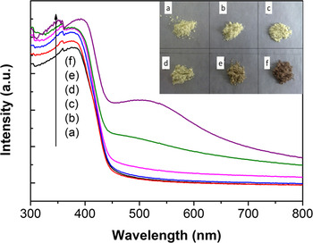
Figure 4 shows the UV-vis DRS of bulk g-C3N4 and Ag/g-C3N4 nanocomposites with various Ag loading. Compared with pure g-C3N4, the Ag/g-C3N4 composites show an additional absorption peak around 550 nm characteristic of the colloidal Ag SPR band, further confirming the formation of AgNPs. Besides, the composites also show increased absorption in ultraviolet and visible region with the increasing Ag content, which can be attributed to the light absorption of AgNPs. Meanwhile, the enhanced light absorption of the samples is in accordance with the color changing from yellow to vandyke red, which is shown in the inset.
UV-vis DRS of the as-prepared samples: (a) pure g-C3N4; (b) Ag(0.5)/g-C3N4; (c) Ag(1.0)/g-C3N4; (d) Ag(2.0)/g-C3N4; (e) Ag(5.0)/g-C3N4; (f) Ag(10.0)/g-C3N4. The inset shows the color of the six sample powders.

B. Photocatalytic test
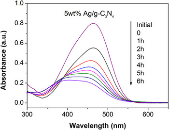
The photocatalytic activities of as-prepared samples were evaluated by the decolorization of organic dye under visible light irradiation. Methyl orange, as one of the most widely used dyes with stable azoic structure and weak adsorption on the photocatalyst surface, was chosen as a representative hazardous dye to evaluate the photocatalyst performance, which showed a major absorption band at 464 nm. The decolorization process of methyl orange was recorded by the temporal evolution of the spectrum and all of the samples processed in the similar procedure. Figure 5 illustrated the variations in the absorbance of methyl orange around 464 nm over Ag(5.0)/g-C3N4. The absorbance of methyl orange obviously decreases with increase of irradiation time. Meanwhile, the position of the peak appears obvious blue shift, which indicates the occurrence of methyl orange decomposition.
The variations in the absorbance of methyl orange around 464 nm over Ag(5.0)/g-C3N4.

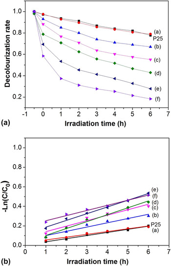
Figure 6(a) showed the faded effect of P25 and the Ag/g-C3N4 composite samples with different Ag contents under visible light irradiation (λ > 400 nm). It demonstrated that the amount of Ag had a great influence on the decolorization effect. As shown in Fig. 6(a), the fade rate was 0.222, 0.210, 0.329, 0.449, 0.571, 0.722, and 0.817, respectively, corresponding to P25, Ag content of 0, 0.5, 1.0, 2.0, 5.0, and 10.0 wt%. The result indicated that the whole decolorization ability of Ag/g-C3N4 enhanced as increasing the Ag loading from 0 to 10 wt%. The Ag/g-C3N4 composite shows more superior decolorization ability than P25.
(a) Decolorization rate of methyl orange under visible light irradiation in the presence of catalysts: (a) pure g-C3N4; (b) Ag(0.5)/g-C3N4; (c) Ag(1.0)/g-C3N4; (d) Ag(2.0)/g-C3N4; (e) Ag(5.0)/g-C3N4; (f) Ag(10.0)/g-C3N4; and P25. (b) MO degradation curves of ln(C 0/C) versus time for different catalysts.

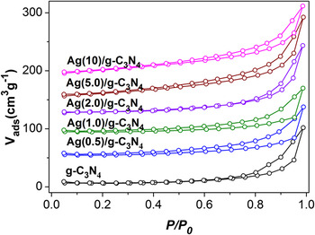
It is important to recognize that the decolorization process includes two parts, i.e., adsorption and degradation, and the photocatalytic activity depends only on degradation process. From N2 adsorption/desorption analyses shown in Fig. 7, we obtain the BET specific surface areas of g-C3N4, Ag(0.5)/g-C3N4, Ag(1.0)/g-C3N4, Ag(2.0)/g-C3N4, Ag(5.0)/g-C3N4, and Ag(10)/g-C3N4, which are 18.5, 15.5, 14.3, 31.6, 46.6, and 51.1 m2g−1, respectively. The surface area of Ag(10)/g-C3N4 is a bit larger than that of Ag(5.0)/g-C3N4 and larger surface area corresponds to stronger adsorption, hence, the degradation effect on MO using Ag(10)/g-C3N4 is nearly the same with Ag(5.0)/g-C3N4 regardless of the impact of adsorption. To further understand the reaction kinetics of MO degradation process, the apparent pseudo-first-order model expressed by Eq. (3) was applied in the experiments:
 $$- \ln \left( {{C \mathord{\left/ {\vphantom {C {{C_0}}}} \right. \kern-\nulldelimiterspace} {{C_0}}}} \right) = {k_{{\rm{app}}}}t\quad {\rm{,}}$$
where k app is the apparent pseudo-first-order rate constant (h−1). Via the first-order linear fit from the data of Fig. 6(b), the calculated k app of P25 and Ag/g-C3N4 samples was 0.031, 0.029, 0.043, 0.056, 0.069, 0.070, and 0.052 h−1, respectively. The value of the k app increases with the increase in the amount of Ag and when the Ag/g-C3N4 ratio reached 5.0% (0.070 h−1), the highest k app was gained. The photocatalytic degradation rate of the Ag(5.0)/g-C3N4 composite was 2.26 times as that of P25. It is well known that high k app value shows a good photocatalytic activity for the catalysts. Thus, the optimal photocatalytic activity is approachable when Ag doping is 5 wt% and the higher Ag content cannot work on improving photocatalytic activity, which just has effect on adsorption for dyes because of its increasing specific surface area.
Nitrogen gas adsorption–desorption isotherms of g-C3N4 and Ag/g-C3N4 materials.

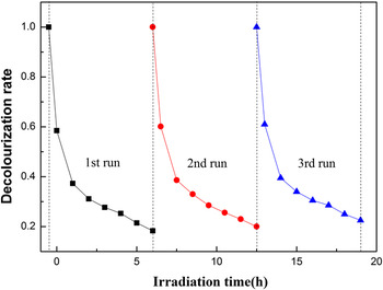
The reusability and stability of photocatalyst are very important issues for its practical applications. So the Ag(5.0)/g-C3N4 photocatalyst was reused in three photodegradation experiments while keeping the experimental condition unchanged. The sample was separated by centrifugation after each cycle. Figure 8 shows the recyclability of the Ag(5.0)/g-C3N4 photocatalyst for the degradation of MO under visible-light irradiation. It can be seen that the Ag(5.0)/g-C3N4 photocatalyst does not exhibit the obvious loss of activity in three photodegradation experiments, and the photocatalytic rates of Rhb for Ag(5.0)/g-C3N4 are 81.7%, 80%, and 77.5% in the first, second, and third runs, respectively. This confirms that the Ag(5.0)/g-C3N4 composite is quite stable during the photocatalytic degradation of MO.
Recyclability of the Ag(5.0)/g-C3N4 photocatalyst for the degradation of MO under visible-light irradiation.

C. Mechanism considerations
According to our results, the photocatalytic activity of g-C3N4 is effectively enhanced after the modification by small amounts of Ag, over which amounts there is a decrease of photocatalytic efficiency. This can be explained from that the more numerous Ag species may conversely behave as a recombination center, encouraging the recombination of charge carriers, or cover the active sites on the g-C3N4 surface and thereby reduce the efficiency of charge separation. Therefore, it is important to make a balance between the active trapping sites favoring the inhibition of the recombination of electron–hole pairs and fewer trapped parts leading to a lower capacity for the separation of interfacial charge transfer.
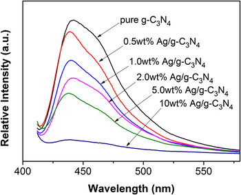
The mechanism can be further concluded from the viewpoint of its physical properties. While the Ag-loaded g-C3N4 photocatalyst is being irradiated, CB-electrons of g-C3N4 easily flow into the metal Ag through the Schottky barrier because the CB of g-C3N4 is higher than that of the loaded metal Ag, which is consistent with the previous study on electron transfer from a semiconductor to a metal. The process of electron transfer is faster than the electron–hole recombination between the VB and CB of g-C3N4. Thus, plenty of CB electrons can be stored in the Ag component. In the present study, the PL spectrum was used to explain this process. Since PL emission arises from the recombination of free carriers, the photoluminescence spectra are widely used to investigate the migration, transfer, and recombination processes of the photogenerated electron–hole pairs in a semiconductor. Figure 9 shows the PL spectra of pure g-C3N4 and Ag/g-C3N4 samples excited upon 393 nm. From the figure, it can be observed that there is a significant decrease in the PL intensity of Ag/g-C3N4 compared with that of pure g-C3N4. A weaker intensity of the peak represents a lower recombination probability of photogenerated charge carriers. Therefore, the Ag dispersed on the surface of g-C3N4 could effectively inhibit the recombination of photogenerated charge carriers, which is helpful for the separation of photogenerated electron–hole pairs in g-C3N4. The significant PL quenching is observed in the Ag/g-C3N4 composite photocatalysts as the content of Ag increases. The quench is due to the photogenerated charge transfer between the Ag and g-C3N4. It can be established that the Ag/g-C3N4 composite photocatalysts are very promising for the photocatalysis with satisfying efficiency.
Photoluminescence (PL) spectra of pure g-C3N4 and Ag/g-C3N4 samples.

IV. CONCLUSIONS
In summary, a simple wet-chemical pathway using silver nitrate as precursor in the presence of dimethylformamide (DMF) as reducing agent and solvent has been proven to be an effective strategy to prepare Ag/g-C3N4 nanocomposites for the first time. The nanocomposites show enhanced photocatalytic performance over bulk g-C3N4 under visible light irradiation, and the optimal photocatalytic activity is approachable when Ag doping is approximately 5 wt% with a considerable k app value of about 0.070 h−1. The enhanced photocatalytic activity is attributed to the Ag particles acting as surface plasmon to increase absorption of light and electron traps to inhibit the recombination of photogenerated electron and hole pair. Our present study is important for the following two reasons: firstly, it is the first demonstration of using dimethylformamide (DMF) as an efficient reducing agent and polyvinylpyrrolidone (PVP) for assisting the nanophasic growth and homogeneous distribution of the plasmonic nanoparticle Ag on g-C3N4 nanosheets; secondly, it provides a new functional material for photocatalytic applications for decoloration of organic dyes.
ACKNOWLEDGMENTS
This work was financially supported by the National Natural Science Foundation of China (No. 21003065). We thank the support of China Council Scholarship.