Major histocompatibility complex (MHC) class I molecules are key players in the internal surveillance function of the immune system that detects the synthesis of abnormal or foreign proteins within cells. Peptides derived from intracellular proteins are expressed in a complex with MHC class I molecules on the surface of all nucleated cells, presenting a picture of the intracellular proteome. Therefore, the MHC presentation system provides the opportunity to target antigens that otherwise would not have been susceptible for recognition by components of the immune system such as antibodies. Viral- or tumour-associated peptides presented by the MHC are recognised by cytotoxic T lymphocytes (CTLs) that act to eliminate malignant or infected cells. CTL recognition is mediated by their clonotypic T-cell receptor (TCR), which binds a specific peptide held within the peptide-binding cleft of the cell surface MHC class I molecule.
Therapeutic antibodies have evolved in the past decade into an effective treatment for cancer. Many of these antibodies target tumour antigens expressed on the tumour cell surface. The number of known antigens expressed exclusively on tumour cells is limited; thus, a great need exists for identifying new cancer-specific markers as targets for therapeutic antibodies. Development of antibodies against disease-associated MHC–peptide complexes might lead to a new class of anticancer therapeutic antibodies. To this end, we and others have developed recombinant antibodies that bind specific MHC–peptide complexes in a peptide-specific, MHC-restricted, TCR-like manner. These antibodies mimic the fine specificity of tumour recognition by T cells but are not susceptible for the regulatory mechanisms in the tumour environment that inhibit the CTL response. Moreover, TCR-like antibodies were demonstrated to be a unique research tool for studying specific antigen presentation on malignant and infected cells. Here, we highlight the recent advances in the development of TCR-like antibodies. We discuss the rationale and strategies for generating these reagents, as well as their clinical implications and different reported applications as therapeutic and diagnostic agents for a variety of cancers and infectious diseases.
T-cell epitopes as immunotherapeutic targets
Tumour epitopes
The past decade saw significant progress in the discovery of well-defined tumour-associated antigens that are expressed in different tumour types and recognised by T cells (Refs Reference Novellino, Castelli and Parmiani1, Reference Rosenberg2). These findings unveiled new targets for immunotherapeutic approaches. Attractive tumour antigen (T-cell epitopes) targets can be classified into three groups. The first group consists of tumour-specific antigens arising from mutated proteins, such as β-catenin in colon cancer (Ref. Reference Morin3). The second group includes differentiation antigens whose expression is restricted to a certain cell lineage and are therefore very specific. Examples include the melanoma-specific targets gp100, MART1 and tyrosinase antigens, which are expressed in the melanocytes (Ref. Reference de Vries4), and the testicular cancer antigens (e.g. NY-ESO-1), which are expressed only by tumour cells and spermatogenic cells from the testis (Refs Reference Sugita5, Reference Nakada6). The third group of potential targets consist of antigens derived from gene overexpression or amplification (e.g. HER2/ neu) (Refs Reference Schechter7, Reference King, Kraus and Aaronson8).
Despite the identification of tumour-associated MHC–peptide complexes (Ref. Reference Shetty9) and the presence of CTLs specific for these complexes in cancer patients (Ref. Reference Coulie10), tumours were demonstrated to develop mechanisms to limit the activity of the CTLs and the antitumour immune response failed to induce tumour regression (Refs Reference Nagaraj and Gabrilovich11, Reference Offringa12, Reference Wang13). A broad spectrum of immunotherapeutic approaches was developed in an attempt to enhance the adoptive cellular immune response to tumours. These approaches are in various stages of clinical development and include adoptive transfer of T cells, active immunisation with tumour antigens (e.g. cancer peptide vaccines and autologous cancer vaccines) and dendritic cell (DC)-based vaccines. The TCR-like antibody approach combines two main advantages of the immune system: the fine specificity of T cells, which use the MHC internal surveillance mechanism to detect cells that express any foreign or abnormal protein, and the biological and pharmacological properties of an antibody that is not susceptible to most tumour regulatory influences that limit T cells.
Downregulation of MHC presentation by tumours is the most relevant tumour environment regulatory mechanism that can also influence the TCR-like antibody-targeting approach. With this limitation in mind, attempts are being made to generate high-affinity TCR-like antibodies that are suitable for detection of the relatively low density of MHC-presented antigens.
Viral epitopes
Cells infected with a replicating virus load viral peptides onto their MHC class I molecules, thereby recruiting the CD8+ CTL response. CD8+ T cells play a critical role in controlling persistent low-level infections and clearing acute viral infections, such as those caused by cytomegalovirus (CMV), Epstein–Barr virus (EBV), hepatitis C virus (HCV) and herpes simplex viruses (HSV) (Ref. Reference Welsh, Selin and Szomolanyi-Tsuda14). Despite the potential for a diverse repertoire of presented MHC–peptide complexes, a more limited number of ‘immunodominant’ epitopes are presented. The reasons for the presentation of only certain viral peptides include the requirement for enzymatic digestion of viral proteins to peptides with high affinity to the autologous MHC alleles, limiting amounts of viral protein, limited stability of viral proteins and timing of protein synthesis during the infection cycle. Many immunodominant epitopes are derived from proteins that are synthesised early in the infection process, before the common phenomenon of MHC downregulation (Refs Reference Boshkov, Macen and Mcfadden15, Reference Miller and Sedmak16). Similar to cancer immunotherapy, a great deal of efforts has been made in the last decade to identify dominant epitopes of several human viruses with the goal of using these epitopes to develop vaccines.
Generation of TCR-like antibodies
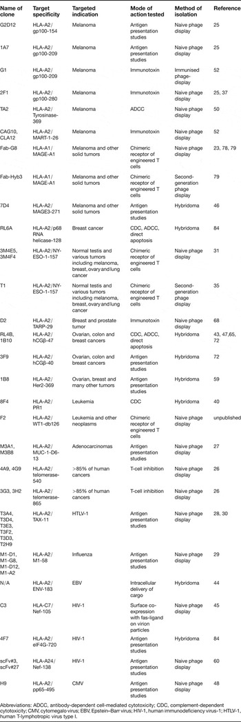
Antibodies with MHC-restricted specificity of T cells have been traditionally rare and difficult to generate. However, over the last few years there has been significant progress in developing such antibodies, because several groups have been able to generate, occasionally in a reproducible manner, TCR-like antibodies directed to a growing repertoire of tumour and viral T-cell epitopes (Table 1). Immunisation strategies, in vitro selection by phage display and combinations thereof have been reported for isolating TCR-like antibodies.
Examples of TCR-like antibodies directed against human CD8+ T-cell epitopes

Abbreviations: ADCC, antibody-dependent cell-mediated cytotoxicity; CDC, complement-dependent cytotoxicity; CMV, cytomegalo virus; EBV, Epstein–Barr virus; HIV-1, human immunodeficiency virus-1; HTLV-1, human T-lymphotropic virus type I.
Obtaining a purified recombinant MHC–peptide complex folded in a native conformation that is recognised by a T cell (Ref. Reference Andersen17) proved to be key for the successful production of TCR-like antibody using either conventional hybridoma or phage-display technology. Recombinant MHC–peptide complexes are efficiently produced in a relatively high amount needed for antibody isolation. These complexes are generated by expressing the extracellular domains of the human leukocyte antigen (HLA) heavy chain and 2-microglobulin inclusion bodies in Escherichia coli followed by their in vitro refolding in the presence of the desired HLA-restricted peptide (Refs Reference Altman18, Reference Denkberg19). The refolded peptide–MHC complexes are very pure, monomeric and folded in the right conformation, as demonstrated by structural (Ref. Reference Garboczi, Hung and Wiley20) and functional studies (Refs Reference Altman18, Reference Denkberg, Cohen and Reiter21). These complexes can be further biotinylated in a site-specific manner, a feature that is utilised for the in vitro selection procedures of TCR-like antibody isolation and for the subsequent step of specificity characterisation.
Phage-display libraries
A major breakthrough in making TCR-like antibodies was achieved when alternative strategies to the conventional hybridoma approach were developed in the mid-1990s. The pioneering work by Andersen and colleagues (1996) demonstrated the ability to use the antibody phage-display approach as a tool to isolate antibodies with a unique specificity (Ref. Reference Andersen17). Phage-display technology includes generating antibody libraries exposed as fusion proteins on the surface of phage particles. Each phage particle displays a unique antibody (a Fab or scFv fragment) encoded by genes present within the same phage particle. Phage particles carrying specific antibodies are purified by repeated rounds of selection on a specific target followed by amplification in bacterial cells of the eluted bound phages. Thus, in contrast to the hybridoma technology, the phage-display procedure involves positive selection for the requested binder (for a review of phage display, see Ref. Reference Hoogenboom22). A phage-display approach enabled the isolation of peptide-specific, MHC-restricted antibodies to murine class I complexes (Ref. Reference Andersen17), and subsequently also to human-disease-related human HLA–peptide complexes (Refs Reference Chames23, Reference Cohen24, Reference Denkberg25, Reference Lev26, Reference Cohen27, Reference Cohen28, Reference Biddison29, Reference Yamano30, Reference Held31).
Naive libraries
TCR-like antibodies were isolated from large naive human phage-display libraries by our group and others (Refs Reference De Haard32, Reference Griffiths33). Several different antibody clones directed to a single T-cell epitope were isolated, in a reproducible manner, from these libraries. The high frequency and high affinities of some of the isolated antibodies suggest that these molecules might be present in the antibody repertoires from the B-cell donors of the phage library; however, an in vivo role for such antibodies remains unclear. To the best of our knowledge, no evidence for B-cell expression of TCR-like antibodies exists. We believe that although the genetic information of TCR-like antibodies exists in the germ line, these molecules are either not produced or are negatively selected. The TCR-like phage-derived Fabs might be the outcome of gene recombination that does not occur naturally. Regardless of the reason for the observed high frequency of antibodies to MHC–peptides, the phage-based approach can be consistently applied to isolate recombinant antibodies with TCR-like specificity to a large variety of MHC–peptide complexes.
Second-generation libraries
The affinity of TCR-like antibodies isolated from a naive phage-display library is not always sufficient for therapeutic purposes. Both random (Ref. Reference Chames34) and rational (Ref. Reference Stewart-Jones35) affinity-maturation strategies were developed to increase the affinity of TCR-like antibodies. Chames and colleagues (2000) reported an 18-fold improved affinity of TCR-like Fab directed to the tumour T-cell epitope HLA-A1-MAGE-A1 using randomised in vitro affinity maturation. Fab G8 with an affinity of 250 nM was used as the platform for the construction of two randomised libraries: L chain shuffling library and H chain complementarity-determining region 3 mutated library. A VH–VL hybrid (clone Hyb3) constructed from clones isolated from these two libraries had an affinity of 14 nM while keeping the fine specificity of the parental antibody (Ref. Reference Chames23).
Rational design for affinity maturation of TCR-like antibodies was demonstrated by Renner and colleagues (Ref. Reference Stewart-Jones35). They analysed the high-resolution structure of TCR-like Fab 3M4E5 bound to the HLA-A2-NY-ESO-1 complex and compared it with the structure of a cognate TCR–pMHC complex. Based on the structural data, they generated a second generation Fab library based on Fab 2M4E5 in which they randomised residues at positions that could optimise peptide interaction without increasing MHC binding. Key residues responsible for direct binding of a peptide motif, as well as residues facing the MHC helices, were left unchanged. This approach resulted in a 20-fold affinity improvement of a new isolated TCR-like Fab to the HLA-A*0201-NY-ESO-1 epitope.
Immunised libraries
In the immunised phage-display approach, which is a strategy complementary to the aforementioned naïve phage-display approach, mice are immunised with the desired recombinant MHC–peptide complexes and phage-display libraries are constructed from the spleens of these mice (Refs Reference Krogsgaard36, Reference Denkberg37). In a syngenic immunisation approach, transgenic mice, which express the desired human HLA allele on a murine MHC knocked out background, are immunised with the recombinant MHC–peptide complexes. The use of specific HLA allele transgenic mice, which have self-tolerance to their transgenic HLA allele, increases the probability of isolating a rare TCR-like antibody rather than pan-reactive anti-HLA antibody with no peptide specificity.
Hybridoma technology
The effectiveness of the immunisation protocol used is key for obtaining TCR-like antibodies using either a phage-display approach with immunised libraries or hybridoma technology. The earliest published studies on hybridoma technology and optimised immunisation and screening strategies used antigen-presenting cells (APCs) presenting strongly immunogenic peptides in their MHC complex as immunogenes (Refs Reference Dadaglio38, Reference Porgador39). It was reported that one to three out of approximately 1000 growth-positive clones tested produced antibodies of the requested specificity, illustrating that peptide-specific, MHC-restricted antibodies are quite rare even under optimal conditions of induction, and underscoring why many attempts have failed (Refs Reference Sergeeva40, Reference Tamminen41, Reference Rubin B42). More recently, several groups have been successful in using a conventional hybridoma approach for isolating TCR-like antibodies directed to tumour (Refs Reference Sergeeva40, Reference Wittman43) and viral (Ref. Reference Sastry44) epitopes. These groups used recombinant MHC–peptide complexes for the immunisation and high-throughput screening of a few thousands of clones for the isolation of rare TCR-like antibodies (Refs Reference Sergeeva40, Reference Wittman43, Reference Sastry44).
Antibodies derived from phage display and hybridomas: pros and cons
The investigator's resources and field of expertise should determine the method of choice for isolating TCR-like antibodies. The most common approach for such isolation published to date is large phage-display libraries (Ref. Reference Herschhorn, Marasco and Hizi45). The major advantage of this approach is the power of high selection towards the desired antigen that is achieved within a relatively short procedure. Other advantages include efficient and reproducible isolation of antibodies directed to different T-cell epitopes from the same source. By contrast, the generation of hybridomas for the isolation of antibodies with such a defined antigenic determinant within the immunogen is somewhat less efficient, and a relatively longer procedure is required for the isolation of rare clones. Furthermore, the isolation of TCR-like antibodies using hybridoma methodology might be limited to stable MHC complexes which bind the peptide with high affinity (Ref. Reference Bernardeau46), because the reduced stability or half-life of MHC and low-affinity peptide complexes might not induce effective immunisation and in vivo IgG maturation.
Advantages of the hybridoma technology include a tendency for isolating antibodies with high-affinity binding to the MHC–peptide complex, because in this approach, the antibodies produced are the result of multiple antigen challenges, leading to in vivo affinity maturation of the antibody clones before their isolation. TCR-like antibodies isolated using hybridoma technology were reported to have relatively high binding affinity (low nanomolar range) (Refs Reference Sergeeva40, Reference Verma47) compared with the moderate average affinity (~50–300 nM) of phage-derived TCR-like antibodies isolated from naïve libraries (Refs Reference Stewart-Jones35, Reference Makler48). Notably, although rare and technically challenging, a few antibodies in the low nanomolar range were successfully isolated from a naïve phage-display library using various screening and selection techniques (Ref. Reference Bronner49). Furthermore, technological improvements such as the aforementioned second-generation libraries and in vitro affinity-maturation procedures improve binding affinities to levels comparable with those observed with hybridoma technology.
TCR-like antibodies isolated from phage-display libraries are in a monovalent antibody form (Fab or scFv fragments) and can be produced in bacterial expression systems with relatively high yields of stable and purified antibody. Monovalent fragments are useful reagents for a variety of TCR-like applications such as epitope presentation (Ref. Reference Michaeli50) and structural (Ref. Reference Denkberg51) studies. In addition, monovalent fragments can be used as targeting agents when conjugated to a cargo protein, (e.g. a truncated form of Pseudomonas exotoxin in the case of immunotoxin) (Ref. Reference Klechevsky52). Notably, one of the limitations of the monovalent antibody form is its reduced binding intensity, which is a major disadvantage when using a TCR-like antibody as a tool to study low-density epitopes (e.g. some tumour-associated MHC–peptide complexes presented by tumour cells). To overcome this limitation, Fab or scFv-tetramers (Ref. Reference Cloutier53), with directly tagged fluorescent probes, can be generated to increase the binding avidity and consequently the binding intensity of specific MHC–peptide complexes (Ref. Reference Cohen27). Fab tetramers are generated by introducing a BirA sequence tag for site-specific biotinylation at the C-terminus of the Fab CL or CH1 domain for tetramerisation by labelled streptavidin reagent. Another approach developed to overcome the reduced binding intensity involves increasing the avidity of the TCR-like antibody molecules by transforming the monovalent fragments into bivalent constructs in the form of IgG molecules. The H and L Fab genes are cloned into IgG antibody vectors carrying secretion signal for expression and secretion in eukaryotic expression systems. This strategy was successfully implemented in our laboratory for several TCR-like Fabs, and for the most part, resulted in the production of TCR-like IgG molecules which kept the fine specificity of the parental monovalent fragment while improving its detection sensitivity and apparent binding affinity significantly (Refs Reference Makler48, Reference Michaeli50). The Fc portion of the IgG is essential when using TCR-like antibodies to target cells for cytotoxic effects by recruiting components of the immune systems through antibody-dependent cell-mediated cytotoxicity (ADCC) or complement-dependent cytotoxicity (CDC). The clinical development of TCR-like IgG antibodies will require humanization of the mouse IgG hybridoma clones to avoid anti-idiotypic host response and to optimise the effector functions and half-life of the antibody.
TCR-like antibodies as research tools for studying antigen presentation
TCR-like antibodies are unique reagents for studying antigen presentation. The growing repertoire of isolated TCR-like antibodies has facilitated the gathering of novel data regarding antigen presentation in various tumour types and viral infections. These antibodies enable direct detection of specific MHC–peptide complexes following the natural intracellular processing of the cognate antigen and can be used for direct in situ visualisation and exact quantification of MHC–peptide complexes presented on the cell surface.
Initial studies confirmed the capability of TCR-like antibodies to detect specific MHC–peptide complexes generated through natural intracellular processing by following cells transfected with certain genes of interest (Refs Reference Cohen28, Reference Porgador39). Subsequent studies were able to achieve a detection of particular MHC–peptide complexes on tumour cells despite the lower density of the MHC–peptide complexes on tumour cells compared with those on peptide-pulsed or transfected APCs. For example, TCR-like antibody isolated against HLA-A2-restricted epitope derived from the melanoma differentiation antigen gp100 reacted very intensely with HLA-A2+ gp100+ melanoma FM3D and YU ZAZ6 cells, but not with HLA-A2-negative melanoma MZ2-MEL3.0 cells or the HLA-A2+ breast carcinoma tumour cell line MCF7 (Ref. Reference Denkberg25).
These TCR-like antibodies also provide new tools to quantify the specific MHC complexes presented on tumour cells. The fluorescence intensity of stained cells was measured by flow cytometry and compared with that of calibration beads with known numbers of fluorophores. Cohen and colleagues (2002) were the first to use this method to quantify the number of tumour-associated T-cell epitopes (Ref. Reference Cohen27). By using TCR-like antibody directed against the mucin epitope (HLA-A2-MUC1-D6), they estimated the number of MUC1-D6-derived complexes on the surface of MUC1 expression tumour cells and found it to be only several hundreds per cell. The availability of multiple TCR-like antibodies against various epitopes facilitates gaining a broader perspective on the antigen presentation of a certain cell type. For example, TCR-like antibodies against three major melanocyte differentiation antigens (gp100, Mart-1 and tyrosinase) demonstrated a defined pattern of presentation hierarchy between these epitopes (Ref. Reference Michaeli50). This study found surprisingly high levels of tyrosinase-derived HLA-A2 complexes expressed on melanoma cells derived from multiple patients. Complexes of HLA-A2 and tyrosinase (369–377) were estimated to account for approximately 20% of the total HLA-A2 complexes on the cell surface with an average of approximately 4000 surface copies in many melanoma cell lines. The other differentiation antigens that were studied have moderate to low expression levels with not more than a few hundreds of complexes per cell. This information about the antigen expression hierarchy and the high presentation levels of the tyrosinase peptide is most valuable when considering immunotherapy targets. Mart-1 and gp100 are immunodominant epitopes and are frequently found in melanoma patients (Refs Reference Kawakami54, Reference Kawakami55, Reference Romero56). By contrast, tyrosinase-specific T cells are hardly detectable among tumour infiltrating lymphocytes and in peripheral blood of the patients (Ref. Reference Kawakami54), possibly because of adaptive tolerance induced to tyrosinase-specific CD8+ T cells upon exposure to high levels of HLA-A2–tyrosinase complexes (Refs Reference Oved57, Reference Taams, Van Eden and Wauben58). Moreover, TCR-like antibody-based evaluations of specific MHC–peptide levels on tumour cells were found to correlate with their susceptibility to CTL killing (Ref. Reference Weidanz59). Thus, TCR-like antibodies proved to be the most accurate and direct tool to detect antigen-presentation and to select CTL epitopes for cancer vaccines.
TCR-like antibodies were generated against CD8+ T-cell epitopes of several viral models including CMV (Ref. Reference Makler48), human T-lymphotropic virus type I (HTLV-1) (Ref. Reference Cohen28), human immunodeficiency virus-1 (HIV-1) (Refs Reference Herschhorn, Marasco and Hizi45, Reference Nunoya60) and hepatitis B virus (HBV) (Ref. Reference Sastry44). The anti-HLA-A2–Tax TCR-like Fab is capable of detecting these specific complexes on the surface of virus-infected cells. Staining pattern of RSCD4 HTLV-1-infected cells revealed two subpopulations with high and moderate presentation of HLA-A2–Tax complexes, displaying 30 000 and several hundreds of complexes, respectively. Moreover, the TCR-like antibodies enabled direct dissection of the phenotype of the different T-cell subpopulations infected by HTLV-1 in patients with HTLV-1-associated myelopathy tropical spastic paraparesis (HAM/TSP). Simultaneous staining with anti-Tax-MHC Fab and specific anti-CD markers revealed that CD4+ CD25+ regulatory T cells show high expression of Tax–HLA-A2 peptide complexes which could be directly correlated to their viral load (Ref. Reference Yamano61). Another example is the use of anti-HLA-A2–pp65-495-503 TCR-like antibody to study the dynamics of antigen presentation during the infection cycle of human-CMV-infected cells (Ref. Reference Makler48). Intracellular and extracellular TCR-like antibody staining at various times post infection demonstrated an accumulation of large intracellular pools of viral-derived HLA-A2–pp65 complexes that migrate slowly to the cell surface. Interestingly, MHC class I downregulation mechanisms, which cause a decrease in the presence of the total MHC class I in infected cells over time, do not affect the increase in the pool of HLA-A2–pp65 complexes (Ref. Reference Makler48). Such studies with TCR-like antibodies contribute to our understanding of some of the mechanisms that control virus pathologies and the balance between viral evasion and the immune response.
Therapeutic applications of TCR-like antibodies
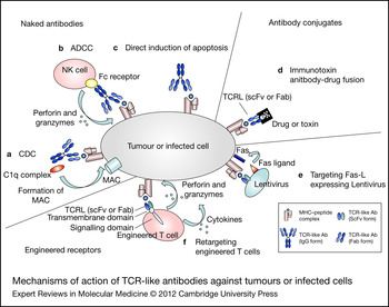
In this section we review the clinical applications of TCR-like antibodies reported to date. Potential applications without supporting published experimental data are beyond the scope of this review. The immunotherapeutic applications of TCR-like antibodies and their mechanisms of action are demonstrated in Fig. 1.
Mechanisms of action of TCR-like antibodies against tumours or infected cells. Most naked TCR-like antibodies interact with components of the immune system through ADCC (a) or CDC (b). Antibodies can also alter signal transduction within the tumour cell, resulting in an induction of apoptosis (c). When fused to drugs or toxins (d), TCR-like antibodies can directly kill tumour cells. Co-expression of TCR-like antibody and Fas ligand on the surface of lentiviruses (e) can mediate killing of cells expressing the cognate T-cell epitope. T-cells engineered to display TCR-like antibody as receptors (f) can re-direct cytotoxic T cells against target cells forming lytic immunological synapse. ADCC, antibody-dependent cell-mediated cytotoxicity; CDC, complement-dependent cytotoxicity; Fab, fragmant antigen binding; MAC, membrane attack complex; MHC, major histocompatibility complex. scFv, single-chain variable fragment; TCRL, T-cell receptor-like.

TCR-like antibodies as anticancer agents
Anticancer monoclonal antibodies (mAbs) are a growing family of agents applied for the treatment of cancer (both solid tumours and haematological malignancies). The biological and pharmacological properties of antibodies make them attractive therapeutic agents (for a review see Ref. Reference Weiner, Surana and Wang62). Over 10 antibody-based anticancer immunotherapeutic approaches have received regulatory approval and many more are under clinical development. Antibody-based immunotherapeutic approaches include the use of naked antibodies as well as antibody conjugates (e.g. immunotoxin, fusion with cytokine molecules, immunoradioisotope labelling and bi-specific antibody therapy).
Presentation of specific MHC–peptide complexes associated with a certain tumour type supports the use of TCR-like antibodies as specific antitumour targeting molecules. Whereas therapeutic anticancer mAbs target membrane proteins, TCR-like antibodies expand the targeting repertoire to the intracellular proteome represented by the MHC surveillance mechanism of the cell. Such specific antibodies present new opportunities for anticancer targeting while transforming the fine cellular specificity of T-cell recognition into an antibody-based immunotherapeutic approach.
Naked TCR-like antibodies
Recruitment of innate immunity is among the proposed mechanisms for unconjugated antitumour-specific mAbs such as rituximab (Rituxan/MabThera, Genentech, South San Francisco, CA) (Ref. Reference Cartron63) and trastuzumab (Herceptin, Genentech) (Ref. Reference Clynes64). Similar to mAbs that target specific tumour antigen, TCR-like antibodies are being used to mediate ADCC and CDC. Weidanz and colleagues (2006) generated a mouse IgG2a mAb RL4B specific to human chorionic gonadotropin (hCG)-β presented by HLA-A2 (Ref. Reference Wittman43) and demonstrated that this mAb prevented and suppressed breast tumour growth in xenograft in vivo models (Ref. Reference Verma47). In vitro studies demonstrated that this mAb induced both ADCC and CDC mechanisms. Interestingly, RL4B was found also to deliver a direct apoptotic signal to cancer cells in a mechanism involving JNK activation and the caspase-dependent pathway (Ref. Reference Verma65). This cytotoxic effect suggests a requirement for antigen-specific TCR-like binding of the antibody to the MHC–peptide complex because such an apoptotic signal was not observed with pan anti-HLA antibodies lacking peptide specificity. The mechanism of such a direct apoptotic signal and its in vivo role compared with immune-mediated mechanisms should be further investigated in the breast tumour model. Furthermore, studies exploring whether such a mechanism exists with other TCR-like antibodies targeting additional tumour types are warranted. TCR-like antibodies should be evaluated for combination treatments with other immunotherapies or cancer treatments. Recently, increased treatment efficiencies were reported for mAbs when tested in combination treatment; for example, US and European guidelines recommend combination chemotherapy with the addition of an mAb for the first-line treatment of metastatic colorectal cancer (Ref. Reference Edwards66).
Immunotoxins
Anticancer mAbs can be used for tumour-specific delivery of drugs. Immunotoxins are chimeric molecules in which a toxin is fused to a recombinant targeting antibody fragment. The targeting moiety can be a Fab fragment or a single-chain Fv molecule (ScFv) composed of the variable domains VH and VL that are covalently linked through a peptide linker. Immunotoxins against a variety of tumour types demonstrated promising anticancer activity and are at different stages of clinical development (Ref. Reference Choudhary, Mathew and Verma67). The major advance of these molecules over using whole antibodies for delivery is their small size, which contributes to their efficient tumour penetration.
We have applied TCR-like antibodies to deliver the truncated form of Pseudomonas exotoxin (PE38KDEL) into tumour target cells. TCR-like immunotoxins have also been generated against HLA-A2-positive melanoma differentiation antigens gp100 (Ref. Reference Denkberg37) and MART-1 (Ref. Reference Klechevsky52) and against the prostate and breast tumour-associated antigen TRAP (Ref. Reference Epel68). These immunotoxins showed in vitro and in vivo cytotoxic activity in an antigen-specific and HLA-A2-restricted manner. They were found to prevent specific tumour growth at nontoxic doses using xenograft models. These data provide a proof of concept for the utility of TCR-like antibodies as targeting agents directed to various cancer types and form the basis for further improvement of TCR-like immunotoxins needed to further evaluate their therapeutic potential in more robust in vivo models. TCR-like immunotoxins, as well as naked TCR-like antibodies that induce apoptosis by direct killing mechanism, might confer a clinical benefit in the treatment of immunocompromised patients who may not respond to agents that require a functioning immune system (e.g. ADCC).
Determining vaccine potency
The ability of TCR-like antibodies to detect and quantify T-cell epitopes makes them a unique tool for evaluating the potency of immunotherapeutic approaches used to augment antitumour cellular immune response (e.g. cancer peptide vaccines, autologous cancer vaccines and DC vaccines). Numerous studies have demonstrated that DC-based vaccines induce antitumour immunity (Refs Reference Berntsen, Geertsen and Svane69, Reference Jähnisch70). Such a presentation of tumour antigens by DCs was demonstrated using specific TCR-like antibodies (Ref. Reference Zehn71), and was found to differ from presentation by other cell types with respect to kinetics and peptide retention on the MHC. This study highlighted the utility of TCR-like antibodies for monitoring MHC–peptide levels on DCs during vaccinations protocols. A more recent study used TCR-like Abs against HLA-A2-hCG-β epitopes in the development of a potency assay for a DC-based vaccine being developed for cancer therapy (Ref. Reference Neethling72). This vaccine targets the hCG-β antigen to DCs using a chimeric molecule composed of the hCG-β antigen and an mAb against the mannose receptor found on DCs (Celldex Therapeutics, Needham, MA). Antibodies directed to two hCG-β epitopes were used and were able to detect specific MHC–peptide complexes on vaccine-treated DCs and specifically block their induction of CTL response, thereby providing a proof of concept for the utility of such antibodies in determining epitope presentation on vaccine-treated DCs.
Studies assessing the correlation between epitope presentation and vaccine potency are warranted and should enable the development of assays that would be highly valuable in quality control procedures and in improving vaccine potency. Furthermore, additional studies might expand the use of these potency assays to other therapeutic modalities including peptide and DNA vaccinations.
Retargeting T cells for adoptive transfer
In the adoptive T-cell transfer therapeutic approach, autologous tumour-infiltrated T cells are isolated, expanded in vitro and re-infused to patients (Refs Reference Topalian and Rosenberg73, Reference Hong74). This approach was demonstrated to be an effective treatment for metastatic melanoma. However, despite the encouraging responses in melanoma, response rates in other common cancers have remained low, partly because of difficulties in isolating and expanding tumour-specific T cells from patients and controlling their affinity and characterisation.
The adoptive T-cell transfer therapeutic approach was improved through genetic modification of T cells using viral transduction systems. Genetic modification for redirection of primary human T cells to a desired tumour antigen was proven to be efficient in vivo based on an engineered receptor in the form of antibody fragments (Ref. Reference Eshhar75) or TCR αβ chains (Ref. Reference Stauss76). In the first approach, the antibody V region responsible for recognition and activation is attached to a cytotoxic T-cell signalling moiety FcɛRI γ chain to mediate tumour-specific killing and cytokine release. Safe and efficient treatment of cancer patients was recently reported using adoptive transfer of genetically modified T cells (Ref. Reference Pule77).
The use of T cells that are genetically transduced to express antitumor TCRs requires the isolation of antigen-specific antitumor T cell clones for cloning the αβ TCR genes or the technically challenging procedures for in vitro selection of recombinant TCR. Therefore, the availability of TCR-like antibody genes constitutes an attractive alternative approach to redirect T cells. Willemsen and colleagues (2001) were the first to use a phage-display TCR-like antibody for retargeting primary human T cells (Ref. Reference Willemsen78). The TCR-like antibody was used to improve the T-cell affinity while keeping the desired T-cell epitope target. Willemsen and colleagues generated engineered T cells carrying a TCR-like antibody specific to HLA-A1-MAGE-A1 as a receptor. The engineered T cells were found to specifically bind HLA–peptide complexes on target cells, leading to the production of cytokines and induction of cytolysis. This strategy provides a new rationale for generating engineered T cells expressing high-affinity chimeric receptors that target tumour T-cell epitopes in an HLA-restricted manner. High-affinity TCR-like Fab directed to HLA-A1-MAGE-A1 was transduced into T cells for comparison with T cells retargeted with moderate-affinity TCR-like antibody carrying the same specificity (14 and 250 nM, respectively) (Ref. Reference Chames34). The introduction of a higher-affinity TCR-like antibody to primary human T cells, in a construct containing the transmembrane γ chain and CD4 as a signalling domain, resulted in an enhanced antitumour activity to HLA-A1-MAGE-A1-positive melanoma cells. A potential drawback of this approach is that the maximal affinity of the TCR-like antibody being used to redirect the engineered T cells seems to be limited. When these two TCR-like antibodies were transducted into T cells using a more potent signalling domain (CD28+γ), the increased potency obtained for the high-affinity TCR-like antibody was associated with loss of specificity because HLA-A1-MAGE-A1-negative cells were also targeted (Ref. Reference Willemsen79). We observed similar findings recently in a study comparing the functionality of T cells retargeted to the same epitope by introducing engineered TCR and TCR-like antibodies carrying different affinities (unpublished results). The increased avidity of TCR-like antibodies expressed as membrane receptors in combination with their high affinity might result in a loss of specificity and crossreactivity to other HLA–peptide complexes. These findings of specificity loss are also supported by a study that compared TCR variants and suggested the existence of maximal receptor affinity thresholds for achieving enhanced T-cell functionality while keeping the fine specificity (Ref. Reference Schmid80). Ultimately, attempts to increase engineered T-cell receptor affinity by using TCR-like antibodies should consider the effects of receptor and co-receptor densities and affinities in the cellular context and carefully re-evaluate the fine specificity determined for the soluble TCR-like antibody form. Optimal high receptor affinity threshold for the use of MHC-restricted chimeric receptors for genetically reprogrammed T cells might limit the clinical benefit of this approach compared with the use of high-affinity non-MHC chimeric receptors. The functional avidity of TCR-like antibody when implanted as a chimeric receptor on the surface of the engineered T cells might play a critical role with respect to the relationships between affinity and avidity. This is particularly important for specificity of engineered T cells compared with the bivalent or monovalent nature of binding when these molecules are being used as soluble targeting agents. More comparative studies should be conducted to shed more light on this issue.
Targeting virus-infected cells
Antibodies have been studied and developed as potential immunotherapeutic approaches and vaccine strategies for viral infections. Neutralising antibodies for inflammatory mediators and their cellular receptors (Ref. Reference Khan81), as well as for viral proteins (Ref. Reference Corti82), are of great interest to immunologists and the medical community at large. Antibodies that specifically target infected cells are an alternative attractive approach that can be applied using TCR-like antibodies targeting specific viral T-cell epitopes.
Bertoletti and colleagues (2011) demonstrated the use of a TCR-like antibody as a delivery agent for targeting HBV-infected cells (Ref. Reference Sastry44). They isolated an antibody specific to a peptide of the envelop protein Env restricted by HLA-A2. The anti-HLA-A2-Env mAb specifically stained infected hepatocytes derived from biopsy specimens from a patient with HLA-A2-positive chronic hepatitis B. Subsequently, they conjugated the TCR-like antibody to a fluorochrome-labelled cargo and incubated the chimeric antibody with cells expressing the HLA-A2-Env epitope. Within 16 h, the labelled TCR-like antibody was internalised and colocalised within the lysosomes of the cells, as required from a vehicle of drug delivery.
Hizi and colleagues (2010) generated lentiviral particles that express TCR-like antibodies directed to HIV-1-derived epitope by isolating an antibody directed to the HLA-C*07-Nef1 complex and coexpressing it with the Fas ligand on the surface of the same virion particle (Ref. Reference Herschhorn, Marasco and Hizi45). Their rationale was to target HIV-1-infected cells that express Fas receptor and to mediate the specific killing of HIV-1-infected cells by Fas–FasR-induced apoptosis. Although dose-dependent killing of Nef-presenting cells was induced by these lentiviruses, it was accompanied by some nonspecific killing of cells presenting a control peptide. Therefore, this approach should be further developed and optimised to fulfil the requirements of a safe, efficient and specific therapeutic agent. As for the engineered T cells, when transforming soluble high-affinity high-specificity TCR-like antibodies into cellular or viral receptors, care should be taken to test whether the TCR-like specificity is retained.
Research in progress and outstanding research questions
The recent advances in generating TCR-like antibodies have resulted in a growing repertoire of antibodies with specificities directed to a large variety of MHC-class-I–peptide complexes. These antibodies constitute valuable tools in molecular immunology for studying antigen presentation and developing new immunotherapy agents. TCR-like antibodies have contributed to the advance in basic research of antigen presentation in various healthy or diseased tissues and cell types, including tumours, virus-infected cells, professional APCs and lymphoid cells. The potential therapeutic and in vivo diagnostic utility of TCR-like antibodies was explored and a proof of concept was demonstrated for several tumour types and infectious diseases. The TCR-like antibody approach is now facing the challenge of transitioning into clinical trials. To this end, a candidate antibody should have high-affinity and high-specificity binding to the TCR ligand. High affinity, in the low nanomolar or picomolar range, implies a longer binding time and increased probability for therapeutic cytotoxic endocytosis or induction of immune-effector mechanisms. TCR-like specificity of the antibodies is key for their utility as specific targeting molecules.
The antibody isolation procedure could benefit from a comprehensive specificity characterisation carried out by mapping the recognition residues within the peptide and the MHC molecule. This procedure would verify the peptide-specific, MHC-restricted, TCR-like binding pattern of the antibody. Crossreactivity to other self-peptides should be excluded by testing binding to a variety of cell types from a range of tissue origins. Another key factor in optimising the TCR-like antibody therapeutic approach is the choice of target antigen from all the T-cell epitopes expressed on malignant cells [studies with anticancer mAbs have demonstrated that the high density of membrane epitopes on target cells is correlated with the therapeutic efficiency of these agents (Ref. Reference Mimura83)]. For example, the exceptionally high expression of the tyrosinase antigen on the surface of melanoma cells makes it an attractive target for TCR-like antibody therapy. Thus, studies evaluating expression levels of therapeutically relevant T-cell epitopes (e.g. the aforementioned study on melanoma T-cell epitopes) can reveal optimal targets for TCR-like antibody therapy. Notably, the TCR-like antibodies are also important tools for such studies, alongside proteomic and mass spectrometry methodologies (Ref. Reference Weidanz84).
TCR-like antibody approaches fit in with the growing field of personalised medicine. These reagents might be used for therapy and diagnosis of HLA-matched patients. The availability of TCR-like antibodies directed to several epitopes associated with a certain tumour type raise the possibility of a cocktail treatment composed of antibodies recognising different antigens. Furthermore, when tumour biopsies are available, the antibody treatment can be personalised to match the tumour antigen presentation hierarchy of a specific patient. A TCR-like antibody cocktail treatment strategy might increase the treatment potency and target malignant cell mutants with the absence or decreased expression of a certain target antigen. Further development of this approach needs to be tested experimentally with the ultimate goal of applying TCR-like antibodies to clinical practice.
Acknowledgements and funding
We thank the peer reviewers for their valuable advice on the manuscript. We acknowledge all the students, post docs and colleagues who over the years contributed to our work on the generation, characterization and applications of TCR-like antibodies. The work in Y.R.'s laboratory on TCR-like antibodies was supported over the years by research grants from the ISF-Israel Science Foundation, the EU 6th framework program, the National Cancer Institute, NIH and the ICRF-Israel Cancer Research Foundation.