INTRODUCTION
Autolytines are small free-living polychaetes, ranging from 1–60 mm in length and 0.1–1.2 mm in width. They are distributed wordwide and inhabit shallow waters, restricted to the shelf areas around the continents. Mostly, they are associated with hydroids and bryozoans, animals which they supposedly feed upon (Okada, Reference Okada1929; Hamond, Reference Hamond1970; Fauchald & Jumars, Reference Fauchald and Jumars1979; Genzano & San Martín, Reference Genzano and San Martín2002; Nygren, Reference Nygren2004). They constitute a well-delineated group of syllid polychaetes, where most of the members are identifiable by a convoluted pharynx, absence of ventral cirri, and presence of simple dorsal chaeta of bayonet type (Nygren, Reference Nygren2004). Syllids in general and autolytines in particular have a bewildering array of reproductive strategies including epigamy as well as different kinds of schizogamy (Franke, Reference Franke, Dorresteijn and Westheide1999; Nygren & Sundberg, Reference Nygren and Sundberg2003). Following Nygren (Reference Nygren2004) and Nygren & Pleijel (Reference Nygren and Pleijel2007) Autolytinae comprises 173 nominal species of which more than 100 are considered valid. These are divided in 11 currently recognized genera (Nygren & Pleijel, Reference Nygren and Pleijel2007), where Imajimaea is one of the least comprehensive containing four species, I. tsugarus (Imajima, 1966), I. japonensis (Imajima, 1966), I. zonatus (Mohammed, 1973) and I. draculai (San Martín & López, Reference San Martín and López2002). Species of Imajimaea are rare and none of the four species has been recorded since their original description, and all of the descriptions except I. japonensis, are based on single specimens. Imajimaea draculai was originally described from off the coast of Portugal based on a single anterior end dredged at 174–180 m depth. There were specimens of Leptometra sp. (Crinoidea: Echinodermata), Munida sp. (Decapoda: Crustacea), and Funiculina quadrangularis (Pallas, 1776) (Pennatulacea: Cnidaria) in the sample, and the authors suspected that their new species was associated with one of these species, although this could not be confirmed. During two expeditions (‘Pandalina IV’ and ‘Pandalina V’) at the Swedish west coast that were initiated by the Swedish Taxonomy Initiative, we came across a number of specimens of this rare species living on F. quadrangularis at 300 m depth. In this paper, we redescribe Imajimaea draculai, assess its phylogenetic position within Autolytinae using mitochondrial COI and 16SrDNA, and nuclear 18SrDNA, and quantify its intraspecific variation with mitochondrial COI, and nuclear ITS1. We also provide information on its reproductive mode, describe its association with F. quadrangularis, and report the species from Scandinavian waters for the first time.
MATERIALS AND METHODS
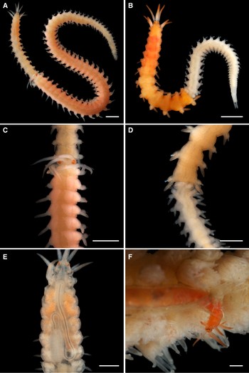
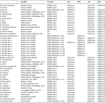
Specimens of I. draculai were collected from F. quadrangularis where the worms were found inside thin mucus tubes in between the polyps of the host (Figure 1F). The red colour and their relatively large size made the worms easy to spot against the grey host. Live specimens were photographed with a Canon EOS 30D connected to a Canon MP-E65/2.8 1-5X macro objective. The specimens were then relaxed with magnesium chloride and then: (1) fixed in formalin (10%), rinsed in fresh water and transferred to 80% alcohol; (2) preserved directly in 90% alcohol for DNA-sequencing (entire or fragment); (3) squeezed between a microscope slide and cover glass while in MgCl2, subsequently transferred to aquamount (Gurr) and saved as permanent slides; or (4) fixed in formalin and directly used for scanning electron microscopy (SEM), critical point-dried, sputter-coated and examined in a Philips XL20 microscope. Origin of specimens, GenBank accession numbers, and deposition of vouchers are detailed in Table 1. Vouchers are deposited at Göteborg Natural History Museum (GNM) and at Swedish Museum of Natural History (SMNH).
Colour photographs of I. draculai. (A) Female with a developing stolon; (B) male with a developing stolon; (C) close-up, developing female stolon; (D) close-up, developing male stolon; (E) anterior end of specimen squeezed between coverslip and microscope slide; (F) specimen partly out of its tube examining the surroundings. A, C and F corresponds to GMN Polychaeta 13172, B corresponds to SMNH 106880, D corresponds to GMN Polychaeta 13171, and E corresponds to GMN Polychaeta 13174. Scale bars 1 mm.

Sequenced taxa, locality, vouchers, and GenBank accession numbers.

1, all vouchers are hologenophores (Pleijel et al., Reference Pleijel, Jondelius, Norlinder, Oxelman, Schander, Sundberg and Thollesson2008).
DNA was extracted using DNAeasy Tissue Kit (Qiagen) following the protocol supplied by the manufacturer. We amplified 658 bp of COI, about 480 bp of 16S rDNA, 1364–1771 bp of 18SrDNA, and 466 bp of ITS1 including flanking regions of 18SrDNA and 5.8SrDNA. We used the same primers as specified in Nygren & Sundberg (Reference Nygren and Sundberg2003) for 16SrDNA, and 18SrDNA, and we used the same primers as specified in Nygren et al. (Reference Nygren, Eklöf and Pleijel2009) for COI and ITS1. PCR mixtures contained 21 µl ddH20, 1 µl of each primer (10 µM), 2µl of DNA template, and puReTaq Ready-To-Go PCR Beads (Amersham Biosciences). The temperature profile was as follows: 96°C/240s–(94°C/30s–48-62°C/30s–72°C/60s)*45 cycles –72°C/480s. PCR products were purified with the E.Z.N.A. Cycle-Pure Kit (Omega Bio-tek). Sequencing was performed at Macrogen Inc. facilities (Seoul, Korea). Overlapping sequence fragments were merged into consensus sequences using Geneious 4.5.5 (Drummond et al., Reference Drummond, Ashton, Cheung, Heled, Kearse, Moir, Stones-Havas, Thierer and Wilson2008). The protein coding COI was trivial to align, and the ribosomal genes were aligned using Clustal X (Thompson et al., Reference Thompson, Gibson, Plewniak, Jeanmougin and Higgins1997) with three different sets of gap/gap length penalties (20/2, 15/7, 30/8). Regions that were not consistent between the alignments, i.e. alignment-ambiguous sites (Gatesy et al., Reference Gatesy, DeSalle and Wheeler1993), were excluded from further analysis. Alignments are available at TreeBase, http://www.treebase.org. Initially we used a number of non-Autolytine syllid sequences from GenBank as outgroups, but as I. draculai was found to be a member of the Procerini-clade with strong support (posterior probability = 1.0), we proceeded with non-Procerini autolytines as outgroups, in order to maximize the number of alignable positions in the ribosomal genes and to increase the phylogenetic signal in the data set. The nuclear and the mitochondrial data sets were analysed separately and combined. We used MrBayes 3.1.2 (Ronquist & Huelsenbeck, Reference Ronquist and Huelsenbeck2003) to conduct Bayesian analyses. The best-fit models were selected using the Akaike information criterion (AIC) in MrModeltest (Nylander, Reference Nylander2004). For COI we used a General Time Reversible model with gamma distributed rate across sites with a proportion of the sites invariable (GTR + G + I) for all three codon positions, for 16SrDNA we used a GTR + G model, and for 18SrDNA we used a GTR + G + I model. The number of generations were set to two millions with four parallel chains (three hot and one cold), sample frequencies were set to 500, and numbers of runs were set to two. A quarter of the samples were discarded as burn in. Parameters were altered in the proposal mechanisms to acquire a span within 20–60% acceptance rates for the moves in the cold chain of each run (Gelman et al., Reference Gelman, Carlin, Stern and Rubin1995). Proposal rates were not changed. The tree files were analysed in AWTY (Are We There Yet) (Wilgenbusch et al., Reference Wilgenbusch, Warren and Swofford2004; Nylander et al., Reference Nylander, Wilgenbusch, Warren and Swofford2007) to visually interpret if the analyses had reached the stationary phase.
SYSTEMATICS
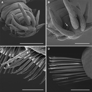
SEM micrographs of I. draculai SMNH 106880. (A) Anterior end, anterolateral view; (B) close-up of anterior end, nuchal epaulettes indicated with an arrow; (C) chaetiger 2, simple unidentate chaetae; (D) chaetiger 10, simple bidentate chaetae. Scale bars A 0.5 mm, B 0.2 mm and C, D 50 µm.

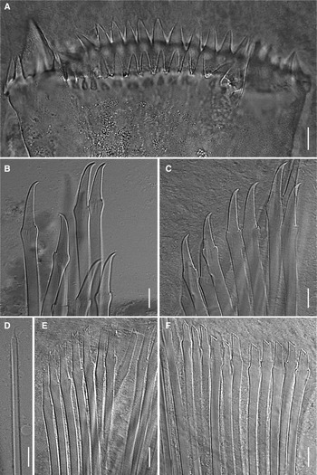
Micrographs of I. draculai GMN Polychaeta 13177. (A) Trepan, dorsal view; (B) chaetiger 1, simple unidentate chaetae; (C) chaetiger 2, simple unidentate chaetae; (D) chaetiger 20, bayonet chaeta of thin type with subdistal denticulation; (E) chaetiger 3, simple bidentate chaetae; (F) chaetiger 4, simple bidentate chaetae. Scale bars 10 µm.

TYPE MATERIAL
Holotype: MNCN 16.01/6095, north-western Spain, off Foz, 43°58.46′N 7°11.8′W, 177 m, sand, 14 June 1991.
NON-TYPE MATERIAL EXAMINED
Sweden: 6 spms preserved in alcohol (GNM Polychaeta 13164–13169), 1 spm preserved in formalin (GNM Polychaeta 13170), Bratten Area, 58°21.144′N 10°33,087′E (start)–58°21.240′N 10°33.338′E (stop), 297–319 m, clay with F. quadrangularis, dredge, coll. M. Berggren & H.G. Hansson, 30 August 2006; 1 spm preserved in formalin (GNM Polychaeta 13171), 1 spm preserved in formalin and subsequently mounted for SEM (SMNH 106880), Bratten Area, 58°11.504′N 10°39.888′E (start)–58°11.769′N 10°40.089′E (stop), 237–277 m, clay with F. quadrangularis, Warén sledge, 12 June 2008; 2 spms preserved in formalin (GNM Polychaeta 13172a, 13173), of which 1 rear end preserved in alcohol (GNM Polychaeta 13172b), 4 spms preserved on slides (GNM Polychaeta 13174a–13176a, 13177), of which 3 rear ends preserved in alcohol (GNM Polychaeta 13174b–13176b), Bratten Area, 58°13.949′N 10°32.679′E (start)–58°14.026′N 10°32.384′E (stop), 271–321 m, clay with F. quadrangularis, Agassiz dredge, 12 June 2008.
DIAGNOSIS
Imajimaea with dorsal cirri absent on chaetigers 2–5, otherwise present; trepan with two lateral large teeth, smaller teeth in a ventral and dorsal arc; only simple chaetae in all chaetigers.
DESCRIPTION
Length 9.6–18 mm for 45–73 chaetigers including stolon (complete specimens only), width at level of proventricle, excluding parapodial lobes, 0.7–0.8 mm. Live specimens yellowish orange to red in anterior non-stolonial segments (Figure 1A, B), most specimens with more distinct red pigmentation from chaetiger 2 to region around proventricle, some specimens distinctly red in all anterior non-stolonial segments; developing female stolons light orange, eggs pink (Figure 1A, C), developing male stolons whitish (Figure 1B, D); antennae, tentacular cirri, and first dorsal cirri with red tips, dorsal cirri in anterior non-stolonial segments sometimes also reddish. Body shape, excluding parapodial lobes, cylindrical in transection, venter flattened. Body of fairly constant width with tapering posterior end. Ciliation present on nuchal epaulettes, prostomium, ventrally on a few anterior segments, and dorsally as poorly developed ciliary rings medially on anterior-most segments (Figure 2B). Prostomium rounded rectangular. Four eyes, with lenses, of about same size (Figure 1A, E); eye spots absent. Palps invisible in dorsal view (Figure 1A, B), present as two ventro-laterally projecting mounds indistinctly separated from prostomium (Figure 2A). Nuchal epaulettes small, present on anterior part of tentacular segment (Figure 2B, indicated with an arrow). Prostomium with three antennae; median antenna inserted medially on prostomium, lateral antennae on anterior margin. Tentacular cirri two pairs. Median antenna reaching chaetigers 2–3. Lateral antennae and first dorsal cirri about equal in length to median antenna. Dorsal tentacular cirri length ~2/3 of median antenna, ventral tentacular cirri length ~1/2 of dorsal pair. Dorsal cirri absent on chaetigers 2–5 (Figures 1A, B, E, 2A). Dorsal cirri from chaetiger 6 of equal length, 1/3–1/2 of body width excluding parapodial lobes. Alternation in direction of cirri not assessed. Cirrophores absent. Antennae, tentacular cirri and first dorsal cirri slightly flask shaped; lateral antennae and tentacular cirri sometimes less so and more cylindrical (Figures 1A, B, F, 2A, B). Dorsal cirri cylindrical (Figure 1A–E) to ovoid, egg shaped. Parapodial lobes rounded. Anterior and median chaetigers with four to seven aciculae, posterior region with one to three aciculae. Chaetigers 1 and 2 with 8–10 unidentate chaetae (Figures 2C, 3B, C) sometimes with minute distal tooth; chaetiger 3 with 13–19 simple bidentate chaetae with rather long ‘blades’ (four to five times longer than wide) (Figure 3E); chaetiger 4 and following chaetigers with 3–30 simple bidentate chaetae with shorter ‘blades’ (two to three times longer than wide) (Figures 2D, 3F), maximum number of chaetae in anterior and median chaetigers. Single thin bayonet chaetae, subdistally denticulate (Figure 3D), beginning on chaetigers 9–20. Pharynx with single sinuation lateral to proventricle (Figure 1E). Trepan in chaetigers 1–4, with two large teeth and 28–34 small teeth, large teeth laterally positioned, alternating with 14–17 small teeth in a dorsal and ventral arc, in single ring (Figure 3A). Thin basal ring present; distinct infradental spines present. Proventricle equal in length to between one and a half, and two and a half segments in chaetigers 5–8 with 39–55 rows of muscle cells (Figure 1E). Anal cirri approximately equal in length to dorsal cirri.
Reproduction
Schizogamous reproduction by anterior scissiparity behind chaetiger 13 (Figure 1A, B). No fully developed stolons examined.
Habitat
Lives inside thin mucus tubes in between the polyps of F. quadrangularis (Figure 1F), at 170–320 m depth. About every tenth Funiculina sampled in the area was found to be infested by between two and four specimens of I. draculai. The red colour and their relatively large size made the worms easy to spot against the grey host.
Distribution
North-east Atlantic, known from the type locality north-west of the Iberian Peninsula, and from the Bratten area in Skagerrak.
Remarks
Contrary to the original description and Nygren (Reference Nygren2004) we found that dorsal cirri were absent on chaetigers 2–5. Re-examination of the type specimen showed that these cirri are absent also in the holotype.
RESULTS
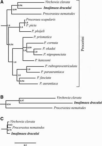
The COI data set consists of 658 characters, of which 311 are parsimony-informative and 42 are variable but not parsimony-informative. Of those, 93 occur in the first position, 42 in the second position, and 218 in the third position. The 16S data set consists of 491–494 positions depending on alignment. Excluding the alignment variable regions leaves 392 characters, of which 155 are parsimony-informative and 33 are variable but not parsimony-informative. The 18S data set consists of 1872–1878 positions depending on alignment. Excluding the alignment variable regions leaves 1686 characters, of which 318 are parsimony-informative and 118 are variable but not parsimony-informative. The results from the Bayesian analyses are presented in Figure 4A, outgroups are not shown. The combined data as well as the separate mitochondrial and nuclear data sets identifies a subclade consisting of I draculai, V. clavata and P. nematodes, but the nuclear data supports a closer relationship between P. nematodes and V. clavata (Figure 4C), while the mitochondrial and the combined data are in preference for I. draculai and V. clavata as sister taxa (Figure 4A, B).
(A) Majority rule consensus tree from the Bayesian analyses from the combined dataset, outgroups are not shown; (B) support values and branch lengths from the mitochondrial data for the V. clavata, I. draculai and P. nematodes node; (C) support values and branch lengths from the nuclear data for the same node as in (B).

DISCUSSION
Autolytines that have been studied in more detail are commonly found to build thin semi-hyaline tubes in which they reside. These tubes are either made in association, or attached directly on to the hydroid colonies on which they prey (Gidholm, Reference Gidholm1967; Fischer et al., Reference Fischer, Mewes and Franke1992; Genzano & San Martín, Reference Genzano and San Martín2002). Not all autolytines seem to secrete tubes, and may instead live within the water channel system of sponges as Proceraea hanssoni (Nygren, Reference Nygren2004), or in induced galls on their hydrocoral host as Proceraea penetrans (Wright & Woodwick, Reference Wright and Woodwick1977), or in induced modified theca of their hydroid host as Proceraea rzhavsky (Britayev & San Martín, Reference Britayev and San Martín2001; but see Nygren, Reference Nygren2004 for a taxonomic discussion of the name).
In their description of I. draculai the authors hypothesized that their new species might live as a parasite in one of the accompanying species in the sample. The reason for this was the absence of compound chaetae, a feature that often is associated with a parasitic life style among polychaetes (Martin & Britayev, Reference Martin and Britayev1988). We found the worms of I. draculai inside tubes situated in between the polyps of the sea pen F. quadrangularis. However, we were unable to find any type of modification of the Funiculina suggesting that I. draculai use their host in any other way than to build a tube on, and presumably also prey and feed upon. Thus, the association between I. draculai and its host does not seem to be any different to what is found between most other autolytines and their hydroid hosts (Fischer et al., Reference Fischer, Mewes and Franke1992; Fischer, Reference Fischer, Dorresteijn and Westheide1999). We can only speculate if the association of I. draculai with F. quadrangularis is obligate but it is possible, and the single specimen that was used for the original description is likely to have come from one of the F. quadrangularis that were present in their sample.
Our molecular data show that Imajimaea is a member of Procerini (Figure 4A), a fact also supported by our observation that I. draculai reproduce with anterior scissiparity where a single stolon is produced at a time from behind chaetigers 13 or 14 (Figure 1A–D), a type of reproduction only found within Procerini (Nygren, Reference Nygren2004). Our data also support a close relationship between the three genera Procerastea, Virchowia and Imajimaea represented here by one species each. The nuclear data are in slight favour for an Imajimaea (Procerastea and Virchowia) clade (Figure 4C), while the mitochondrial data and the combined data support a Procerastea (Virchowia and Imajimaea) clade (Figure 4A, B). In Nygren (Reference Nygren2004) Imajimaea was found to be the sister taxon to all other Procerini, hence a view not supported here. The results from Nygren (Reference Nygren2004), that were based on a combination of morphological characters for all taxa and molecular characters for some taxa, were only weakly supported. Molecular data from more Procerini taxa, especially representatives from the five genera not represented here, are desirable in order to establish a more robust phylogenetic hypothesis. No intraspecific genetic variation was found in the examined specimens of I. draculai, probably due to the fact that the individuals were sampled within a radius of 20 km. Specimens from other localities in Skagerrak and the North Atlantic would allow us to tell more about the population structure of I. draculai, and now that we know where to look for the worms, this might not be an impossible mission.
ACKNOWLEDGEMENTS
We would like to thank Hans G Hansson and Matz Berggren for collecting the specimens during the ‘Pandalina IV’ expedition. Thanks also to Daniel Martin for an excellent review of the manuscript. Financial support was obtained from the Swedish Species Information Centre (A.N., grant numbers 140/07 1.4, dha 166/08 1,4) and the Magnus Bergvall Foundation.