Introduction
Laryngeal dysplasia is a recognised pre-cancerous condition with an estimated annual incidence of 10.2 per 100,000 in men and 2.1 per 100,000 in women.Reference Bouquot and Gnepp1 The clinical management of these lesions is challenging; insufficient intervention can lead to a significant risk of malignant transformation, while excessive intervention risks scarring and permanent vocal changes. This complexity has driven the development of various monitoring and treatment protocols.Reference Zeitels2 However, comprehensive studies examining the effectiveness of these transitions and identifying key factors that accelerate the progression from dysplasia to cancer are limited. This study aims to evaluate the rate of dysplasia progression to invasive cancer and explore associated risk factors through 13 years of single-center data.
Materials and methods
Ethics committee approval was obtained for this retrospective data review (I5-308-20). Patient records from 1 January 2005 to 31 December 2017 were screened, including individuals who underwent direct laryngoscopy and whose histopathological results indicated dysplasia. All subjects were adults (at least 18 years of age). Patients who were subsequently diagnosed with invasive cancer within 3 months of the initial biopsy were excluded from the analysis, based on the assumption that the initial biopsy may have been too superficial to identify an already existing invasive carcinoma. Similarly, individuals were excluded from the study if their pathology reports indicated at least carcinoma in situ, if they had a history of radiation therapy for head and neck cancer, or if they had previously undergone laryngeal surgery with pathology results showing dysplasia or invasive carcinoma. These exclusions were implemented to ensure the study cohort comprised only patients with primary laryngeal dysplasia or carcinoma in situ, as initially intended.
Demographic data, history of smoking and alcohol use, follow-up period, treatment type and evidence of cancer progression were extracted and analysed. The site of biopsy, treatment modality post-diagnosis and follow-up outcomes, including malignancy transformation, were compared. Following the discussion of the biopsy pathology results at our in-clinic meeting, which includes all members of the ear, otorhinolaryngology, head and neck department, cases of severe dysplasia and carcinoma in situ are referred to the Radiation Oncology Clinic for additional consultation. The treatment plan is then developed with careful consideration of the meeting outcomes, the patient’s preferences, co-morbidities, and, if available, the results of the radiation oncology consultation, ensuring a personalised and comprehensive approach to care.
Statistical Analysis
Categorical data were expressed as frequency and percentage, while continuous variables were presented as mean plus or minus standard deviation (SD) or median with interquartile range (IQR) depending on normality, assessed via the Shapiro–Wilk test. Categorical variables were analysed using Pearson’s chi-squared and Fisher’s exact tests. The Mann–Whitney U-test and independent t-test were used for continuous data comparison. Kruskal–Wallis test was used for non-parametric comparison of continuous variables according to dysplasia severity, and Bonferroni correction was used in post-hoc tests for significant cases. A univariate logistic regression was conducted to identify independent risk factors for cancer progression, presented with odds ratios (OR) and 95 per cent confidence intervals. Analyses were performed using IBM SPSS 23.0 software package (IBM Corp., Armonk, NY), with a p-value less than 0.05 considered statistically significant.
Results
A total of 221 patients were included, with an average age of 55.8 plus or minus 11.2 years. The cohort predominantly comprised males (88.2 per cent). The median smoking history was 30 packs/year, and 19 per cent reported alcohol use. The individuals were categorised according to their alcohol consumption status. The quantity of alcohol consumption was not considered. Hypertension (27.1 per cent), diabetes mellitus (17.2 per cent) and coronary artery disease (16.4 per cent) were the most common co-morbidities. Dysphonia was the predominant symptom (94.2 per cent). Based on the World Health Organization (WHO) 2005 classification of laryngeal dysplasia, the incidence of mild dysplasia was higher among patients (39.5 per cent). Treatment approaches varied, with biopsy followed by cold excision (42.6 per cent) and biopsy with observation (40.4 per cent) being the most frequent. The rate of observation or subsequent excision following pathology is closely associated with the severity of the initial dysplasia. One key factor contributing to the high observation rates after biopsy is the predominance of mild dysplasia. Information about the general characteristics of the patients included in the study and the treatment methods applied are given in Table 1.
Patient demographics, pathological classification of laryngeal dysplasia and applied treatment modalities

Findings are presented as mean ± SD, median (IQR), or n (%).
Abbreviations: BPH = benign prostate hyperplasia; COPD = chronic obstructive pulmonary disease; IQR = interquartile range; SD = standard deviation; WHO = World Health Organization.
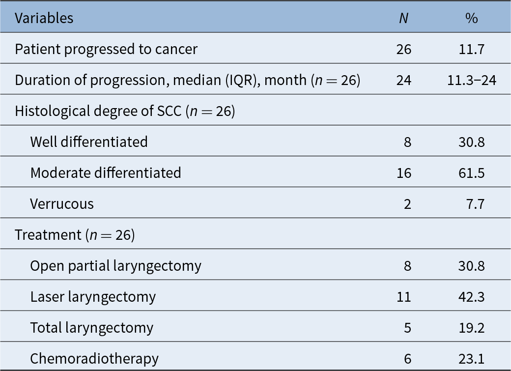
Cancer progression was identified in 26 patients (11.7 per cent), with a median time to progression of 24 months (IQR: 11.3–24). Among these, moderately differentiated cancer was most common (61.5 per cent). Surgical interventions included laser laryngectomy (42.3 per cent) and partial laryngectomy (30.8 per cent). All cases that progressed to cancer were classified as glottic T1 and T2 lesions. These patients were referred for consultation with the radiation oncology department, and treatment decisions were made based on a combination of the consultation results, the patient’s co-morbidities and their preferences. For patients with difficult exposure during direct laryngoscopy (D/L), partial surgical approaches, such as vertical laryngectomy or cordectomy, were preferred. In contrast, laser laryngectomy was chosen for patients with more favorable exposure. Descriptive statistics of follow-up findings of patients who progressed to cancer are given in Table 2.
Follow-up outcomes and treatment approaches for patients progressing to invasive cancer

Abbreviations: IQR = interquartile range; SCC = squamous cell carcinoma.
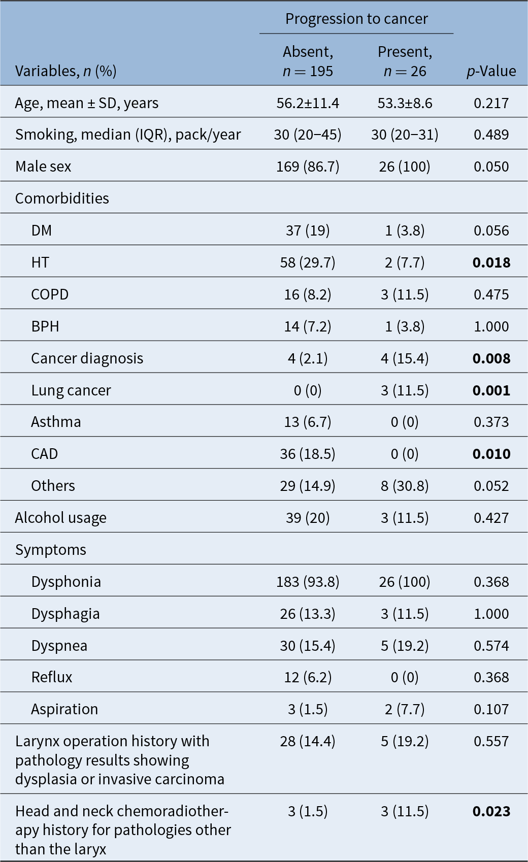
No significant age difference was found between progressing and non-progressing patients (p = 0.217). All patients included in the study were smokers, and no significant correlation was found between the amount of cigarette consumption and progression to cancer (p = 0.489). Co-morbidities such as hypertension and coronary artery disease were more common in the non-progressing group. Cancer history (15.4 per cent vs. 2.1%; p = 0.008), history of chemoradiotherapy for head and neck cancers originating from sites other than the larynx, including the oral cavity, oropharynx, nasopharynx and thyroid (11.5 per cent vs 1.5 per cent; p = 0.023) and lung cancer incidence (11.5 per cent vs. 0 per cent; p = 0.001) were higher in the progressing group. General characteristics of patients with progressive disease is given in Table 3.
Comparison of general characteristics between patients with and without progression to invasive cancer

Findings are presented as mean ± SD, median (IQR) or n (%). *Bold values indicate statistically significant results (p < 0.05).
Abbreviations: BPH = benign prostatic hyperplasia; CAD = coronary artery disease; COPD = chronic obstructive pulmonary disease; CRT = chemoradiotherapy; DM = diabetes mellitus; HT = hypertension; IQR = interquartile range.
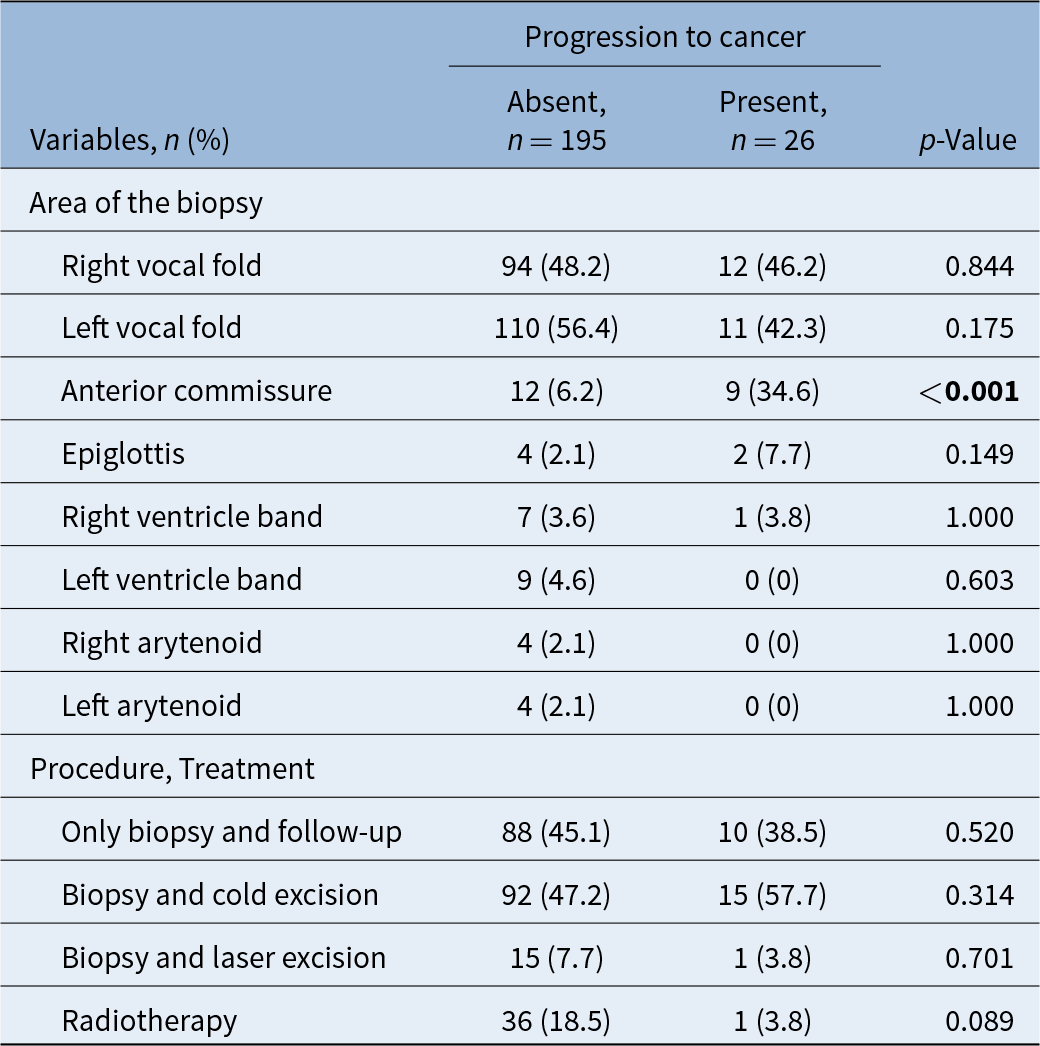
Progression was notably linked to the site of the initial biopsy, with anterior commissure involvement showing a significantly higher risk (34.6 per cent vs. 6.2 per cent; p < 0.001). The odds ratio of anterior commissure involvement in patients with progression to invasive cancer was 8.074 (95 per cent CI: 2.979–21.879). Radiotherapy as an initial treatment was more common in non-progressing cases (18.5 per cent vs. 3.8 per cent), but this was not statistically significant (p = 0.089) (Table 4).
Comparison of biopsy site and treatment approaches between patients with and without progression to invasive cancer

Findings are given as n (%). Pearson chi-squared test. Fisher’s exact test. Results that were significant in post-hoc tests were shown with different lowercase letters. *Bold values indicate statistically significant results (p < 0.05).
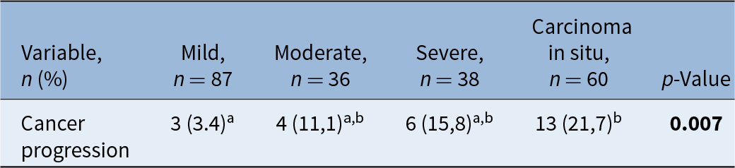
The cancer progression rates have been analysed in relation to the initial severity of dysplasia among patients. The findings indicate that the incidence of cancer progression is notably higher in patients diagnosed with carcinoma in situ, at a rate of 21.7 per cent, compared to a mere 3.4 per cent in those with mild dysplasia, with a statistically significant p-value of 0.007 (Table 5).
Relationship between cancer progression and dysplasia severity

Findings are given as n (%). Fisher’s exact test. Results that were significant in post-hoc tests are shown with different lowercase letters. *Bold values indicate statistically significant results (p < 0.05).
Discussion
Approximately 90 per cent of malignant laryngeal tumours originate from pre-cancerous lesions.Reference Hakeem, Hakeem and Pradhan3 Literature on long-term follow-up of laryngeal dysplasia and its progression to invasive cancer shows considerable variability. Reported cancer progression rates from dysplasia vary widely, ranging from 2 per cent to as high as 74.3 per cent.Reference Minni, Barbaro, Rispoli, Diaferia, Bernardeschi and Filipo4, Reference Sampedro, Alvarez, Suarez and Nistal5 For example, a 2008 review from the University of Wisconsin followed 3,107 biopsy results and found a malignant transformation rate of 8.2 per cent. When analysed according to the severity of dysplasia, the rate of malignant transformation was significantly higher in cases of severe dysplasia and carcinoma in situ (18.1 per cent).Reference Isenberg, Crozier and Dailey6 Similarly, a 2011 study from Paisley, Scotland, which analysed 131 patients over a 10-year period, reported a malignant transformation rate of 16 per cent, with an average time to malignancy of 43 months.Reference Montgomery and White7 Other retrospective study conducted in Baltimore assessed 107 patients over 20 years and found a progression rate of 8.4 per cent, with no significant association between the severity of dysplasia and cancer progression.Reference Karatayli-Ozgursoy, Pacheco-Lopez, Hillel, Best, Bishop and Akst8 A 2010 meta-analysis by Weller et al., which included nine studies encompassing 940 patients, highlighted the variability in sample sizes across existing research, ranging from 26 to 116 patients. This meta-analysis reported a 14 per cent overall progression rate and a threefold increased risk of progression to carcinoma in situ. The temporal range for progression varied widely across studies, from 11 months to 14 years, with insufficient data to make precise predictions about the timing of malignant transformation.Reference Weller, Nankivell, McConkey, Paleri and Mehanna9 The present study, which includes a cohort of 223 patients followed over 13 years, provides valuable additional insights. The progression rate in this cohort was 11.7 per cent, with an average time to malignancy of 24 months. Notably, patients with carcinoma in situ exhibited significantly higher malignant transformation rates compared to those with mild dysplasia, consistent with the findings of the meta-analysis of Weller et al.Reference Weller, Nankivell, McConkey, Paleri and Mehanna9
The anterior commissure has been a focal point of debate due to its anatomical and diagnostic complexities.Reference Hendriksma and Sjögren10, Reference Alonso, González, Teno, Durbán, Pallàs and Mestre11 Defined by Olofsson as the ventral region between the vocal cords with an extension of 2–3 mm including the vocal ligament’s elastic condensation,Reference Olofsson12 its involvement often poses diagnostic and therapeutic challenges. The 1996 study on embryological development by Rucci et al. and the 2017 comparative study of Prades et al.Reference Rucci, Gammarota and Cirri13, Reference Prades, Peoc’h, Petcu, Karkas, Dumollard and Gavid14 highlighted unique characteristics of the anterior commissure, emphasising its propensity for deep tissue extension and potential for diagnostic underestimation, often contributing to poor prognosis.Reference Zohar, Rahima, Shvili, Talmi and Lurie15, Reference Chone, Yonehara, Martins, Altemani and Crespo16 This study found that dysplastic lesions involving the anterior commissure had an approximately 8.2-fold increased risk of progression to invasive cancer compared to those without such involvement, underscoring the need for further research on tailored follow-up and treatment protocols for these cases.
Limitations
This study’s retrospective nature limits the ability to control for all potential confounders. Future research should focus on prospective analyses to verify these findings and further elucidate the mechanisms behind the observed progression risks.
• This study determined an 11.7 per cent progression rate of laryngeal dysplasia to cancer
• A significant association was found between cancer progression and initial biopsies obtained from the anterior commissure
• Lesions involving the anterior commissure carried an approximately 8.1-fold increased risk of progression
Conclusion
This study identified an 11.7 per cent progression rate of laryngeal dysplasia to invasive cancer. The most critical risk factors for malignant transformation were anterior commissure involvement and the histological severity of dysplasia. Dysplastic lesions involving the anterior commissure posed an approximately 8.2-fold higher risk of progression, highlighting the importance of targeted management. Further prospective studies are warranted to refine monitoring and treatment strategies for laryngeal pre-cancerous lesions.
Statement of grant or other support
The authors have no conflicts of interest to disclose. The authors have no sources of financial support or funding to disclose.
Statement of ethics approval
The study was conducted in accordance with the Declaration of Helsinki and was approved by the Ankara University Human Research Ethics Committee (No. I8-539-20) on 2 September 2020, with the need for written informed consent is waived.