INTRODUCTION – BASIC BIOLOGY OF EIMERIA
The Eimeria are obligate, intracellular parasites belonging to the phylum Apicomplexa, a group of organisms characterized morphologically by a number of specialized organelles, most notably those from within the apical complex. These generally include micronemes, rhoptries and dense granules, as well as a conoid and polar rings that appear to provide the structural stability (Hu et al. Reference Hu, Johnson, Florens, Fraunholz, Suravajjaia, DiLullo, Yates, Roos and Murray2006) required for the apical organelles to complete their roles in host cell attachment, invasion and remodelling (Plattner and Soldati-Favre, Reference Plattner and Soldati-Favre2008; Cowper et al. Reference Cowper, Matthews and Tomley2012; Kemp et al. Reference Kemp, Yamamoto and Soldati-Favre2013).
Over 1000 species of Eimeria have been named, the majority of these parasitizing the intestinal epithelia of vertebrates. They include species infecting horses, domestic dogs and cats, and wildlife, as well as economically significant species that infect rabbits, cattle, sheep, pigs, turkeys and chickens. Individual species show a high degree of host and site specificity, generally parasitizing a single host species and targeting specific cell types within that host. Although some species are known to infect unrelated hosts, in these cases they have been found to exhibit attenuated life cycles and do not cause significant levels of disease. The reasons for this specificity are unclear, however, it is assumed to be due to a combination of genetically determined factors and specific host immune responses (Rose, Reference Rose and Soulsby1987). Similarly, it is not understood why, in the normal host, the parasite invades and develops within specific sites although, in the laboratory, Eimeria species have been induced to complete the typical life cycle in other than the usual cell types (Fernando, Reference Fernando and Long1990). Improved understanding of the specificity of surface, microneme and rhoptry proteins is, however, shedding light on this question (Cowper et al. Reference Cowper, Matthews and Tomley2012).
Life cycle of Eimeria
The life cycle of Eimeria is complex but can be conveniently viewed as occurring in three distinct stages – sporogony, schizogony and gametogony. Under suitable environmental conditions of oxygen supply, humidity and temperature, the free-living stage of the organism – the oocyst – undergoes sporogony to form a sporulated oocyst. Sporulated oocysts of Eimeria contain four sporocysts, each of these containing two sporozoites. Following ingestion of the sporulated oocyst by the host, the microenvironment of the host's digestive tract stimulates excystation of the oocyst, resulting in the release of motile sporozoites. In the chicken, the effects of bile, enzymes and the grinding action of the gizzard are known to assist excystation (Davis, Reference Davis, Rose, Payne and Freeman1981).
Subsequent to their release, the sporozoites enter the host by penetrating intestinal epithelial cells. Depending on the individual species, further development occurs either at the site of entry or in other areas of the intestinal mucosa. Of the species of Eimeria parasitizing the domestic fowl, Eimeria acervulina, Eimeria maxima, Eimeria necatrix and Eimeria tenella develop in crypt epithelial cells, while Eimeria brunetti and Eimeria praecox develop at the site of entry (Fernando, Reference Fernando and Long1990).
Schizogony (or merogony) is the process of asexual reproduction that gives rise to merozoites following the entry of sporozoites into the appropriate host cells. Mature merozoites are superficially similar to sporozoites and, following their release from infected cells, move to reinfect other cells. This usually occurs in cells near to those from which the merozoites originally emerge; however, in the case of E. necatrix, second-generation merozoites travel from the small intestine to the caecum to continue development (Fernando, Reference Fernando and Long1990).
Following what is generally a fixed number of cycles of schizogony (two to four generations in most Eimeria species), released merozoites enter suitable cells and undergo gametogony (see Walker et al. Reference Walker, Ferguson, Miller and Smith2013 for a review), forming either a microgamont (male) or a macrogamont (female). Macrogamonts develop into a single macrogamete, while microgamonts give rise to many microgametes via cellular division similar to that occurring in schizogony. Microgametes are motile by means of flagella and, after their release, travel to host cells containing macrogametes. The microgamete penetrates and fuses with the macrogamete forming a zygote. A wall develops around the zygote, giving rise to the oocyst, which is released into the intestinal tract and shed in the feces.
Characteristics of coccidiosis
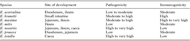
The clinical effects of disease caused by Eimeria vary widely according to the level of infection, the strain of host (Bumstead and Millard, 1992), the age and immune status of the host, the site of infection and the species and strain of Eimeria, which vary widely in their degree of pathogenicity (Rose, Reference Rose and Soulsby1987). Table 1 lists the seven species of Eimeria infecting the chicken, shows the normal site of development as well as the pathology and immunogenicity associated with each. As a generalization, overt symptoms of coccidiosis can include weakness, listlessness, loss of appetite, poor weight gain due to low feed conversion, diarrhoea that may be accompanied by mucus or blood and, in extreme cases, haemorrhaging and death (Marquardt et al. Reference Marquardt, Demaree and Grieve2000). These are believed to be due to physical damage caused to the gut mucosa during development and proliferation of the parasite and, therefore, severity of illness is largely due to the magnitude of the infecting dose and the degree of reinfection. Essentially the disease is self-limiting as all merozoites develop into gametes following the completion of schizogonic cycles and, under natural conditions involving low doses, a relatively harmless balance between the parasite and host is maintained. However, in intensive rearing facilities, where birds are potentially exposed to heavily contaminated litter, a damaging level of transmission can occur.
Site of development, relative pathogenicity and relative immunogenicity of the seven species of Eimeria parasitic in chickens

CONTROL OF COCCIDIOSIS BY CHEMOPROPHYLAXIS
Practices of good animal husbandry are essential to control coccidial infection and prevent transmission of disease via the fecal/oral route; however, in the intensive rearing conditions found within the commercial poultry industry, where birds are continually exposed to contaminated litter, such practices alone are insufficient. Thus, for the last 60–65 years, control of the disease has relied primarily upon the use of anticoccidial chemicals included in the feed; these drugs are convenient to administer and have proven to be cost effective. Within the broiler industry in the USA, for example, it has been reported that a vast majority of complexes use anticoccidial drugs (Sangster et al. Reference Sangster, Batterham, Chapman, Duraisingh, Le Jambre, Shirley, Upcroft and Upcroft2002; Chapman et al. Reference Chapman, Barta, Blake, Gruber, Jenkins, Smith, Suo and Tomley2013).
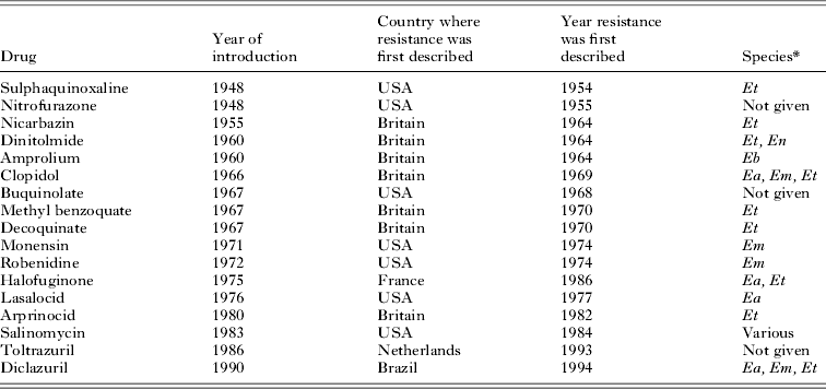
The extensive use of in-feed medication to control coccidiosis began in the late 1940s following a ‘landmark publication’ (see Chapman, Reference Chapman2009) by Grumbles et al. (Reference Grumbles, Delaplane and Higgins1948) showing that it was possible to control coccidiosis by the inclusion of relatively low concentrations of sulphaquinoxaline in feed. Since that time, large numbers of anticoccidials have been introduced into the poultry industry. Unfortunately, resistance has eventually developed to them all (Stephan et al. Reference Stephan, Rommel, Daugschies and Haberkorn1997; Table 2).
Summary of reported resistance to anticoccidials in field strains of Eimeria (adapted from Chapman, Reference Chapman1997)
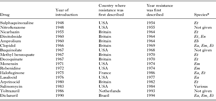
* Species: Ea, E. acervulina; Eb, E. brunetti; Em, E. maxima; En, E. necatrix; Et, E. tenella.
Resistance, as opposed to naturally occurring differences in sensitivity due to parasite strain or species, is considered to arise following extended exposure to chemicals and can be defined as, ‘the ability of a parasite strain to multiply or survive in the presence of concentrations of a drug that normally destroy parasites of the same species or prevent their multiplication’ (Chapman, Reference Chapman1997). Little is known regarding the molecular mechanisms involved in the development of resistance to anticoccidials in Eimeria species, although some information on the mode of action of some compounds and their effect on biochemical pathways is available. They include compounds affecting membrane function, mitochondrial function and co-factor synthesis (Chapman, Reference Chapman1997). The most widely used anticoccidials are the ionophorous antibiotics, which include lasalocid, maduramicin, monensin, narasin and salinomycin, and are known to affect cellular processes involving cation transport through the cell membrane. Other anticoccidials affect co-factor uptake and synthesis, including amprolium, which inhibits thiamine uptake, and antifolates such as dihydrofolate reductase inhibitors and sulphonamide compounds, which prevent dihydrofolate synthesis. The quinolone anticoccidials, such as decoquinate and methyl benzoquate, affect mitochondrial function and are used less frequently today because of the relatively rapid development of resistance to them that arose not long after their introduction in the 1970s (Chapman, Reference Chapman1997). They act to block electron transport in the mitochondrion and, so inhibit respiration (Wang, Reference Wang1976) although how this occurs is not fully understood.
Cross-resistance and multiple resistance to anticoccidial medications has also been noted and complicates potential control via the rotation of different drugs. Cross-resistant strains exhibit resistance to compounds sharing a similar mode of action and may arise from exposure to a single anticoccidial of a particular type, while multiple resistant strains are resistant to compounds having different modes of action and arise following sequential exposure to the compounds involved (Chapman, Reference Chapman1993). In a study of resistance to anticoccidials in ten Eimeria field isolates from northern Germany, nine out of the ten isolates showed resistance to the compounds tested, with seven out of the ten having developed multiple resistance (Stephan et al. Reference Stephan, Rommel, Daugschies and Haberkorn1997). Similar results have been shown in isolates from broiler farms in the UK (Chapman, Reference Chapman1993) and further indicate the degree of resistance to anticoccidials and its widespread occurrence.
Despite the fact that, ‘Most [anticoccidial] drugs are no longer as effective as when they were first introduced due to the development of drug resistance’ (Chapman et al. Reference Chapman, Barta, Blake, Gruber, Jenkins, Smith, Suo and Tomley2013), there is little evidence that use of in-feed, anticoccidial medication has declined, nor that management of the control of coccidiosis has become particularly problematic as a result. An important paper by Chapman (Reference Chapman1999) probably explains why. It is clear from this extensive, careful study that most drugs actually continue to be effective in industrial settings precisely because they only partially interfere with the eimerian life cycle, allowing entire flocks of chickens to acquire natural immunity through exposure to the relatively low, albeit significant, number of parasites that do not succumb to chemoprophylaxis. As Chapman (Reference Chapman2009) notes, this was actually foreseen by Grumbles et al. in 1948!
CONTROL OF COCCIDIOSIS BY VACCINATION WITH LIVE PARASITES
Vaccination with controlled doses of live, virulent oocysts
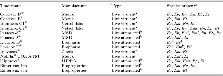
The fact that at least part of the explanation for the effectiveness of in-feed, anti-coccidial medication is via the development of natural immunity has encouraged the belief that vaccination with controlled doses of live, fully virulent parasites is a genuine alternative means to control coccidiosis. As early as the 1920s, it was recognized that Eimeria infections of chickens are self-limiting, with sexual development beginning after a fixed number of cycles of asexual division; and young chickens could be infected and immunized readily, including via addition of small (and, therefore, non-pathogenic) doses of oocysts into their feed (Johnson, Reference Johnson1927, Reference Johnson1932; Tyzzer, Reference Tyzzer1929; Tyzzer et al. Reference Tyzzer, Theiler and Jones1932). Over the years, this has inspired interest in the potential of immunoprophylactic means of control and has led to the introduction of a number of commercially available, live oocyst vaccines directed against coccidiosis (Table 3). The first of these, Coccivac®, was introduced in the USA in 1952 and is similar to Immucox®, which was released during the 1980s. Both utilize controlled doses of virulent oocysts administered to young chickens, usually during the first 2 weeks after hatching. Although having met with some success, it is sometimes difficult to control the infection rate when vaccinating with live parasites, and the potential exists for infectious cycling and the ingestion of large numbers of oocysts by susceptible birds, leading to disease (Chapman et al. Reference Chapman, Barta, Blake, Gruber, Jenkins, Smith, Suo and Tomley2013), notwithstanding the development of several regimens to better standardize mass dosing of farmed poultry (Williams, Reference Williams2002).
Examples of live oocyst vaccines against coccidiosis in chickens (adapted, in part, from Cornelissen and Schetters, Reference Cornelissen and Schetters1996; Vermeulen et al. Reference Vermeulen, Schaap and Schetters2001)

* Species: Ea, E. acervulina; Eb, E. brunetti; Em, E. maxima; Emi, E. mitis; En, E. necatrix; Ep, E. praecox; Et, E. tenella.
a Virulent parasites (low dose).
b Precocious strains.
c Two strains present.
d Egg-adapted lines.
e Delivered by in ovo inoculation of oocysts into embryonated eggs using proprietary machinery.
f Ionophore resistant virulent parasites (low dose).
Vaccination and chemotherapy in combination
The alternation of drug treatment programmes and immunization with live, virulent oocysts as a means of controlling coccidiosis has been practised for many years. It is possible that such regimens have helped to slow the development of resistance and extend the life of anticoccidials (Chapman, Reference Chapman1994; Peek and Landman, Reference Peek and Landman2006; Chapman et al. Reference Chapman, Jeffers and Williams2010; Jenkins et al. Reference Jenkins, Klopp, Ritter, Miska and Fetterer2010). The concept has even led to the release of the live, virulent vaccine, Nobilis® COX ATM, which comprises ionophore-resistant strains of E. acervulina, E. maxima and E. tenella. Following delivery of the vaccine, birds are reared with ionophores in the feed, aiming to reduce the risk of serious infection by field strains during the natural development of immunity (Vermeulen et al. Reference Vermeulen, Schaap and Schetters2001).
Vaccination with irradiated oocysts
To overcome the potential problems associated with vaccinating flocks of birds with virulent oocysts, extensive studies have been conducted investigating the effects of ionizing radiation on Eimeria in an attempt to develop an attenuated line suitable for use as a vaccine in chickens. For example, Sokolic et al. (Reference Sokolic, Movsesijan, Tanielian and Abu Ali1976) showed that oral administration of irradiated oocysts of E. tenella, E. brunetti and E. necatrix could significantly decrease the symptoms of coccidial infection when compared with non-immunized control birds. Following challenge with large doses of infective oocysts of the three species, 70% of control chickens died while all birds previously treated with irradiated oocysts survived.
The difficulty in assessing this result, and similar early experiments investigating irradiation, was that conditions were not standardized. Due to this, conclusions were drawn suggesting that the effects of irradiation were merely to destroy a proportion of the treated oocysts and, therefore, administration of large numbers of irradiated oocysts was, in reality, equivalent to vaccination with small doses of virulent oocysts (Long and Rose, Reference Long, Rose, Taylor and Muller1980). However, work in the 1990s with E. tenella indicated that, while exposure to radiation has no effect on excystation of oocysts or the invasive capabilities of sporozoites, it greatly reduces subsequent schizogonic development (Jenkins et al. Reference Jenkins, Augustine, Danforth and Barta1991). Gilbert et al. (Reference Gilbert, Fuller, Scott and McDougald1998) demonstrated that, following exposure of between 50–200 Gy of gamma-irradiation, a reduction in the numbers of developing E. tenella sporozoites of between 77–94% could be achieved. The effects seen on the development of the parasite are believed to be due to the generation of free radicals, and the fragmentation of chromosomes and large macromolecules (Gilbert et al. Reference Gilbert, Fuller, Scott and McDougald1998). In order to fully determine the effects of ionizing radiation and the protective potential of vaccines incorporating irradiated oocysts, a great deal more work is required that, until now, has not been taken up as either an academic or commercial pursuit. It seems unlikely that irradiated oocysts will be used in a commercial vaccine as no evidence has been made available indicating that the attenuation of virulence caused by radiation is a genetically stable trait (Shirley, Reference Shirley, Barta and Fernando1993).
Vaccination with attenuated parasite vaccines
In contrast to irradiation, precocious development does appear to be a selectable and genetically stable trait for various species of Eimeria (McDonald and Shirley, Reference McDonald and Shirley2009). Precocious lines of Eimeria are characterized by a life cycle reduced in the number and size of schizogonous stages and, thus, a considerably reduced reproductive potential. For example, in a study involving infection with E. maxima, an infective dose of 100 precocious line oocysts led to the release of 4×10⁵ progeny oocysts, while the same dose of the virulent parent strain produced 2·4×107 oocysts (Shirley and Bedrnik, Reference Shirley and Bedrnik1997). Additionally, precocious lines are significantly less pathogenic than the parent wild-type strains from which they are developed, but retain the ability to induce protective immunity effective against subsequent infection (reviewed recently by McDonald and Shirley, Reference McDonald and Shirley2009; Sharman et al. Reference Sharman, Smith, Wallack and Katrib2010). Hence, a number of vaccines utilizing live, attenuated parasites have been introduced to protect flocks of poultry against coccidiosis.
The selection of a precocious line of Eimeria was first demonstrated in E. tenella by Jeffers (Reference Jeffers1975), who discovered a resulting attenuation in virulence following the serial passage of the first oocysts released during the course of infection. Precocious lines were subsequently developed for all economically significant strains of Eimeria infecting chickens (Shirley and Long, Reference Shirley, Long and Long1990) and form the basis of the commercially available vaccines Paracox-8® and Livacox®. Paracox-8® contains precocious lines of the seven Eimeria species infecting chickens, including two different E. maxima lines, while Livacox® contains precocious lines of E. acervulina and E. maxima plus an egg-adapted line of E. tenella (Table 3).
Egg-adapted lines of Eimeria also display abbreviated life cycles in chickens and are developed by inoculating the allantoic cavity of embryonating chicken eggs with sporozoites. Ensuing development of the parasite occurs within the chorioallantoic membrane and following long-term, serial passages (over 100 for the Livacox® E. tenella line), a line of parasite results that is less pathogenic than the wild-type parent strain from which it was derived. Eimeria lines developed in this manner are generally characterized by having diminished numbers of sporozoites and/or merozoites (Bedrnik et al. Reference Bedrnik, Hiepe, Mielke, Drossigk, Eckert, Braun, Shirley and Coudert1995). However, some problems may exist with the use of egg-adapted lines; reversion to virulence following passage through chickens or significant loss in immunogenicity have been observed (Shirley, Reference Shirley, Barta and Fernando1993).
While having been accepted worldwide, vaccines utilizing attenuated parasites have some limitations. Their relatively high expense to produce has precluded their extensive use in vaccinating broiler birds and, in addition, it has been estimated that the number of oocysts required to vaccinate the world's broiler market cannot be produced (Cornelissen and Schetters, Reference Cornelissen and Schetters1996). Furthermore, the performance of birds vaccinated with live oocyst vaccines has not always equalled that of birds treated with anticoccidials (Danforth, Reference Danforth1998). This is believed to be due, in the main, to immunological variability, as the strain of E. maxima included in Immucox®, for example, has been shown to stimulate variable levels of protective immunity in birds challenged with field variants of E. maxima (Lee, Reference Lee, Barta and Fernando1993). This variability, often seen in parasites isolated from different geographical regions (Martin et al. Reference Martin, Danforth, Barta and Fernando1997), is able to be controlled by the inclusion of variant strains in the vaccine preparation (Danforth, Reference Danforth1998). As mentioned, the formulations of Paracox® vaccines available commercially contain attenuated lines of two different E. maxima strains in an attempt to combat this problem. Hence, for a combination of reasons, attenuated vaccines have mainly been used in the breeding and egg laying industries though some versions of attenuated vaccines (including one designated Paracox-5®) are comprised of fewer species (including, minimally, E. acervulina, E. maxima and E. tenella), are cheaper to produce and are, therefore, more acceptable to broiler farmers.
CONTROL OF COCCIDIOSIS VIA MATERNAL IMMUNIZATION
The almost inescapable lesson learned from the history of use of both medicated feed and live vaccines is that reduction rather than eradication of infection is the key to successful control of coccidiosis, with both approaches resulting in the same fundamental outcome – flocks of birds become solidly immune via exposure to relatively low numbers of oocysts, thereby limiting the damage caused by the asexual stage of the eimerian life cycle. This shared experience from these two means of parasite control suggests that an alternative approach, namely transmission-blocking mediated by maternal immunization, may be feasible.
Maternal immunity in chickens
The transfer of protective antibodies is recognized in all species of birds and mammals and, while the route of transmission varies between different species, the mechanism by which antibody transfer occurs is essentially similar. The protective effects elicited by maternal immunity are believed to be largely due to the transfer of IgG (or IgY in birds) class antibodies, mediated by Fc receptors. In the chicken, Fc receptors exposed to the maternal circulation localize in pit regions of the oocyte plasma membrane. Following binding to the receptors by ferritin-conjugated maternal IgY, the pit regions invaginate to form vesicles that are transported across the plasma membrane where the IgY is released into the egg yolk (Loeken and Roth, Reference Loeken and Roth1983). In a similar fashion, the IgY is then transported by Fc receptors on the yolk sac endoderm and released into the circulation of the developing embryo (Kowalczyk et al. Reference Kowalczyk, Daiss, Halpern and Roth1985). The levels of IgY transferred in this fashion correlate directly with the amount of antibody produced by the hen; the more produced the greater the amount transported into the egg yolk and, subsequently, delivered to the embryo.
There is a good deal of evidence suggesting that IgY antibodies are the primary effectors of maternal immunity in chickens. IgY antibodies are found in milligram quantities in egg yolks (Rose and Orlans, Reference Rose and Orlans1981; Losch et al. Reference Losch, Schranner, Wanke and Jurgens1986), and these levels have been shown to correlate strongly with immunity to Eimeria infection (Smith et al. Reference Smith, Wallach, Miller, Braun and Eckert1994a ). IgA and IgM antibodies are found only in small quantities in the egg white and do not react specifically with Eimeria antigens (Smith et al. Reference Smith, Wallach, Miller, Braun and Eckert1994a ). Additionally, gamma-livetin (egg yolk immunoglobulin) fractions taken from hens infected with E. maxima and injected into susceptible chickens, provide protection against challenge infection (Rose, Reference Rose1972). It is unlikely that other immune effectors such as cytokines would play a role in maternally derived immunity, given that they have a short half-life, mitigating against their persistence in the egg for the duration of the incubation period (Wallach et al. Reference Wallach, Smith, Braun and Eckert1995b ).
Potential of maternal immunity to control coccidiosis
It has been known for decades that maternally derived immunity can be used to control other infectious diseases of poultry, most notably Gumboro or infectious bursal disease. Relatively low levels of antibody are required to control infectious bursal disease and a maternally delivered vaccine utilizing killed virus particles was developed many years ago to protect broilers for the duration of their commercial lifetime – approximately 6–7 weeks (Box, 1985). Studies have also indicated the potential for maternal transfer of antibodies to control avian encephalomyelitis virus (Box, 1985) and Newcastle disease (Stone et al. Reference Stone, Brugh and Xie1992). Additionally, experiments involving vaccination of hens with an avirulent strain of Salmonella typhimurium have shown that maternal antibodies can prevent colonization of progeny by wild type Salmonella strains (Hassan and Curtiss III, Reference Hassan and Curtiss1996), and maternal transfer of IgY has been shown to afford significant protection against infection with Cryptosporidium baileyi (Hornok et al. Reference Hornok, Bitay, Szell and Varga1998).
The protective potential of maternally transmitted immunity against coccidiosis was first noted by Rose (Reference Rose1971), who attempted to transfer immunity to E. maxima by injecting chicks with sera taken from infected birds at different times following a single inoculation. Using oocyst production as a measure of infection, the results showed that sera taken from birds between 3 and 4 weeks after an initial infection were able to passively protect susceptible chicks against infection with E. maxima. Following up on this work, Rose (Reference Rose1972) compared the degree of infection in progeny of infected birds with that of progeny of uninfected birds and demonstrated maternal immunity to E. maxima; oocyst output in progeny hatched from eggs laid between 3 and 4 weeks after inoculation of hens was reduced by greater than 50% when compared with control birds. A perceived limitation of the effectiveness of maternal immunity, as indicated by these results, though, was that protection declined in progeny from eggs laid after the third week following inoculation of the hens and, by the sixth week, the protection afforded was negligible. However, it was subsequently shown that heavy infection with E. maxima can result in transfer of protection over a more prolonged period.
In a series of experiments (Smith et al. Reference Smith, Wallach, Miller, Braun and Eckert1994a ), hens were infected with large doses of E. maxima oocysts (20 000 per inoculum) in order to stimulate production of high levels of antibodies. In progeny challenged subsequently with E. maxima, a greater than 90% reduction in oocyst production was observed in hatchlings from eggs laid between 3 and 4 weeks after inoculation of the hens, and in hatchlings from those laid 8 weeks post-infection, an average reduction of 68% was observed. These levels of protection were further enhanced by the injection of hens with the adjuvant, Arlacel A, prior to inoculation with oocysts. A greater than 90% reduction in oocyst numbers was seen in offspring from eggs collected at 8 weeks post infection of hens, and progeny from eggs collected 19 weeks post infection showed a 60% reduction. These results imply that protection conferred by maternally derived antibodies can occur over extended periods and serve to affirm the potential of maternal immunity as a means of controlling coccidiosis.
A further potential advantage of a maternally delivered vaccine lies in the ability of antibodies raised against one species of Eimeria to recognize antigens of other species (Smith et al. Reference Smith, Wallach, Petracca, Braun and Eckert1994c ). Thus, maternal transfer of antibodies resulting from infection with E. maxima can confer protection in progeny chickens against infection with E. tenella. Hatchlings from eggs collected between 28 and 39 days following infection with E. maxima were challenged with E. tenella and showed a significant decrease in oocyst production of up to 62% when compared with controls. The mechanism by which cross-reactivity occurs is not fully understood, although it appears that different species may share conserved antigenic epitopes (Smith et al. Reference Smith, Wallach, Petracca, Braun and Eckert1994c ).
Identification of antigenic proteins and the development of a maternal vaccine comprised of gametocyte antigens
Knowledge that maternally derived IgY antibodies are the main effectors of maternal immunity enabled the development of ELISAs to assess antibody responses and identify putatively protective antigenic proteins. ELISA analyses of yolk from hens infected with E. maxima and of sera from their offspring showed that antibody responses occur to both asexual and sexual stages of Eimeria and that the responses observed change over time (Smith et al. Reference Smith, Wallach, Miller, Braun and Eckert1994a , Reference Smith, Wallach, Miller, Morgenstern, Braun and Eckert b ). High IgY titres were found in the egg yolk in the period 3–5 weeks following infection of hens and reacted with antigen from all developmental stages of Eimeria, while in later stages the antibodies were of lower titre and primarily recognized asexual stages (Smith et al. Reference Smith, Wallach, Miller, Braun and Eckert1994a ). Western blot analyses of the later stage, lower titre antibodies from egg yolk and progeny sera have consistently identified two proteins, a 250 kDa asexual stage protein (originally designated, incorrectly, as a 230 kDa protein) and an 82 kDa gametocyte (sexual stage) protein.
Limited work has been undertaken on characterizing the immunodominant 250 kDa asexual stage antigen from E. maxima due partly to a difficulty in isolating the protein in large quantities (Smith et al. Reference Smith, Wallach, Miller, Morgenstern, Braun and Eckert1994b ). In a study designed to indicate its protective effects, crude E. maxima merozoite extract was electrophoresed by SDS-PAGE and a 250 kDa protein band excised, emulsified in Freund's adjuvant and injected into hens. Progeny of the hens from between 28 and 39 days post-infection and challenged with E. maxima excreted 54% fewer oocysts than control birds (Smith et al. Reference Smith, Wallach, Miller, Morgenstern, Braun and Eckert1994b ). However, a rapid waning of protection was seen in the following 2-week period to the point where the benefits of maternally derived immunity were negligible. A possible explanation for this offered by the authors is that very small quantities of the 250 kDa merozoite cut-out were used to stimulate an immune response and would, therefore, be less likely to induce immunity over a more protracted period. The significant reduction in oocyst numbers observed, though, indicates the potential of the 250 kDa merozoite protein as a subunit vaccine candidate especially since similar levels of protection were seen in a preliminary study where birds were vaccinated with a ∼45 kDA recombinant fragment of this protein (Witcombe, Reference Witcombe2002).
The 250 kDA asexual stage protein from E. maxima was later identified as a member of the thrombospondin-related anonymous protein (TRAP) family and designated EmTFP250 (Witcombe et al. Reference Witcombe, Belli, Wallach and Smith2003). It was shown to localize to the micronemes of the parasite (Witcombe et al. Reference Witcombe, Ferguson, Wallach and Smith2004) and confirmed to be the orthologue of the E. tenella microneme protein, EtMIC4 (Tomley et al. Reference Tomley, Billington, Bumstead, Clark and Monaghan2001). The advantages of an immune response directed at proteins such as this, expressed during the asexual developmental stages of the parasite, are clear. Given that pathogenesis in coccidial infection is a result of physical damage caused by asexual replication within the intestinal mucosa, targeting asexual stages would not only block transmission of the parasite via the fecal-oral route, but would inhibit endogenous cycles of replication and, therefore, reduce resulting injury to the host. Further investigation of this, and other asexual-stage antigens, in the context of maternal immunization, appears warranted.
The 82 kDa gametocyte antigen, along with gametocyte proteins of 230 and 56 kDa, had been identified previously by Western blotting using protective sera taken from chickens 14 days after infection with E. maxima (Wallach et al. Reference Wallach, Mencher, Yarus, Pillemer, Halabi and Pugatsh1989). It was later demonstrated that these belong to a family of proteins involved in oocyst wall formation (Ferguson et al. Reference Ferguson, Belli, Smith and Wallach2003; Belli et al. Reference Belli, Smith and Ferguson2006). The protective potential of these antigens was indicated by the ability of a monoclonal antibody developed against the 56 kDa antigen to significantly reduce oocyst production in naive chickens challenged with E. maxima (Wallach et al. Reference Wallach, Pillemer, Yarus, Halabi, Pugatsh and Mencher1990). Similarly, transfer of immune sera raised against affinity-purified 56 and 82 kDa gametocyte antigens conferred protection to susceptible chickens (Wallach et al. Reference Wallach, Pillemer, Yarus, Halabi, Pugatsh and Mencher1990). In addition, affinity purified forms of E. maxima gametocyte proteins, when injected into hens with Freund's adjuvant, induced the transfer of maternal antibodies leading to a reduction in oocyst numbers excreted from E. maxima-challenged progeny of between 60 and 80% (Wallach et al. Reference Wallach, Halabi, Pillemer, Sar-Shalom, Mencher, Gilad, Bendheim, Danforth and Augustine1992, Reference Wallach, Smith, Petracca, Miller, Eckert and Braun1995a ). Furthermore, the affinity purified gametocyte antigens (APGA) have also been shown to provide significant levels of protection in offspring challenged with E. acervulina and E. maxima (Wallach et al. Reference Wallach, Smith, Petracca, Miller, Eckert and Braun1995a ).
These findings ultimately led to the development and release of the commercial maternal vaccine, CoxAbic®, which contains APGA and is injected in a water-in-oil adjuvant into the breast muscle of broiler breeder hens shortly before they begin producing eggs. Laboratory and floor pen trials showed that immunization with APGA provides maternal protection leading to a 50–80% reduction in total oocysts excreted by broiler birds challenged with E. maxima, E. acervulina and E. tenella (Wallach, Reference Wallach1997, Reference Wallach2002). In addition, a series of very large, multinational field trials of CoxAbic® demonstrated that performance of maternally protected broilers, as measured by egg production, body weight, feed conversion and mortality, is at least as good as that of control broilers fed anticoccidials or vaccinated with live, attenuated vaccines (Wallach et al. Reference Wallach, Ashash, Michael and Smith2008).
Potential problems associated with maternal immunization with purified gametocyte antigens
There are two potentially problematic features associated with the current transmission-blocking, maternally delivered vaccination strategy for coccidiosis embodied by CoxAbic®. The first is that the vaccine, like live vaccines, relies on growing parasites in strict coccidia-free conditions in specific, specialized facilities, plus carries an additional burden that gametocyte proteins then need to be purified from parasites within infected intestines of chickens. This is laborious and places a significant limit on production capacity. The production of recombinant versions of the key components of CoxAbic® is, arguably, a solution to this problem and, indeed, recombinant versions of the 56 and 82 kDa proteins have been produced and demonstrated to possess many of the same antigenic features of the native proteins (Belli et al. Reference Belli, Mai, Skene, Gleeson, Witcombe, Katrib, Finger, Wallach and Smith2004). Unfortunately, these recombinant proteins have never been tested for their maternal vaccine potential.
The second potential problem that can be envisaged for any vaccine utilizing maternal immunity is that of the observed waning in protection over time due to physiological breakdown of the antibodies and a dilution effect caused by rapid growth in broiler chickens (Smith et al. Reference Smith, Wallach, Miller, Braun and Eckert1994a ). At 2 weeks of age, high levels of protection are observed but by 3 weeks immunity to infection is negligible (Smith et al. Reference Smith, Wallach, Miller, Braun and Eckert1994a ). It has been estimated, however, that a 2–3 week period of protection would be sufficient to prevent an increase in peak oocyst numbers contaminating floor litter (Wallach et al. Reference Wallach, Smith, Braun and Eckert1995b ). Supporting evidence comes from floor-pen trials using the progeny of hens immunized with APGA. Peak oocyst litter counts from progeny of vaccinated birds, even at 7 weeks of age, showed an average reduction of 60–70% in comparison to negative control birds (Wallach, Reference Wallach1997, Reference Wallach2002). More recent, more extensive investigations showed that the reduction in oocyst excretion in progeny of CoxAbic®-vaccinated hens compared with control birds rose from 63% if birds were exposed to infectious oocysts at 4 days of age, to 85% by 39 days of age, up to 98% when they reached 57 days of age (Wallach et al. Reference Wallach, Ashash, Michael and Smith2008). This demonstrates that, just as has been observed in flocks fed on medicated feed or immunized with live vaccines, natural immunity improves throughout the life of maternally immunized birds and this holds the key to the successful management of coccidiosis.
CONCLUSION: THE FUTURE FOR CONTROL OF COCCIDIOSIS – RECOMBINANT VACCINE DEVELOPMENT?
Control of coccidiosis in the immediate future will continue to rely heavily on the use of anticoccidials but, if parasite resistance to prophylactic drugs increases and/or consumer concerns about the use of chemicals in food production grows, live vaccines and CoxAbic® might be expected to carve out larger niche markets. However, as noted already, both these approaches have inherent production limitations as well as labour and, therefore, cost issues.
Arguably, the best alternative for controlling coccidiosis in the future may lie with the development of a recombinant vaccine(s) that would potentially be easier to produce and more cost-effective to administer than vaccines utilizing live oocysts or purified native proteins. Indeed, substantial work has been undertaken in this field for several decades, centred on the selection of protective antigens that, in genetically modified form, would be included as subunits in such a vaccine. Many of the proteins studied to date have been identified as vaccine candidates on the basis of their cellular location and biological role in the development of the parasite, and include proteins associated with micronemes, rhoptries, dense granules and refractile bodies, which mediate a series of complex and specific molecular interactions between parasite and host (see Mercier et al. Reference Mercier, Adjogble, Daubner and Delauw2005; Plattner and Soldati-Favre, Reference Plattner and Soldati-Favre2008; Cowper et al. Reference Cowper, Matthews and Tomley2012; Kemp et al. Reference Kemp, Yamamoto and Soldati-Favre2013 for expert reviews). These vaccine candidates have been thoroughly and expertly reviewed recently (Blake and Tomley, Reference Blake and Tomley2014) so details will not be repeated here. Invariably, each individual antigen, whether tested as protein-, DNA- or vector-delivered vaccines (again, expertly reviewed by Blake and Tomley, Reference Blake and Tomley2014) has conferred partial protection against coccidiosis.
There has been, historically, a tendency to dismiss partially protective candidates from further consideration in the quest for a subunit vaccine against coccidiosis. This may have been a mistake. First, because we now know that individual recombinant components of CoxAbic® (e.g. the 56 kDA or the 82 kDa antigen), used as either recombinant protein or DNA vaccines, give only partial protection (Jang et al. Reference Jang, Lillehoj, Lee, Lee, Park, Cha, Lillehoj, Subramanian, Sriraman and Srinivasan2010; Xu et al. Reference Xu, Zhang and Tao2013), yet the cocktail of antigens that is CoxAbic® does something more. Perhaps in combination, some of these asexual-stage proteins may indeed be effective. And, second, as emphasized throughout this review, the three control strategies that have so far enjoyed some success against coccidiosis in the field – chemoprophylaxis, live vaccines and transmission-blocking maternal immunization – share one crucial thing in common, namely that they confer sufficient protection against infection with multiple species of Eimeria, early in the life of chickens, to control disease but not eradicate the parasite. This allows natural immunity to develop in flocks of birds, complementing and enhancing the protection afforded by these three quite different control strategies. We neglect or forget this fact at our peril. Indeed, as Chapman (Reference Chapman2009) reminds us, in the 1970s, several highly effective new drugs were introduced and these almost completely controlled Eimeria, the result being that flocks were extremely vulnerable to infection during the drug-withdrawal period prior to slaughter. Furthermore, the development of resistance to these drugs by the parasite was rapid and widespread.
Whilst we can, and should, be optimistic about the future development of effective subunit vaccines against coccidiosis, there are (at least) two major practical issues that require consideration: first, how may such vaccines be delivered in a cost-effective way; and, second, how might they be delivered to young birds whose immune system is still developing and is naïve? We may already have some of the answers – the vaccination of breeder hens is potentially extremely economical given that each hen produces between 100 and 150 offspring in her commercial life, and advantageous in that each of these chicks is protected by their mothers’ antibodies from the moment they hatch.