Introduction
Significant progress has been made with nanotechnologies which are applied in many fields of the food and pharmaceutical industries and in environmental technology, science, and energy, etc. (Bayda et al., Reference Bayda, Adeel, Tuccinardi, Cordani and Rizzolio2020). Interest in tubular nanoformulations has grown in recent years, due to their potential application in various fields of nanomedicine (Patra et al., Reference Patra, Das, Fraceto, Campos, del Pilar Rodriguez-Torres, Acosta-Torres, Diaz-Torres, Grillo, Swamy, Sharma, Habtemariam and Shin2018; Liu et al., Reference Liu, Fakhrullin, Novikov, Panchal and Lvov2019). Many nanomaterials are available naturally or are produced artificially. Among these, halloysite nanotubes (HNTs) are abundantly available in nature (economic and sustainable deposits) or can be produced synthetically (Jin et al., Reference Jin, Fu, Yang and Ouyang2015). In nature, HNTs are present in soils and rocks exposed to atmospheric agents typical of humid tropical and subtropical areas, and particularly in volcanic ash and tephra in a variety of climates (Massaro et al., Reference Massaro, Colletti, Lazzara, Milioto, Noto and Riela2017). Natural deposits of HNTs are found in Japan, New Zealand, China, the United States, Korea, Japan, Turkey, Brazil, and France. Halloysite clay is available, therefore, in large quantities (many thousands of tons) from various mines, at low cost, making halloysite an extremely competitive alternative to carbon-based nanomaterials (Jawaid et al., Reference Jawaid, Qaiss and Bouhfid2016). The physical properties of the HNTs, such as length, tube-wall thickness, internal radius, dispersion, and zeta potential, are influenced by the charatcteristics of the deposit, however (Yuan et al., Reference Yuan, Tan and Annabi-Bergaya2015). Inhomogeneous HNTs with lengths between 100 nm and 2 mm are available commercially. Long nanotubes have been shown to act as inducers of inflammation and cell damage (Wang et al., Reference Wang, Gong, Rong, Gui, Hu and Xu2018). The size of the nanoparticles (NPs) has an effect on how the body’s cells ‘see’ them and, therefore, determine their distribution, toxicity, and ability to reach the desired site. For example, in order to overcome the blood-brain barrier (BBB) for the administration of therapeutic agents to the Central Nervous System, NPs between 2 and 200 nm in size accumulate more efficiently in the brain according to Persano et al. (Reference Persano, Batasheva, Fakhrullina, Gigli, Leporatti and Fakhrullin2021). In addition, the size of the NPs influences their biological fate, i.e. NPs with a size of >200 nm activate the lymphatic system, with consequent rapid removal from circulation (Maisel et al., Reference Maisel, Sasso, Potin and Swartz2017). Consequently, NPs with a diameter of <200 nm are more suitable as nano-platforms for the administration of therapeutic agents; such particles undergo endocytosis to a much greater extent than larger NPs (Behzadi et al., Reference Behzadi, Serpooshan, Tao, Hamaly, Alkawareek, Dreaden, Brown, Alkilany, Farokhzad and Mahmoudi2017). Heterogeneity in terms of the size of HNTs does not hinder their application, because smaller-diameter nanotubes can be produced by ultrasonic treatment of larger HNTs (Fig. 1) (Rong et al., Reference Rong, Xu, Zhu, Li, Wang and Tang2016).
TEM images of HNTs obtained by ultrasound for 600 s at a 100 W, b 300 W, c 500 W, and d 700 W, respectively. e Effect of ultrasonic power on the average length of HNTs. f Effect of ultrasonic power on the yield of HNTs. (Reprinted from Rong et al. (Reference Rong, Xu, Zhu, Li, Wang and Tang2016); with the permission of Chemical Engineering Journal)

Halloysite particles occur in various forms, from short tubular clay structures to spheroidal structures and even flat and other nanostructures, but the most common are elongated tubular structures (Hillier et al., Reference Hillier, Brydson, Delbos, Fraser, Gray, Pendlowski, Phillips, Robertson and Wilson2016). HNTs are produced by winding on themselves of flat kaolinite layers (aluminosilicate, empirical formula Al2Si2O5 (OH)4˙ nH2O), 15–20 layers thick, giving rise to a hollow, porous tubular structure, with a large length/diameter ratio, a lumen diameter of between 10 and 40 nm, and an external (total) diameter between 40 and 70 nm (Daou et al., Reference Daou, Lecomte-Nana, Tessier-Doyen, Peyratout, Gonon and Guinebretiere2020). HNTs typically range between 0.2 and 2 μm long (Fig. 2a). Some authors claim that HNTs have a two-layered structure (Chen et al., Reference Chen, Yang and Wang2018). In its pure form, halloysite is white in color, but sometimes the presence as impurities of traces of transition metal ions, which replace the Al and Si atoms, give rise to different colors ranging from yellow to brown and sometimes green (Saif et al., Reference Saif, Asif and Naveed2018). Almost pure HNTs have been identified in Utah (USA) while most natural HNTs have some impurities, such as quartz, kaolin, chlorite, illite, gibbsite, feldspar, salts, and metal oxides (e.g. iron oxide, copper oxides, oxides of titanium, and calcium oxides). These impurities can have a significant impact on the biocompatibility of HNTs, e.g. copper oxide can cause damage to DNA; a purification step is essential, therefore. One way to remove metal oxides is to treat the HNTs with hydrochloric acid (Pasbakhsh et al., Reference Pasbakhsh, Churchman and Keeling2013). In HNTs the Al is exposed on the inner surface, in an octahedral gibbsite sheet (Al–OH); siloxane groups (Si–O–Si) are exposed on the outer surface, the water of the intermediate layer interacts with the surface groups Al–OH (Ouyang et al., Reference Ouyang, Mu, Zhang and Yang2018) (Fig. 2b). This configuration gives a positive charge to the internal lumen, consisting mainly of aluminum hydroxide, and a negative charge to the external surface, consisting mainly of silicon dioxide (Yah et al., Reference Yah, Takahara and Lvov2012). In addition, compared to platelet-like clays (such as kaolinite and montmorillonite), an important advantage of HNTs is that such tubular clays do not need an exfoliation process and can be dispersed easily as single particles in polar polymers and water, forming stable dispersions in water and can be redispersed easily after sedimentation, making a good filler for biopolymers (Wu et al., Reference Wu, Zhang, Ju, Yan, Huang and Tan2019). Uncontaminated HNTs have a smaller zeta potential than pure silica particles. The HNTs, because of a negative zeta potential of ~–30 mV over a wide pH range (between 3 and 8), can form stable suspensions for up to 2–3 h (Katana et al., Reference Katana, Takács, Csapó, Szabó, Jamnik and Szilagyi2020). Furthermore, the external and internal charges of the HNTs are different, which allow selective immobilization of charged molecules (such as drugs, DNA, and proteins) on the outside or inside (Lvov et al., Reference Lvov, Wang, Zhang and Fakhrullin2016). The immobilization on the internal lumens and on the external surface of the HNTs is driven by electrostatic adsorption. Following drug loading, especially negatively charged drugs, the lumen charge is neutralized and the zeta potential of the HNTs increases to ~–60 mV, with resulting colloidal formulations stable even for months (Lisuzzo et al., Reference Lisuzzo, Cavallaro, Parisi, Milioto and Lazzara2019a).
a Structure of HNTs. b Crystal structure of HNTs

Several studies have shown a prolonged release of drugs, proteins, DNA, antiseptics, etc. for HNTs, with a water release profile of ~5–12 h. In addition, sealing the ends of the nanotubes or even incorporating the nanotubes loaded with the therapeutic agents into the bulk polymers allows an even slower release up to days, weeks, and even months (Lisuzzo et al., Reference Lisuzzo, Cavallaro, Milioto and Lazzara2019b).
A classification of HNTs is based on their state of hydration. One group is represented by hydrated HNTs characterized by a crystal structure with a basal spacing of 10 Å; the second group is represented by dehydrated HNTs with a basal (d 001) spacing of 7 Å. After dehydration, the d 001 spacing of the HNTs changes from 10 to 7 Å and this change is irreversible (Xia et al., Reference Xia, Rubino and Auras2019). Several characteristics make HNTs excellent candidates for various biomedical applications: a large surface area (up to 184.9 m2/g) and a large pore volume (up to 0.353 cm3/g) make HNTs effective as nano-platforms for the administration of therapeutic agents (Du et al., Reference Du, Guo and Jia2010). The size of the pores of the HNTs is in the range 2–50 nm, making it possible to refer to them as mesoporous materials (Joo et al., Reference Joo, Sim, Jeon, Lee and Sohn2013). The main chemical and physical properties of HNTs are listed in Table 1.
Chemical and physical properties of HNTs

HNTs are generally characterized using transmission electron microscopy (TEM) (Fig. 3a), scanning electron microscopy (SEM) (Fig. 3b,c,d), Fourier-transform infrared spectroscopy (FTIR), and X-ray diffraction (XRD) (Hou and Wu, Reference Hou and Wu2020). The presence of functional groups at the surfaces of the HNTs facilitates the loading of negatively charged biomacromolecules (such as DNA and RNA) at the lumen of the positive nanotube (Satish et al., Reference Satish, Tharmavaram and Rawtani2019). The interaction between DNA and HNTs has also been exploited for the study of DNA damage through the use of HNTs-gold nanoparticle (AuNPs) and HNTs-silver nanoparticle (AgNPs) composites (Massaro et al., Reference Massaro, Noto and Riela2020). As seen, the HNTs are characterized by a negative surface charge on the outside and a positive charge on the internal lumen over a range of pH settings, allowing for various changes (Bretti et al., Reference Bretti, Cataldo, Gianguzza, Lando, Lazzara, Pettignano and Sammartano2016). This characteristic, together with improved biocompatibility and reduced cytotoxicity, has allowed important progress in their applications in various fields, including biomedical sciences, i.e. the creation of platforms for the delivery of drugs and gene material, the development of scaffolds for tissue engineering, the production of medical devices for wounds, in the isolation of cancer cells, and for improved adhesion of human cells (Mantha et al., Reference Mantha, Pillai, Khayambashi, Upadhyay, Zhang, Tao, Pham and Tran2019).
a SEM image of HNTs. b, c, TEM images of HNTs, longitude-section. d TEM image of HNTs cross-section. (Reprinted from Vergaro et al. (Reference Vergaro, Abdullayev, Lvov, Zeitoun, Cingolani, Rinaldi and Leporatti2010); with the permission of Biomacromolecules)

The pore size of HNTs, on mesoporous scales, greatly exceeds that of many other synthetic porous materials, including carbon nanotubes. This property allows HNTs to be used in a wide range of applications, including application as a substrate on a nanoscale for the trapping of various functional molecules (Setter and Segal, Reference Setter and Segal2020). The internal diameter of the HNTs allows not only the loading of small drug molecules but also of nucleic acids (DNA and RNA) and proteins (Shi et al., Reference Shi, Tian, Zhang, Shen and Jia2011). Two types of hydroxyl groups characterize HNTs, inside and outside, and can be used as active sites for functionalization and loading of drugs by modification (Bediako et al., Reference Bediako, Nyankson, Dodoo-Arhin, Agyei-Tuffour, Łukowiec, Tomiczek, Yaya and Efavi2018). Various classes and various types of drugs can be trapped in the internal lumen of the HNTs. In addition, various studies have confirmed that HNTs represent a new potential material for the administration of genetic material and anticancer drugs in the treatment of cancer, such as curcumin and adriamycin, as reported by Liu et al. (Reference Liu, Zhang, Li and Wang2016), and for improved antitumoral action (Vikulina et al., Reference Vikulina, Voronin, Fakhrullin, Vinokurov and Volodkin2020).
The characteristics of HNTs strengthen their role as promising nano-platforms for the trapping and controlled release of various therapeutic agents, including drugs, nucleic acids (DNA and RNA), peptides and proteins, agents with antibacterial (antifouling) action and biocide, and chemical agents such as polymers with self-healing and anticorrosive capabilities (Li et al., Reference Li, Liu, Zhang, Correia, Mäkilä, Salonen, Hirvonen and Santos2017a).
The HNTs have a number of disadvantages, however, including a tendency to form surface hydrogen bonds (with deterioration of the affinity in the formation of agglomerates or other materials) and polydispersity (Ma et al., Reference Ma, Wu, Higaki and Takahara2018). Surface modification is required, therefore, to modify their surface in order to increase their hydrophilicity and biocompatibility. Modification of HNTs can be grouped into two main categories: (1) external modification can include the alkaline etching process, the grafting of organosilanes, NPs, surfactants, and polymers on the external surface; and (2) while internal modification may include acid etching and the grafting of surfactants, biocomposites, polymers, and organosilanes on the internal surface (Tan et al., Reference Tan, Yuan, Liu, Du, Yuan, Thill and Bergaya2016). The modification of the HNTs increases their stability in corrosive environments, as well as conferring electrical and thermal properties. The alkaline and acid attack can cause a partial alteration of the shape of the HNTs, making them more compatible with various chemical agents (Joshi et al., Reference Joshi, Abdullayev, Vasiliev, Volkova and Lvov2013). In this regard, organosilanes can increase the versatility of HNTs, while surfactants can improve their stability, resulting in their greater dispersion in aqueous media. Furthermore, biomolecules can determine different properties, such as the manifestation of zwitterionic nature at different pH values and an increase in the mechanical and thermal properties of polymers (Lo Dico et al., Reference Lo Dico, Semilia, Milioto, Parisi, Cavallaro, Inguì, Makaremi, Pasbakhsh and Lazzara2018). Similarly, modification of the internal space of the HNTs can be achieved, allowing the controlled loading and release of various drugs (Venkatesh et al., Reference Venkatesh, Clear, Major, Lyons and Devine2019). Modification of the surfaces of the HNTs, moreover, plays a key role in their study, as it allows the use of HNTs in multiple environmental, catalytic, and, above all, biomedical applications.
Biocompatibility of HNTs (in vitro and in vivo studies)
One of the key characteristics in the use of HNTs as a nanobiomaterial in the treatment of cancer, as a system for the prolonged release of biologically active molecules, in medical, pharmaceutical, and personal care devices, is biocompatibility (Dionisi et al., Reference Dionisi, Hanafy, Nobile, De Giorgi, Rinaldi, Casciaro, Lvov and Leporatti2016). The application of HNTs in biomedicine requires a scrupulous evaluation of the biocompatibility of this material. Recently conducted toxicity studies confirmed the significant biocompatibility of HNTs (Liu et al., Reference Liu, Zhang, Wu, Xiong and Zhou2012). The topical route, however, remains the most promising for application of HNTs, as this material is highly biocompatible but at the same time not biodegradable (Tully et al., Reference Tully, Yendluri and Lvov2016). Toxicity is often associated with synthetic materials. In this regard, the search for new, natural, green materials, such as nanoclays, is extremely promising (Rashid et al., Reference Rashid, Chetehouna, Cablé and Gascoin2021). Compared to carbon nanotubes, the main advantage of HNTs is represented by the fact that carbon-based materials typically require surface modification before their use in biomedical applications, while HNTs can be used directly, after washing and sterilization (Cho et al., Reference Cho, Yasir, Jung, Willcox, Stenzel, Rajan, Farrar and Prusty2020). Several studies have confirmed the biocompatibility of HNTs for a variety of cell cultures and microbial and animal models. Microbial cells (such as bacteria, yeast, and algae) and protozoa can tolerate HNTs at relatively high concentrations (1 mg/mL) (Fakhrullina et al., Reference Fakhrullina, Akhatova, Lvov and Fakhrullin2015). Various human cell lines have been treated with HNTs, either uncontaminated or surface-modified; the clay nanotubes have been determined to be non-toxic, and simple surface modification of the HNTs can attenuate their bioavailability and toxicity (Madani et al., Reference Madani, Mandel and Seifalian2013). For various types of human tissues and cells, HNTs are safe up to a concentration of 0.2 mg/mL, resulting in inorganic inclusions which are probably safe. Indeed, for freshwater protists, the safe concentration reaches 10 mg/mL (Massaro et al., Reference Massaro, Cavallaro, Colletti, Lazzara, Milioto, Noto and Riela2018). In addition, the HNTs are apparently able to stimulate processes associated with cell proliferation and growth. Through the study of the differential expression of proteins, HNTs have been shown to improve the cellular response to injuries, infections, and irritations (Sandri et al., Reference Sandri, Aguzzi, Rossi, Bonferoni, Bruni, Boselli, Cornaglia, Riva, Viseras, Caramella and Ferrari2017). Finally, in vivo, the HNTs were found to be biocompatible for Caenorhabditis elegans nematodes. The toxicological profile of HNTs has been studied, mainly in vitro, with few in vivo studies (Kurczewska et al., Reference Kurczewska, Pecyna, Ratajczak, Gajęcka and Schroeder2017). A brief overview of the studies conducted on HNT toxicity in vitro and in vivo is given below.
In vitro Biocompatibility of HNTs
HNTs are a natural ‘green’ material with no associated significant biological hazards. In recent years, HNTs have been studied extensively in relation to possible use in various technological applications, including biomedicine (representing a cost-effective alternative to carbon and lipid nanotubes) (Cheng et al., Reference Cheng, Song, Zhao and Zhang2020). Biocompatibility studies conducted on various cell lines (such as dermal fibroblasts and the epithelial adenocarcinoma cell line) support the biocompatibility of HNTs (Lai et al., Reference Lai, Agarwal, Lvov, Pachpande, Varahramyan and Witzmann2013). Reduced cytotoxicity was also confirmed with studies conducted on animal cells and microbial cells. When HNTs are conjugated with iron oxide NPs (Fe3O4), the HNTs are able to reduce the cytotoxicity of iron oxide NPs by modifying their surface properties and suppressing their intrinsic cytotoxicity toward bacterial cells (Setter and Segal, Reference Setter and Segal2020). Only a few studies have been conducted in vitro on tumor cell lines (e.g. HeLa, A549, MCF-7, and NIH-3T3) with the aim of evaluating the cytotoxicity of HNTs; all had promising results (Patil et al., Reference Patil, Adireddy, Jaiswal, Mandava, Lee and Chrisey2015). Pure HNTs were found to be almost non-toxic to living organisms. HNTs, in the concentration range of 0.5–2.5 mg/mL, have no effect on the vitality and proliferation of yeast cells and bacterial cells (Rozhina et al., Reference Rozhina, Ishmukhametov, Batasheva, Akhatova and Fakhrullin2019). Higher concentrations led to minimal growth reduction which may be linked to mechanistic effects on cells, related to adsorption on cell walls or mechanical damage to the cell membrane (Huffer et al., Reference Huffer, Clark, Ning, Blanch and Clark2011). Cell viability tests based on cell morphology and enzyme activity were used to show the viability of different types of cells treated with HNTs (Kamalieva et al., Reference Kamalieva, Ishmukhametov, Batasheva, Rozhina and Fakhrullin2018). Surface modification of Saccharomyces cerevisiae (yeast) cells with polyelectrolyte and halloysite multilayers did not lead to toxic effects; ~99% of the cells coated with HNTs maintained their viability (Wang et al., Reference Wang, Schuman, Vuppalapati and Chandrashekhara2014). Two human cell lines, MCF-7 (breast cancer cells) and HeLa (epithelial adenocarcinoma cells), were used to study the toxicity and cellular uptake of HNTs (Fig. 4) (Vergaro et al., Reference Vergaro, Abdullayev, Lvov, Zeitoun, Cingolani, Rinaldi and Leporatti2010). A lung cancer cell line (A549) and a hepatoma cell line (Hep3B) were used to study the absorption and cytotoxicity of dextrin-coated HNTs. The coated HNTs showed reduced cytotoxicity toward human cell lines for concentrations not exceeding 100 μg/mL. The dextrin-modified HNTs penetrate mainly through the cell membrane of A549 cells and concentrate mainly in the vicinity of the nuclei, while for Hep3B cells, a low absorption rate and localization of the nanotubes at the level of the microvilli network has been detected (Naumenko and Fakhrullin, Reference Naumenko and Fakhrullin2019). A greater level of cytotoxicity toward A459 cells (lung cancer cells) was demonstrated for the polyelectrolyte-modified HNTs compared with the unmodified nanoclay, which resulted in decreased cell viability, changes in nuclear morphology, and disruption of the cytoskeleton. The biocompatibility of a polymer/HNT composite material was evaluated; in particular, the HNTs were modified on the surface with a polycathione (such as poly (ethylenimine), poly (allylamine) or poly (diallyldimethylammonium)). To study the toxicity of polycathione-modified HNT, 2D and 3D cultures of human lung-cancer cells (A549) were used as models of cellular and nuclear changes (Tarasova et al., Reference Tarasova, Naumenko, Rozhina, Akhatova and Fakhrullin2019). A recent study (Rozhina et al., Reference Rozhina, Panchal, Akhatova, Lvov and Fakhrullin2020) evaluated the cytocompatibility of HNTs coated with octadecyl-trimethoxysilane (ODTMS). The study was conducted on human A549 lung cancer cells, revealing that HNT-ODTMS tubes are able to penetrate model cells without causing membrane damage. In addition, HNTs-ODTMS do not induce cellular apoptosis and do not cause an increase in the enzymatic activity of NO-synthase. The cytotoxicity mechanisms of HNTs have remained unclear thus far, however (Khodzhaeva et al., Reference Khodzhaeva, Makeeva, Ulyanova, Zelenikhin, Evtugyn, Hardt, Rozhina, Lvov, Fakhrullin and Ilinskaya2017). The study of protein expression on human colorectal adenocarcinoma cells and human colon epithelial cells (Caco-2/HT29-MTX) treated with co-cultured HNTs was performed by Lai et al. (Reference Lai, Agarwal, Lvov, Pachpande, Varahramyan and Witzmann2013); variations induced by exposure to HNTs, for 4081 proteins, were noted by the authors. Significant changes in protein expression were observed for cells treated with high concentrations of HNTs (~100 mg/mL) (Lai et al., Reference Lai, Agarwal, Lvov, Pachpande, Varahramyan and Witzmann2013). Human umbilical vein endothelial cells (HUVEC) and MCF-7 cells exposed to HNTs at concentrations ≤200 μg/mL showed reduced cytotoxicity, good biocompatibility, and the ability to be absorbed by cells (Long et al., Reference Long, Wu, Gao, Zhang, Ou, He and Liu2018b). Finally, the hemocompatibility of HNTs was studied for high concentrations of HNTs (up to 0.5 mg/mL); aggregation and changes in red blood cells (RBC) in PBS were observed. In addition, HNTs can determine the activation of the complement system and influence coagulation processes in a non-severe way (Wu et al., Reference Wu, Feng, Jiao and Zhou2017). In another study (Can et al., Reference Can, Demirci, Yildrim, Çoban, Turk and Sahiner2021), the interaction with blood of HNTs modified with alkyl halides of various lengths, bromoethane (BrE), bromodecane (BrD), and bromooctadecane (BrOD), was evaluated. While HNT-BrE and HNT-BrD were shown to be non-hemolytic at a concentration of 1 mg/mL, HNT-BrOD slightly exceeded the hemolytic safety limit with induction of hemolysis of 6.6 ± 0.2%. The hemolytic ratio of the modified HNTs decreases with increase in the length of the alkyl chain of the halides, with a consequent significant antithrombogenic character (Can et al., Reference Can, Demirci, Yildrim, Çoban, Turk and Sahiner2021). The HNTs in one study were processed with anticoagulated rabbit blood to evaluate their hemocompatibility. The hemolysis test showed that hemolysis ratios were <0.5% lower, confirming a non-hemolytic effect of the HNTs (Liu et al., Reference Liu, Du, Zhao and Tian2015c). Furthermore, the HNTs caused a reduction in the plasma recalcification time (in a dose-dependent manner), due to the activation of the platelets, showing procoagulant activity (Liu et al., Reference Liu, Du, Zhao and Tian2015c). Further studies on the interaction of HNTs with blood components are needed in order to provide important guidance in terms of the design of HNT-based nanoformulations applied to clinical research and biomedicine.
Viability of a HeLa and b MCF-7 cells treated with increasing concentrations of HNTs for 24, 48, and 72 h. (Reprinted from Vergaro et al. (Reference Vergaro, Abdullayev, Lvov, Zeitoun, Cingolani, Rinaldi and Leporatti2010); with the permission of Biomacromolecules)

In vivo Biocompatibility of HNTs
In vitro studies on the biocompatibility of HNTs have shown a reduced level of cytotoxicity; in vivo toxicity studies can provide important information on the mechanism of action of HNTs within the body, however (Liu et al., Reference Liu, Du, Zhao and Tian2015a). Organisms of various evolutionary levels have been used to conduct a limited number of studies during the past 5 y. The protozoa, Paramecium caudatum, showed positive chemotaxis (~70%) toward HNTs (and other clay nanoparticles), unlike silica and bentonite which induced overall negative chemotaxis (~80%) at a concentration of 10 mg/mL, while graphene oxide rejected the protozoa even at a concentration of 1 mg/mL (Kryuchkova et al., Reference Kryuchkova, Danilushkina, Lvov and Fakhrullin2016). Furthermore, the lowest reduction per cell in the number of food vacuoles was observed for HNTs. Escherichia coli cells modified with HNT/polyelectrolytes (nematode food source) were used to determine the effects of HNTs in free-living nematodes, such as Caenorhabditis elegans. The absorption of the HNTs did not affect the viability of the nematodes, with a localization of the nanotubes exclusively in the intestine, while the reproductive organs (spermatheca, ovaries, and uterus) were devoid of HNTs. In another study, the toxicity of HNT was evaluated using C. elegans as a model organism. The results obtained showed a negative effect of HNTs on the reproduction of C. elegans without affecting life span or other phenotypes, such as locomotion capacity, suggesting that HNTs do not have long-term toxic effects (Zhao et al., Reference Zhao, Wan, Fu, Meng, Ou, Zhong, Zhou and Liu2019a). These reduced toxic effects of HNTs compared to other nanoformulations (such as graphene oxide, TiO2 NPs, and carbon nanotubes) can be explained by a relatively low absorption of the nanotubes by the intestinal cells and with an extremely limited transport to other districts (tissues and organs) of nematodes (Fakhrullina et al., Reference Fakhrullina, Akhatova, Lvov and Fakhrullin2015). The biocompatibility of HNTs in vivo was also tested, by analyzing the early embryonic development of zebrafish. No significant changes were observed in the survival rate of zebrafish larvae and embryos at different stages of development when exposed to HNTs (concentrations between 0.25 and 10 mg/mL) (Fig. 5). In addition, HNTs induce hatchability of zebrafish embryos and have no effect on morphological development for a concentration of ≤25 mg/mL. In this case the HNTs were also located mainly in the gastrointestinal tract of the zebrafish larvae (as for the nematodes) (Long et al., Reference Long, Wu, Gao, Zhang, Ou, He and Liu2018b). For the toxicity of HNTs in vivo for mammals, only very few studies have been done. Among these, Wang et al. (Reference Wang, Gong, Rong, Gui, Hu and Xu2018) estimated the hepatic toxicity of pure HNTs administered orally in mice, observing that HNTs are able to stimulate their growth at low doses (5 mg/kg) without inducing liver toxicity. The HNTs inhibited the growth of the mice at half (50 mg/kg) and high (300 mg/kg) doses, however. For large doses of HNTs, hepatic aluminum accumulation and significant oxidative stress were observed, leading to hepatic dysfunction and histopathological changes. In addition, HNTs in mice are potential inducers of subchronic toxicity after inhalation. A block of autophagic processes with consequent accumulation of p62 was observed (with induction of apoptosis, oxidative stress, and inflammatory responses). An inversion of the results was observed following oral administration of trehalose to mice, with a reduction in p62 levels, which favored autophagy and led to lower toxicity related to HNTs (Ryman-Rasmussen et al., Reference Ryman-Rasmussen, Cesta, Brody, Shipley-Phillips, Everitt, Tewksbury, Moss, Wong, Dodd, Andersen and Bonner2009). These results confirm that HNTs represent one of the safest clays for biomedical applications.
Acute toxicities of HNTs in zebrafish. The survival rate was evaluated following treatment with various concentrations of HNTs at various time intervals: a 24 h, b 48 h, c 72 h, d 96 h, and e 120 h. (Reprinted from Long et al. (Reference Long, Wu, Gao, Zhang, Ou, He and Liu2018b); with the permission of the Royal Society of Chemistry)

Applications of Halloysite Nanotubes in Biomedicine
HNTs have many biological and related applications. The present review aimed to list and discuss the main goals achieved by HNTs in biomedicine/nanomedicine in the recent past and the future prospects of this emerging nanomaterial in the biomedical sciences. The main applications of HNTs in clinical settings and in the development of new vectors for the delivery of therapeutic agents are highlighted here (Zhao et al., Reference Zhao, Zhou and Liu2020). Several studies have focused on the trapping and controlled release of substances of clinical interest: drugs, including nifedipine, dexamethasone, furosemide, and resveratrol; biopharmaceutical agents including enzymes (lipase, laccase, and glucose oxidase) and nucleic acids; natural compounds (vitamins); and biosensors for imaging and contrast agents (Homayun and Choi, Reference Homayun and Choi2020). In addition, HNTs have also found application in tissue engineering and regenerative medicine with the development of targeted self-healing nanocomposites (for bone and dental cement, wound healing, and strengthening of microvascular networks) and tissue scaffolds. In addition, HNTs have found use in oral and topical drug administration (Bonifacio et al., Reference Bonifacio, Gentile, Ferreira, Cometa and De Giglio2017). The main applications of HNTs in biomedicine are summarized schematically in Fig. 6.
Clinical applications of HNTs

Drug Delivery
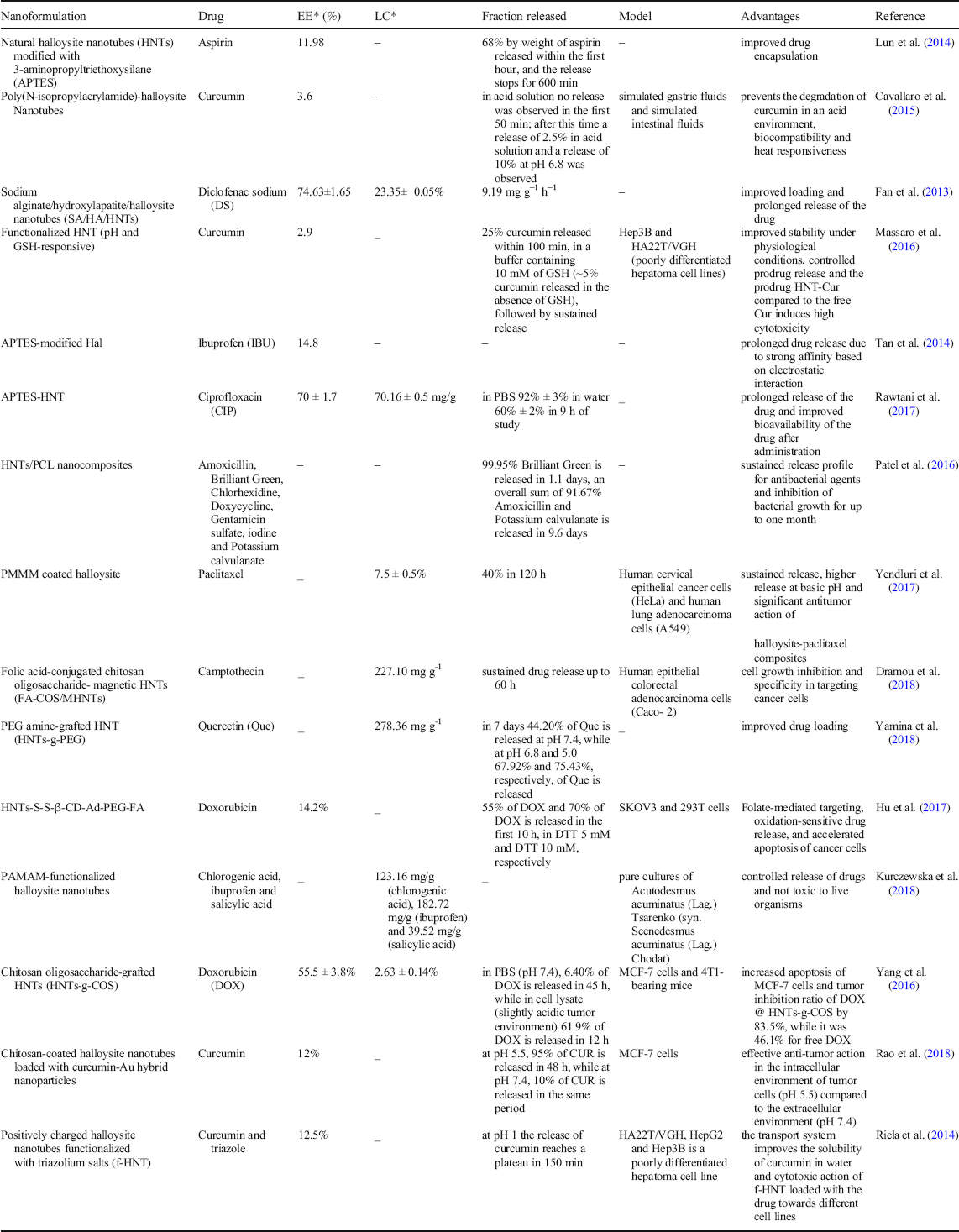
HNTs have been examined in many studies as nanosystems for drug delivery as well as for targeted drug delivery. Several strategies are used for the trapping of drugs within the lumen or on the surface of the HNTs, including intercalation, adsorption, and tubular trapping (vacuum strategy) (Hanif et al., Reference Hanif, Jabbar, Sharif, Abbas, Farooq and Aziz2016). Usually, the HNTs used as vectors for the administration of drugs first of all undergo modifications, which affect both the external surface and the lumen. This is a consequence of the fact that, very often, natural HNTs show weak interaction with drugs, not allowing prolonged release (Pan et al., Reference Pan, Li, Hong, Tang, Zheng, Weng, Zheng and Huang2017). In a study by Abdullayev and Lvov (Reference Abdullayev and Lvov2011), natural HNTs were employed as nuclei for layer-by-layer (LbL) entrapment; an increase in payload and continuous drug release for up to 100 h were noted, with the formation of caps at the end of the nanotube which helped to extend drug-release time. Natural HNTs were used to design a biocompatible vector for the administration of rabeprazole sodium (RAB) in order to counteract the acid degradation of this drug in the stomach. The results showed improved bioavailability and prolonged drug release (Khatoon et al., Reference Khatoon, Chu and Zhou2020). Generally, before the trapping of drugs on HNTs, the latter are subjected to various surface-modification strategies. One of these strategies involves modification of the surface of the HNTs with APTES (3-(aminopropyl)triethoxysilane) which is an organosilane, and this approach is a renowned functionalization method for both ease of use and reduced toxicity. Organosilane acts as an intermediate medium for binding the desired molecules. With APTES, silanol groups are introduced that can bind (through hydrogen bonding) to the hydroxyl groups present on the surface of the HNTs (Tian et al., Reference Tian, Wang, Wang, Komarneni and Yang2015). APTES-functionalized HNTs were employed as aspirin transporters; the results obtained showed an increase in loading efficiency, with an 11.8% weight load of aspirin, compared to 3.84% weight without functionalization (Lun et al., Reference Lun, Ouyang and Yang2014). The release profiles of ibuprofen (IBU) loaded in both APTES-modified HNT (APTES-HNT) and unmodified HNT were investigated. The results revealed greater loading efficiency and a better release profile for APTES-HNT (Tan et al., Reference Tan, Yuan, Annabi-Bergaya, Liu, Wang, Liu and He2014). In another study, chitosan-modified HNTs (now an emerging drug-delivery microsphere) were loaded with aspirin and the drug-release profile was investigated. The trapping efficiency of aspirin was 42.4% by weight for porous microspheres, ~20 times greater than that of uncontaminated HNTs (2.1% by weight) (Li et al., Reference Li, Ouyang, Yang and Chang2016). Furthermore, HNTs demonstrated a high trapping efficiency of camptothecin, while the results for an in vitro release study confirmed a much higher outflow of camptothecin from the nanosystem at pH 5 than that detected for higher pH values (6, 8, and 7.4). This study highlighted a pH-dependent release, which is vital considering that the pH of the tumor microenvironment in lysosomes and endosomes is acidic. For toxicity assessment, analyses of 3-(4,5-dimethylthiazol-2-yl)-2,5-diphenyltetrazolium bromide (MTT) showed that the HNT loaded with camptothecin showed greater inhibition of cell growth against colon cancer cells (Dramou et al., Reference Dramou, Fizir, Taleb, Itatahine, Dahiru, Mehdi, Wei, Zhang and He2018). A recent study by Sharif et al. (Reference Sharif, Abbas, Hanif, Bernkop-Schnürch, Jalil and Yaqoob2019) showed microcomposites coated with chitosan for long-term drug release. The microcomposites produced showed a size of 0.151 ± 0.49 μm. Metoclopramide hydrochloride was used as a model drug, with drug release ranging from 35.14 ± 1.5 to 53.97 ± 5.23%. Furthermore, these microcomposites (chitosan-coated HNTs) have been shown to be pH responsive, with a 25 h release of 66.8% and 46.7% at pH 1.2 and 5.5, respectively. It follows that these microcomposites can be used as carriers for the long-term release of drugs in the treatment of cancer (Sharif et al., Reference Sharif, Abbas, Hanif, Bernkop-Schnürch, Jalil and Yaqoob2019). The functionalization of HNTs with an opposite matrix is a technique that has recently been used widely for the realization of a system for a controlled release of drugs. In another work, HNTs were integrated into electrospun poly-and-caprolactone (PCL) scaffolds with the aim of improving structural coherence. The HNT inserted into the scaffold acted as a nanocarrier for various antibacterial agents such as amoxicillin, brilliant green, chlorohexidine, doxycycline, iodine, gentamicin sulfate, and potassium calvulanate. These nanocomposites were able to inhibit bacterial growth for up to one month, showing promising potential in applications for sutures and surgical dressings (Lepoittevin et al., Reference Lepoittevin, Devalckenaere, Pantoustier, Alexandre, Kubies, Calberg, Jérôme and Dubois2002). A potential selective antimicrobial formulation was developed by Fakhrullina et al. (Reference Fakhrullina, Khakimova, Akhatova, Lazzara, Parisi and Fakhrullin2019) for pathogenic microflora, based on curcumin-loaded HNTs and externally coated with dextrin (DX); its efficacy was tested in Caenorhabditis elegans (nematodes). In vitro HNT+Curc/DX was able to inhibit effectively the growth of Serratia marcescens enterobacteria. In addition, in vivo treatment of S. marcescens infected nematodes with HNTs+Cur/DX resulted in improved fertility and restoration of longevity of C. elegans (Fakhrullina et al., Reference Fakhrullina, Khakimova, Akhatova, Lazzara, Parisi and Fakhrullin2019). Curcumin (thermosensitive) release kinetics studies were conducted for HNTs modified with biocompatible poly (N-isopropylacrylamide). The in vitro tests were aimed at simulating the gastrointestinal transit of the HNT nanoformulation loaded with curcumin (natural anticancer molecule). The results demonstrated a targeted release of the active molecule in the intestine (Cavallaro et al., Reference Cavallaro, Lazzara, Massaro, Milioto, Noto, Parisi and Riela2015). Doxorubicin (DOX), another anticancer drug, has been encapsulated in multifunctional HNTs, and prolonged and targeted release of the drug has been investigated. In vitro studies of non-targeted and targeted HNTs revealed that targeted HNTs were able to accelerate tumor cell death by apoptosis. In addition, the in vivo studies have reinforced the fact that the targeted HNTs have no side effects in tumor-bearing mice, unlike free DOX which has induced tissue damage previously (Yang et al., Reference Yang, Wu, Shen, Zhou, Li, He and Liu2016). To obtain an improved loading and a prolonged release of quercetin (a drug with reduced solubility in water), HNTs with grafted (polyethylene glycol)-amine (PEG) functionalized with carbon dots (to confer fluorescent properties) were made. Quercetin has antioxidant properties, and the goal was to facilitate targeting of tumor tissues. To improve targeting and enhance cellular uptake, biotin was conjugated to the free amine groups of the PEG (Yamina et al., Reference Yamina, Fizir, Itatahine, He and Dramou2018). A more recent strategy that has shown very promising results in drug delivery involves functionalization with dendrimer. In a study by Kurczewska et al., (Reference Kurczewska, Cegłowski, Messyasz and Schroeder2018), the synthesis of functionalized HNTs with polyamide dendrimers (PAMAM) and 3-aminopropyltrimethoxysilane for the administration of drugs such as chlorogenic acid, salicylic acid, and IBU (as model drugs) was demonstrated. The dendrimer functionalized HNT revealed a better entrapment capacity for the three drugs compared to unmodified HNTs and APTES-HNTs. In addition, a very slow release rate was reported for chlorogenic acid and salicylic acid from dendrimer-modified HNTs while the release of IBU was similar to that recorded for nanotubes functionalized with APTES (Fig. 7). Because in vivo biocompatibility studies have not shown damage to living organisms by HNTs functionalized with dendrimers, it follows that the main advantage of functionalization of HNTs with dendrimer is the improved biocompatibility, resulting in the promising hybrid nanotubes for use in biomedical applications (Kurczewska et al., Reference Kurczewska, Cegłowski, Messyasz and Schroeder2018).
Release kinetics of a CHLG, b IBU, and c SAL by: ● HNT, ■ APTES-HNT, and ♦ PAMAM-HNT. (Reprinted from Kurczewska et al. (Reference Kurczewska, Cegłowski, Messyasz and Schroeder2018); with the permission of Applied Clay Science)

In conclusion, HNTs, both in pure and modified form (e.g. compressed with polymers), represent promising natural nanosystems for the trapping and controlled and prolonged release of drugs, both with reduced water solubility (such as aspirin, curcumin, IBU, and DS) and normal water solubility (such as amoxicillin), with satisfactory results, underlining the versatility of HNTs as nanocontainers for a large variety of drugs. The applications of HNTs in drug delivery with relative advantages are summarized in Table 2.
HNTs in drug delivery

*EE Encapsulation efficiency, LC Load capacity
Vaccine and Gene Delivery
Recently, due to advantageous properties such as reduced cytotoxicity (for bacterial and human cells), good biocompatibility, and remarkable mechanical strength, HNTs are emerging as potential non-viral nanosystems for the delivery of vaccines and genes (Guo et al., Reference Guo, Wang, Muhammad, Qi, Ren, Guo and Zhu2012). In one study, HNTs together with multi-walled carbon nanotubes functionalized with carboxylic groups (COOH-MWCNT) were used in the analysis of antigen release in order to improve the immune response to a recombinant LipL32 protein (rLipL32). The results obtained showed that immunization by HNTs and COOH-MWCNT increased significantly the IgG antibody titer specific to rLipL32 in golden Syrian hamsters with leptospirosis disease (Teixeira et al., Reference Teixeira, Fernandes, Cavenague, Takahashi, Santos, Passalia, Daroz, Kochi, Vieira and Nascimento2019). With regard to gene therapy, several disadvantages are associated with the use of viral vectors; so, increasing attention is paid to new non-viral nanocarriers for the release of genes. APTES-functionalized HNTs were used by Zhang et al. (Reference Zhang, Men, Zhang, Zhang, Yang and Duan2019) as vectors for the administration of gene material, and in particular the antisense oligodeoxineuclotide as a therapeutic gene was bound on the outer surface of functionalized HNTs with the aim of targeting survivin (a protein belonging to the family of apoptosis inhibitors) (Shi et al., Reference Shi, Tian, Zhang, Shen and Jia2011). In another study (Long et al., Reference Long, Wu, Gao, Li, He and Liu2018a), modification of HNTs with polyethyleneimine (PEI) was shown to be effective at the release of plasmid DNA (pDNA) and short interference RNA (siRNA). These PEI-modified HNTs were employed for the intracellular delivery of antisurvivine therapeutic siRNA. Western blot analysis confirmed that siRNA administration via PEI-HNTs reduced target protein levels in PANC-1 cells with significant reduction in survivin gene expression, reinforcing its therapeutic potential in cancer treatment (Long et al., Reference Long, Wu, Gao, Li, He and Liu2018a). In the treatment of genetic diseases and cancer, gene therapy has shown considerable potential; gene-transport systems must meet extremely important requirements, however, such as biocompatibility and cytotoxicity, which continue to hinder this treatment strategy (Christensen et al., Reference Christensen, Ashmead and Choy2019). HNTs have shown promising results for application as nanocarriers in gene therapy due to their easy availability, significant biocompatibility, remarkable mechanical strength, and various structural advantages.
Cancer Therapy and Cancer and Stem-Cell Isolation
Several strategies have been examined for the targeted delivery of anticancer drugs via HNTs. In a study by Mirzapur et al. (Reference Mirzapur, Khazaei, Moradi and Khazaei2018), HNTs were loaded with resveratrol and drug delivery to cancer cells was investigated. The viability study with MTT assay was carried out using a tumor cell line model (MCF-7) which showed that the nanotubes loaded with resveratrol were able to increase cytotoxic action significantly, resulting in cell death by apoptosis. In another study, multicomponent HNT (FA-Fe3O4 HNT) was analyzed as a targeted delivery system of DOX (anticancer drug) in cancer cells. The results confirmed that the drug loaded in FA-Fe3O4 HNT is able to induce apoptosis of HeLa cells (model cells) (Luo et al., Reference Luo, Zhang, Wu, Yang, Kuang, Li, Li, He and Liu2020a). Curcumin is a natural polyphenol with antioxidant properties that has found application because of its potential anticancer activity; several strategies have been used for loading it into HNTs ( Abdullayev et al., Reference Abdullayev, Price, Shchukin and Lvov2009). In one of these, positively charged HNTs were functionalized with trizolium salts. This HNT-based delivery system was employed for the delivery of curcumin in several cell lines. HNTs functionalized with curcumin-loaded trizolium salts were shown to be active in several cancer cells (Massaro et al., Reference Massaro, Noto and Riela2020). In another work, by Massaro et al. (Reference Massaro, Noto and Riela2020), biopolymer-modified HNTs were used for the targeted delivery of anticancer drugs. In particular, HNTs modified with chitosan (HNT-g-CS) were studied for the administration of curcumin in cancer cells. Curcumin-loaded HNT-g-CSs showed cytotoxicity toward various tumor cell lines, such as HeLa, HepG2, SV-HUC-1, MCF-7, EJ, and Caski; among these the EJ cell line showed an increase in the apoptotic process. In addition, the amount of reactive oxidative species (ROS) induced by curcumin encapsulated in HNT-g-CS is greater than that of non-encapsulated curcumin, making this platform a promising system for the administration of anticancer drugs (Massaro et al., Reference Massaro, Noto and Riela2020). HNTs modified with chitosan oligosaccharide have been employed for the release of another anticancer drug, DOX, for the treatment of breast cancer using the MCF-7 cell line. DOX-loaded HNT-g-CS was internalized by MCF-7 cells, triggering mitochondrial damage and attacking nuclei (Yang et al., Reference Yang, Wu, Shen, Zhou, Li, He and Liu2016). In another study, by Li et al. (Reference Li, Liu, Zhang, Correia, Mäkilä, Salonen, Hirvonen and Santos2017a), the release efficacy of anticancer drugs by modified HNTs was tested, with a new-design nanotube-microsphere, in which the HNTs are enclosed in a polymeric shell (hydroxypropylmethylcellulose acetate succinate) sensitive to pH. Atorvastatin and celecoxib were used as model drugs, as they have different physicochemical properties, and demonstrated colon cancer inhibition. In that study, the pH-reactive polymer/HNT composite system hindered premature drug release at pH 6.5, allowing for quicker release and increased drug permeability at pH 7.4 (Li et al., Reference Li, Liu, Zhang, Correia, Mäkilä, Salonen, Hirvonen and Santos2017a).
An important advance in the diagnosis and treatment of cancer is linked to the possibility of capturing and isolating rare circulating cancer cells from the blood of patients, for the development of personalized therapies. With this in mind, HNT surfaces were coated with a layer of poly-1-lysine and functionalized through a selective recombinant human protein. This strategy resulted in an increase in the capture capacity of leukemic cells (under flow) by HNTs (Saif and Asif, Reference Saif and Asif2015). In another study, a rapid, effective, and economical manufacturing procedure was employed to coat large, raw HNTs by thermal spraying of HNT dispersions in ethanol. All this led to improved surface communication between the HNTs’ coating and tumor cells, with effective capture of the tumor cells compared to normal cells (except for HeLa cells). Coated HNTs have also been shown to be efficient in capturing cancer cells in patient blood samples (with metastatic breast cancer) and in artificial blood (He et al., Reference He, Liu, Shen, Liang, Liu and Zhou2018). This effective ability to capture tumor cells constitutes a potential for early diagnosis and offers an important window into the progression and feasibility of personalized anti-cancer therapy. In addition to recent consideration of the potential of HNTs in the capture and isolation of cancer cells, their use in the isolation of stem cells was also considered. Research (Luo et al., Reference Luo, Humayun and Mills2020b) has shown that a three-dimensionally printed polylactic acid (PLA) pattern functionalized with a layer of polydopamine helped to bind the HNTs effectively on the surfaces of the PLA pattern in order to orient the guided cells. The HNTs improved significantly the hydrophilicity and roughness of the PLA pattern, and in vitro studies with human mesenchymal stem-cell cultures confirmed that the PLA pattern with HNTs has a different ability to induce cell orientation based on different widths of the strips (Luo et al., Reference Luo, Humayun and Mills2020b). Antitumor therapy and the study of stem cells are an important and delicate field of research in biomedicine and the use of HNTs widely available in nature could represent an important turning point, but with many points yet to be explored.
Tissue Engineering
HNTs have been used in the development of nanocomposites as supportive matrices for the controlled release of various drugs for various targets, thus finding a role as a delivery system in tissue engineering (Schmitt et al., Reference Schmitt, Creton, Prashantha, Soulestin, Lacrampe and Krawczak2015). An example is the incorporation of alkaline phosphatase (ALP) in HNTs in bone repair. In this system, the HNT acted as a heat sink resulting in an increase in the thermal stability of the ALP. In addition, a significant improvement in the activity of the enzyme (ALP) was recorded, with the promotion of the biomineralization process, which was studied in vitro with the use of calcium glycerophosphate as a substrate. This bioactive nanocomposite could also be incorporated into biomaterials applied as scaffolds in tissue engineering (Liu et al., Reference Liu, Zhang, Wu, Xiong and Zhou2012; Satish et al., Reference Satish, Tharmavaram and Rawtani2019).
In tissue engineering, the creation of a scaffold, sufficiently suitable and competent in supporting the three-dimensional formation of tissues, is essential. These scaffolds should be able to meet specific basic requirements such as: (1) an adequate pore size (in order to facilitate nutrient diffusion and seeding) with high porosity; (2) lower rate of degradation and biodegradability (the scaffold must be absorbed by the surrounding tissue and at the same time favor the formation of new tissue); and (3) high mechanical strength for a suitable support for the growth of the new tissue (Chan and Leong, Reference Chan and Leong2008). HNTs aligned in parallel strips on a solid support represent a promising platform capable of promoting the proliferation of human bone mesenchymal stem cells (HBMSC), promoting osteogenic differentiation in the absence of growth factors (Zhao et al., Reference Zhao, Zhou, Lvov and Liu2019b).
The use of HNTs in tissue engineering is a very recent challenge, however; only a few promising studies have been carried out over the past decade. The basic idea is to couple HNTs with various compounds with the aim of creating potential folds capable of promoting the growth of bones and tissues (Naumenko et al., Reference Naumenko, Guryanov, Yendluri, Lvov and Fakhrullin2016). Chitosan-HNT nanocomposite scaffolds are characterized by high compressive strength, Young's modulus, and significantly improved thermal stability compared to pure chitosan scaffolds. Although the HNTs particularly affected the porosity and porous structure of the scaffolds, they did not induce any toxicity on the cells. In addition, the cells were reported to adhere to and to develop optimally on the scaffold. Chitosan-HNTs produced by electrospinning, with 0, 2, and 5 wt.% HNT, led to nanocomposites that demonstrated increased tensile strength and increased Young's modulus with increasing HNT concentration. The chitosan-HNTs represent ideal membranes for bone-tissue engineering due to their thermal stability and improved mechanical properties (Liu et al., Reference Liu, Wu, Jiao, Xiong and Zhou2013). Another compound coupled with the HNTs (to make scaffolds) is alginate, which demonstrates reduced water absorption, high compressive strength, and reduced degradation rate. The HNTs also resulted in an improvement in the thermal stability of the alginate scaffolds, in addition to an increase in cell adhesion and improved biocompatibility. In one study, the incorporation of HNT made it possible to improve the physical properties of a sodium alginate scaffold and the composite was cross-linked with calcium ions. With low loading of HNTs, the composite scaffold demonstrated increased cell adhesion and proliferation in cultured preosteoblasts (MC3T3-E1), useful in bone-tissue engineering (Liu et al., Reference Liu, Dai, Shi, Xiong and Zhou2015b). In one paper (Qi et al., Reference Qi, Guo, Shen, Cao, Zhang, Xu, Yu and Shi2010), metronidazole-loaded HNT grafted into poly (caprolactone)/gelatin microfibers through electrospinning as a membrane for sustained drug release and guided bone regeneration was developed. Once again, scaffolds of poly (L-lactic acid) nanofibers were reinforced with the use of unidirectionally aligned HNTs in order to increase Young's modulus, tensile strength, and fracture stress. A porous antibacterial membrane for bone regeneration was produced, with multilayer polylactic acid (PLA)/HNT encapsulated with gentamicin (an aminoglycoside antibiotic). The studies carried out have shown a satisfactory antibacterial efficacy against both Gram-positive (Staphylococcus aureus) and Gram-negative (Escherichia coli) bacteria, resulting in a promising tool in the prevention of infections during bone regeneration (Qi et al., Reference Qi, Guo, Shen, Cao, Zhang, Xu, Yu and Shi2010; Berton et al., Reference Berton, Porrelli, Di Lenarda and Turco2020). In one study (Utech and Boccaccini, Reference Utech and Boccaccini2016), a composite chitosan-gelatin-agarose hydrogel grafted to HNT demonstrated increased water absorption and improved mechanical strength and thermal properties. These scaffolds implanted in rats showed excellent resorption even at 6 weeks. Furthermore, the newly formed connective tissue located in the vicinity of the scaffold showed the complete restoration of blood flow thanks to neovascularization (Utech and Boccaccini, Reference Utech and Boccaccini2016). In another work in gellan rubber matrices, HNTs were integrated with the aim of creating composite hydrogels with modular physical properties. These systems showed good biocompatibility with human dermal fibroblasts, when the cells were seeded on the surface of the gels or encapsulated within the composite matrix. In addition, the fibroblasts seeded on the hydrogel with HNT showed high metabolic activity due to the improved mechanical and topographic characteristics (Venugopal and Ramakrishna, Reference Venugopal and Ramakrishna2005; Alkatheeri et al., Reference Alkatheeri, Palasuk, Eckert, Platt and Bottino2015). These scaffolds, therefore, prove suitable in soft-tissue engineering applications (such as liver, pancreas, chondral, and skin regeneration). The group of Huang et al., Reference Huang, Liu and Zhou2017 incorporated the HNTs in a methacrylate gelatin hydrogel in order to evaluate the potential of the composite hydrogel to promote bone-tissue regeneration. In vitro hDPSC cells (human dental pulp stem cells) incubated with the hydrogel revealed increased expression of genes related to the osteogenesis process and subsequently improved in vivo tissue-regeneration rates in rats with cranial defects. Though few studies on the role of HNTs as bionanocomposites in tissue engineering are found, the current results confirm an important potential of HNTs in tissue engineering (Ou et al., Reference Ou, Huang, Fu, Huang, Fang, Gu, Wu and Wang2020). Overall, these studies demonstrated that the incorporation of HNTs into agarose, chitosan and gelatin bioscaffolds provides structural stability, improved adhesion, and cell growth. In vivo studies on rats showed the ability to promote neoangiogenesis in the vicinity of the scaffold without inducing an inflammatory reaction. In addition to the use of HNTs to supplement scaffolds based on alginate, polycaprolactone and gellan gum, in which HNT silicate ions have been used to stimulate osteoblasts in collagen secretion.
Wound Healing
HNTs are characterized by high mechanical strength, good biocompatibility, and hemostatic properties which make them suitable for applications in the development of medical devices. In addition, several studies have evaluated the possible use of HNTs as biocompatible nano-platforms in the controlled and prolonged release of antiseptic and antibacterial drugs, for application in wound healing (De Silva et al., Reference De Silva, Dissanayake, Mantilaka, Wijesinghe, Kaleel, Premachandra, Weerasinghe, Amaratunga and de Silva2018). In a study by Patel et al. (Reference Patel, Jammalamadaka, Sun, Tappa and Mills2016), copper-benzotriazole-coated HNTs were loaded with an antiseptic drug, Bright Green, and the release kinetics were investigated, detecting sustained release, over the period 50 to 200 h, from the nanotube. In addition, controlled release of iodine and amoxicillin by HNT took place (Patel et al., Reference Patel, Jammalamadaka, Sun, Tappa and Mills2016). In another study by Huang et al. (Reference Huang, Liu and Zhou2017) flexible three-dimensional chitosan composite sponges were manufactured with the addition of HNT, with improved toughness, compressive strength, and elastic modulus. The integration of HNTs resulted in an improvement in the blood coagulation capacity of chitosan. In vivo studies demonstrated the biocompatibility and improved the wound-healing properties of composite sponges, with improved skin reorganization and re-epithelialization compared to HNT or chitosan used separately. All this is linked to the induction of the activity of repairing inflammatory cells, stimulated by the prolonged release of chitosan oligosaccharides (homo- or hetero-oligomers of N-acetylglucosamine and D-glucosamine) making this nanocomposite a promising medical device in wound repair. (Huang et al., Reference Huang, Liu and Zhou2017). In another study, HNTs were used to deliver vancomycin in an alginate-based medical device. The resulting dressing exhibited high stability and neutrality in relation to living organisms used in biological tests, resulting in a potential parameter-optimized dressing for effective long-term wound treatment (Satish et al., Reference Satish, Tharmavaram and Rawtani2019). In a similar study by Xue et al. (Reference Xue, Niu, Gong, Shi, Chen, Zhang and Lvov2015), ciprofloxacin (antibiotic) was dispersed in a gelatin-based matrix in which the HNTs loaded with polymyxin B sulfate (antibiotic) were dispersed uniformly. The combined use of the two antibiotics within the nanocomposite demonstrated synergistic antimicrobial activity. In addition, this bio-nanomaterial has been shown to have effective properties such as significant water absorption, reduced toxicity, regulation of biodegradation, and high elasticity, optimal characteristics for applications in wound healing (Xue et al., Reference Xue, Niu, Gong, Shi, Chen, Zhang and Lvov2015).
The grafting of the HNTs has also made it possible to strengthen an elastic nano-fibrous material formed by PCL and gelatin, for the development of a device that could be used in wound dressing with a prolonged drug release. The drug was administered using HNTs modified with silane and loaded with metronidazole; these were inserted into electrospun polycaprolactone/gelatin microfibers, creating membranes for guided tissue regeneration, which contained ~25 wt.% metronidazole. These membranes were biocompatible and able to inhibit the internal growth of fibroblasts, as well as colonization by the anaerobic Fusobacterium nucleatum thanks to the prolonged release of metronidazole, up to 20 days (Shi et al., Reference Shi, Niu, Gong, Ye, Tian and Zhang2018). In addition, the HNTs have been integrated into a wound-healing powder formulation. First of all, a nanocomposite based on HNTs and chitosan oligosaccharide was created. The resulting powder was applied to burns on the backs of rats for 7 days, with evident improvement in the healing process (Sandri et al., Reference Sandri, Aguzzi, Rossi, Bonferoni, Bruni, Boselli, Cornaglia, Riva, Viseras, Caramella and Ferrari2017). HNTs loaded with antibiotics and incorporated into different types of biopolymer matrices demonstrate the extraordinary potential for wound-dressing applications with integrated anti-inflammatory and antimicrobial activities.
Biosensing
Interest is growing in the application of nanoformulations as potential future contrast agents (CAs) in molecular ultrasound imaging, especially if functionalized on the surface with specific biological recognition elements (e.g. ligands) for targeted delivery and release of therapies (Andreou et al., Reference Andreou, Pal, Rotter, Yang and Kircher2017). Nanomaterials have numerous advantages that make them useful as CAs including their optical and magnetic properties that can be modified and adapted by manipulating their composition, size, structure, and shape (Kumar et al., Reference Kumar, Mazinder Boruah and Liang2011). Parametric evaluation of the efficiency of HNTs as a scatterer in safe ultrasound-based molecular imaging has been demonstrated in a published work by Soloperto et al. (Reference Soloperto, Conversano, Greco, Casciaro, Casciaro, Ragusa and Lay-Ekuakille2013). The results obtained confirmed the possible use of a clinically readily available ultrasound system for the detection of ultrasonic backscatter generated by different concentrations of HNTs using sonication frequencies of 5.7 and 7 MHz (Soloperto et al., Reference Soloperto, Conversano, Greco, Casciaro, Casciaro, Ragusa and Lay-Ekuakille2013). The key to creating a highly sensitive electrochemical enzymatic biosensor is to be able to obtain a direct transfer of electrons between the electrode surface and the enzyme, and this can be achieved through an improved immobilization of the enzyme with high load capacity (Grieshaber et al., Reference Grieshaber, MacKenzie, Vörös and Reimhult2008). In one paper, by Cheng et al. (Reference Cheng, Song, Zhao and Zhang2020) HNTs were used in enzymatic immobilization for glucose detection. A hybrid nanocomposite consisting of silver nanoparticles, with an average diameter of 10 nm, was immobilized on the surface of HNTs, modified with silane obtained by in situ chemical reduction of silver ions (Ag+). The hybrid nanocomposite thus produced constituted a platform for the immobilization and electrical wiring of the redox enzyme, glucose oxidase (GOx), which catalyzes the oxidation of glucose into D-gluconolactone with the production of H2O2. The immobilization of the enzyme resulted in the improvement of the direct transfer of electrons with the formation of a highly sensitive and stable device for the detection of glucose in the monitoring of diabetic patients (Cheng et al., Reference Cheng, Song, Zhao and Zhang2020). In another study, a nanocomposite based on HNTs functionalized with polyaniline (PANI) was used as an extremely sensitive biosensor for the detection of ascorbic acid (AA). A composite sensitivity to AA of ~ 826.53 mA mM–1 cm–2 over a linearity range of 0.005–5.5 mM and a lower detection limit of 0.21 mM was reported by Shao et al. (Reference Shao, Wang, Yang, Wang, Tian, Ji and Zhang2017). The high sensitivity of the biosensor can be attributed to its porous structure with the formation of efficient detection channels, which lead to an improvement in electron transport and the interaction between PANI and analyte (AA) (Shao et al., Reference Shao, Wang, Yang, Wang, Tian, Ji and Zhang2017). The HNTs were used to make a sensor for the detection and quantification of tumor biomarkers. In a study by Li et al. (Reference Li, Khan, Tian, Liu, Hu, Fan, Cao and Wei2017b), based on a core–shell structure, particles of palladium (Pd) were grafted onto the surface of HNTs coated with polypyrrole (PPy) (HNT@PPy-Pd). This hybrid nanocomposite was fabricated as an analytical signal label for the quantitative detection of the tumor marker, prostate specific antigen (PSA), a prostate cancer biomarker. The sensor exhibited acceptable reproducibility, selectivity, and stability, with a lower detection limit of 0.03 pg/mL. The development scheme of the PSA biosensor (Li et al., Reference Li, Khan, Tian, Liu, Hu, Fan, Cao and Wei2017b) is shown in Fig. 8.
Development phases of the PSA biosensor

In conclusion, despite the limited number of studies conducted on the use of HNTs in the construction of biosensors, characteristics such as high biocompatibility, easy availability, selective modification for specific targeting, and dimensions in the order of nanometers offer potential applications for nanotubes in this area.
FUTURE DIRECTIONS AND CONCLUSIONS
The main properties of HNTs have been described, including cytocompatibility, considerable surface area, tubular structure, and nanometric dimensions, and are useful for those working on the construction of nanodevices. In addition, the small cost, the wide availability in nature, and the ability to improve the mechanical characteristics have led to growing interest in materials based on HNTs as a multifunctional biomaterial in biomedical applications. The hollow lumen is an essential feature for the encapsulation of bioactive molecules and for their gradual and controlled release near or within a desired cell; and finally, the high aspect ratio of the tubular nanosystem favors penetration through the cell membrane. In addition, HNTs are formed by laminated layers of aluminosilicate which create a structure with a positively charged hollow lumen and a negatively charged external surface, all of which lead to good dispersibility in alcohol, water, and polar polymers. The inner lumen with a diameter of 10–40 nm allows for 10–20 wt.% drug and protein load, with a prolonged release of the therapeutic agent up to 10–20 h, which can be extended to 100 h with the termination of the nanotube or with its inclusion in a polymer matrix. In addition, the opposite distribution of charge between the external and internal surfaces allows selective immobilization of the therapeutic agents. To all this is added the functional versatility of HNTs, which includes modification to silanes and LbL self-assembly, as well as functionalization with targeted fractions (for safer and more efficient drug release), including folic acid, biotin, and materials which respond to specific enzymes (e.g. end caps of dextrins). This allows for selective cellular internalization and triggered drug release for anticancer applications.
Several biologically active molecules have been loaded into HNTs. Such pharmaceutical applications include the entrapment of anticancer, anti-inflammatory, antibiotic, analgesic, antihistamine, corticosteroid, and cardiovascular drugs. In addition, HNTs have shown themselves to be promising nano-platforms for the immobilization of enzymes and nucleic acids in clinical diagnostics and therapy. Entrapment in HNTs preserves the activity of enzymes, providing protection from degradation by proteases. The HNTs have resulted in an improvement in the use of enzymes in colon, cervical, and breast cancer. In addition, HNTs have emerged as non-viral gene-delivery agents (less toxic than carbon nanotubes and cationic-nucleic acid polymer complexes) for DNA, siRNA, and oligonucleotides, showing high transfection efficiency and gene silencing which confirm promising application of HNTs as vectors in gene therapy.
The development of scaffolds from HNTs and polymers is an exciting direction in the design of emerging materials in biomedicine. HNTs represent a promising material in the structuring of polymer matrices. The grafting of just 3–6% by weight of HNTs leads to significant changes in the physicochemical characteristics of the resulting materials. The known properties of HNTs such as solubility, hydrophilicity/hydrophobicity, and mechanical strength are manipulated for their application in various fields such as biosensing, wound healing, and hydrogel fillers in tissue engineering. The application of HNTs in regenerative medicine and tissue engineering has been very successful, particularly in bone and dental repair, and in hair surface engineering. The realization of biomimetic architectures has led to improved proliferation, adhesion, and differentiation of stem cells. The grafting of HNTs allows improvement in the mechanical characteristics of the scaffolds, in addition to the absorption of water. In addition, the encapsulation of bioactive molecules in the lumen of HNTs allows the development of functional matrices or membranes which release antibiotics to inhibit infections and promote faster wound healing. Likewise, the grafting of antibiotic-loaded HNTs at the bone cement level has shown important potential in clinical applications in orthopedic replacement surgery. The use of HNTs in the immobilization of enzymes associated with the bone biomineralization process is a further step forward in bone repair. Halloysite nanotubes have extraordinary potential, therefore, in the production of multifunctional, high-performance HNT-polymer nanocomposites, showing improved mechanical, thermal, and flame-retardant properties, as well as a prolonged drug-release capacity and good hemocompatibility/cytocompatibility. Other possible biomedical applications of HNT-polymer nanocomposites, such as biosensors and cancer diagnosis (bioimaging), remain to be developed, however. In addition, the toxicology of HNTs should be investigated thoroughly; a weakness is the absence of comprehensive studies evaluating the pharmacokinetics, toxicity, and safety of HNTs, in humans, rather than in vitro or in vivo in animals. The toxicological evaluation of HNTs is in its infancy and must be further investigated; because HNTs are natural nanomaterials, their properties vary according to their origin, with different toxicological profiles. Future research will elucidate fully the long-term effect of HNTs (uncontaminated and modified) on human health. An important aspect for an advance in the application of HNTs in biomedicine is related to the safety of this material which usually has different impurities with which different toxic effects can be associated. It will be essential to introduce new technologies that enable greater purity of the materials. Furthermore, hybrid clay-drug nanosystems are very effective in vitro, though some of them fail in vivo due to complications with real physiological conditions. The most valid formulations seem to be topical cosmetic formulations. The focus on topical application is related to the fact that this nanomaterial is not readily biodegradable in the blood, limiting its use in intravenous formulations, due to the risk of thrombosis.
ACKNOWLEDGMENTS
FP, GG, and SL are grateful to the Tecnopolo per la medicina di precisione (TecnoMed Puglia)–Regione Puglia: DGR no. 2117 del 21/11/2018, CUP: B84I18000540002 and Tecnopolo di Nanotecnologia e Fotonica per la medicina di precisione (TECNOMED)–FISR/MIUR-CNR: delibera CIPE no. 3449 del 7-08-2017, CUP: B83B17000010001.
Funding
Funding sources are as stated in the Acknowledgments.
Declarations
Conflict of Interest
The authors declare that they have no conflict of interest.