Ductal stents are emerging as alternatives for surgical shunts to palliate neonates with pulmonary hypoperfusion. Reference Sagar, Sivakumar and Umamaheshwar1 Despite preprocedural aspirin and adequate heparinisation, an acute stent thrombosis may complicate the procedure if there is a mal-deployment, mal-apposition or bend in the stent, or interaction between multiple stents. Reference Promphan and Qureshi2 Thrombolytic agents are used to avoid an emergent surgery but lead to bleeding around vascular sheaths and other sites. Reference Promphan and Qureshi2 Intraprocedural acute thrombosis of coronary artery stents is often managed with platelet targeted therapy, rather than thrombolysis. Reference Kirtane and Stone3 However, experience is sparse in neonates and not reported after ductal stents. Reference Emani, Pereira and Piekarski4–Reference Bhatt, Anil and Sivakumar7
Case report
A 5-month-old infant weighing 5.5 kg with corrected transposition, ventricular septal defect, and pulmonary atresia presented with critical hypoxia and oxygen saturations of 50%. The narrow arterial duct from the undersurface of right aortic arch was tortuous and measured 14 mm in length. Heart team favoured an emergent ductal stent in view of critical hypoxia instead of shunt due to logistic delay.
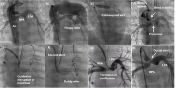
After one dose of oral aspirin, informed parental consent, femoral arterial access, and adequate heparinisation, aortography confirmed the ductal anatomy (Fig 1). As the tortuosity of the duct persisted despite use of an additional buddy coronary wire, it was exchanged to Ironman wire (Abbott Vascular, Santa Clara, California, United States of America) over a Finecross microcatheter (Terumo corporation, Tokyo, Japan). The straightened duct was stented using 4 mm x 18 mm Resolute Onyx (Medtronic, Santa Rosa, California, United States of America) resulting in the improvement of oxygen saturations to 95%. A bend occurred in the stent after the removal of the extra-support guidewire leading to sluggish blood flows, thrombus formation, and recurrence of hypoxia despite adequate activated clotting time from heparinisation. Passages of floppy coronary guidewires to disintegrate the thrombus as well as a similar attempt from a second coaxial right axillary access failed to disrupt the thrombus (Supplementary video 1). An attempt to deploy an additional reinforcing stent to straighten the bend did not succeed as the second stent could not manoeuvre the bent portion. As the thrombus progression worsened the hypoxia despite optimising the ventilator settings, two boluses of 180 µg of eptifibatide per kilogram body weight were administered at 30-minute interval along with rapid vigorous saline flushes from the axillary sheath resulting in dissolution of the thrombus and recovery from hypoxia (Supplementary video 2). A dose of aspirin and clopidogrel was administered through the nasogastric tube to sustain the antiplatelet effect. The femoral and axillary arterial sheaths were safely removed after few hours when the activated clotting time reduced to 200 seconds without any complications.
Aortogram ( a ) showed a vertical tortuous arterial duct from right aortic arch filling right (RPA) and left (LPA) pulmonary arteries. When floppy wires failed to provide adequate support due to the tortuosity ( b ), an extra-support wire straightened the duct ( c ) before a stent implantation. Removal of wire led to a bend in the stent causing thrombus formation ( d ) that warranted a right axillary access to provide a coaxial support. Repeated passages of guidewires ( e ) failed to disrupt the thrombus and a second reinforcing stent ( f ) could not manoeuvre the bend. When thrombus further progressed into the branch pulmonary arteries ( g ), eptifibatide boluses caused dissolution of the clots ( h ).

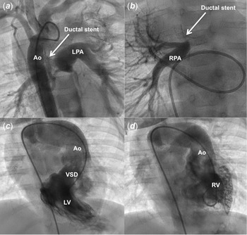
After maintaining dual antiplatelet therapy with aspirin and clopidogrel for 4 months, an elective cardiac catheterisation showed a patent stent and oxygen saturation of 80%. Even though the ductal insertion showed stenosis, the hilar right and left pulmonary arteries measured 10 mm. A bidirectional Glenn shunt and reconstruction of confluence was performed at 9 months of age as the first preparatory step before an one-and-half ventricular anatomical repair, planned for the future (Fig 2).
Aortogram at 4-month follow-up ( a ) showed patent ductal stent and adequate sizes of the left (LPA) and right (RPA) pulmonary arteries despite ductal site coarctation of the RPA ( b ) seen on a right pulmonary vein wedge angiogram. Left (LV) and right (RV) ventriculogram ( c , d ) show corrected transposition, routable subaortic ventricular septal defect (VSD) filling the left malposed aorta (Ao).

Discussion
Intraprocedural ductal stent thrombosis is a morbid complication warranting emergent surgery. Reference Mack, Wheeler and Hirsch6 In the absence of clear guidelines, expert reviews suggest thrombolysis but warn about bleeding around the vascular access sites as well as during emergent operation if the lysis fails. Reference Promphan and Qureshi2 Acute platelet-mediated thrombus during coronary stent angioplasty is often managed with bolus infusions of glycoprotein IIb/IIIa receptor inhibitors such as abciximab, eptifibatide, or tirofiban followed by oral dual antiplatelet therapy suggesting platelets as a potential target. Reference Kirtane and Stone3 These drugs are occasionally used after aortopulmonary shunts in young cardiac patients as a bridge to oral aspirin. Reference Emani, Pereira and Piekarski4,Reference Emani, Kaza and Almodovar5
On a head-to-head comparison, eptifibatide produced a more sustained early (minutes) and late (24 hours) inhibition of platelet aggregation than the other two agents during coronary interventions. Reference Bhatt, Anil and Sivakumar7 Moreover, the dosing of eptifibatide is more convenient with two bolus doses of 180 µg/kg bodyweight administered at 30-minute interval, which allows safe removal of vascular sheaths without local bleeding. Reference Alwi8 The effect of these antiplatelet agents can be monitored by platelet aggregometry, available largely in research laboratories. Reference Bhatt, Anil and Sivakumar7
We used eptifibatide as a salvage option for acute stent thrombosis during the intervention. The prompt dissolution of thrombus facilitated by rapid saline flushes through the stent lumen indicated the potential utility of an antiplatelet strategy during these neonatal stent procedures. Other anecdotal use of eptifibatide also supports its utility in neonates. Reference Batchelor, Tolleson and Huang9 Abciximab may be preferred in neonates when a prolonged antiplatelet action is desired. Reference Giugliano, White and Bode10
Supplementary material
For supplementary material accompanying this paper visit https://doi.org/10.1017/S1047951121003474
Acknowledgements
The authors thank Monica Rajendran for providing images.
Financial support
This research received no specific grant from any funding agency, commercial, or not-for-profit sectors.
Conflicts of interest
None.
Ethical standards
The authors assert that all procedures contributing this work comply with the ethical standards of the Indian Council of Medical Research and with the Helsinki Declaration and have been approved by the institutional committees of Madras Medical Mission, India.