Case report
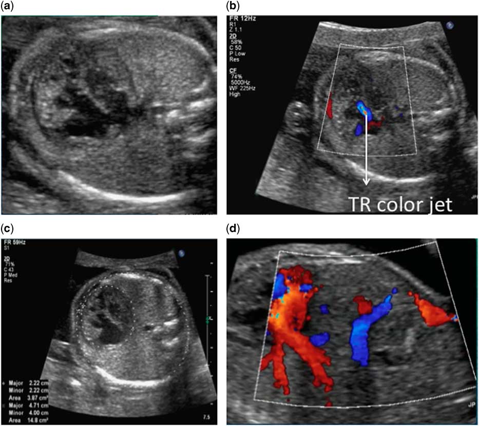
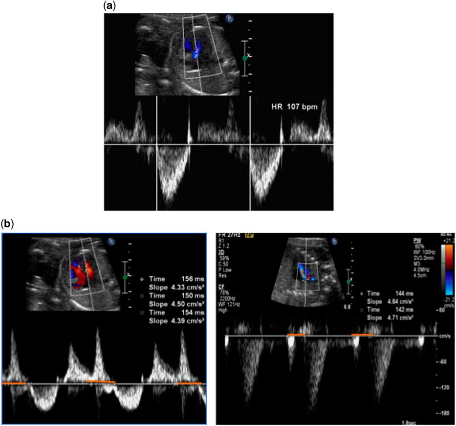
A 29-year-old G2P1 woman was referred for foetal echocardiogram at 21 weeks of gestational, given concern of an enlarged right atrium and bradycardia. The patient was healthy with no significant family history and did not take any medication. She and her partner were non-consanguineous. Foetal echocardiography was performed at a gestational age of 21 weeks. The foetal heart was normally connected and was found to have left ventricular non-compaction, fulfilling the Jenni criteria,Reference Jenni, Oeschlin, Schneider, Attenhoffer Jost and Kaufmann 1 with preserved systolic function (Fig 1, Supplementary video clips 1 and 2). The cardiothoracic ratio was 26%. Ebstein’s anomaly of the tricuspid valve (apical displacement 4.5 mm) was seen with mild tricuspid regurgitation in addition to a small mid-muscular ventricular septal defect. The right atrium and right ventricle appeared mildly enlarged. The ductus venosus was absent, and the umbilical vein drained into the portal system (Fig 1). There was foetal bradycardia (Fig 2); first-degree atrioventricular block was seen with atrioventricular intervals of 142–144 ms by superior caval vein/aortic Doppler and 150–156 ms by mitral valve/aortic Doppler (Fig 2).Reference Wojakowski, Izbizky, Carcano, Aiello, Marantz and Otano 2 Premature atrial contractions and ventricular contractions were also observed. No pericardial or pleural effusions, ascites, or skin oedema were observed. Close follow-up was arranged at 24, 28, 32, and 36 weeks of gestational, as there was concern for potential in utero demise; however, there was no change in the findings. At the time of the foetal diagnosis, the family was counselled about the overall potential poor prognosis for this foetus related to the left ventricular non-compaction with the additional arrhythmias and agenesis of the ductus venosus. The family was offered genetic testing, which was declined.
( a ) Two-dimensional four-chamber view of the foetal heart showing left ventricular non-compaction and Ebstein’s anomaly of the tricuspid valve. There is normal biventricular systolic function (M-mode SF 41%, EF 79%). ( b ) With colour Doppler, there is mild tricuspid valve regurgitation. ( c ) Two-dimensional image showing CT area ratio of 26%. ( d ) Colour Doppler showing absence of the ductus venosus. The umbilical vein drains directly into the portal system of the liver. No superficial umbilical vein is seen draining into the right atrium. EF=ejection fraction; SF=systolic function; TR=tricuspid regurgitation.

( a ) Pulsed Doppler images showing bradycardia. ( b ) Mitral valve/aortic and superior caval vein/aortic Dopplers showing prolonged 1:1 atrioventricular conduction in keeping with first-degree heart block. A–V interval is highlighted with a red line. HR=heart rate.

A female neonate was delivered vaginally at 39 weeks of gestation with a birth weight of 3.11 kg. Postnatal echocardiogram confirmed all the antenatal findings (Supplementary video clips 3 and 4). An electrocardiogram showed sinus bradycardia with first-degree atrioventricular block (P-R interval of 168 ms). Abdominal ultrasound showed a normal portal venous system and inferior caval vein to the right atrium. The neonate was discharged after 48 hours of observation and has been clinically asymptomatic with normal ventricular systolic function at 8 months of age.
Discussion
Left ventricular non-compaction is an uncommon cardiomyopathy associated with increased risk of arrhythmia, cardiac dysfunction, and sudden death both prenatally and postnatally.Reference Arunamata, Punn, Cuneo, Bharati and Silverman 3 , Reference Brescia, Rossano and Pignatelli 4 In a case series of foetally diagnosed left ventricular non-compaction, 81% of foetuses that survived to delivery died or required transplant postnatally. Left ventricular non-compaction is associated with structural cardiac defects, genetic syndromes, and conduction abnormalities.Reference Arunamata, Punn, Cuneo, Bharati and Silverman 3 The combination of left ventricular non-compaction, agenesis of the ductus venosus, and first-degree atrioventricular block has, to our knowledge, not been reported so far. Agenesis of the ductus venosus in the presence of other anomalies has been shown to have a poor prognosis.Reference Thomas, Petersen, Cincotta, Lee-Tannock and Gardener 5 We had expected this foetus to develop progressive cardiac dysfunction in utero and had counselled the family accordingly. We postulate that because there was preserved ventricular systolic function throughout pregnancy, the foetus had a stable postnatal outcome. In addition, there was only mild Ebstein’s anomaly allowing for adequate growth of foetal lungs and there was no progression to a higher degree atrioventricular block, allowing for a better postnatal outcome. When left ventricular non-compaction is diagnosed in the foetus, care should be taken to evaluate the foetus for other cardiac anomalies, conduction abnormalities, and extracardiac anomalies such as agenesis of the ductus venosus. Families should be counselled about the often poor prognosis; however, the foetal cardiologist should be aware that some foetuses with left ventricular non-compaction can have an unremarkable antenatal and postnatal course, at least in the short-term.
Acknowledgements
None.
Financial Support
This research received no specific grant from any funding agency, commercial, or not-for-profit sectors.
Conflicts of Interest
None.
Supplementary Material
To view supplementary material for this article, please visit http://dx.doi.org/10.1017/S1047951115000773