INTRODUCTION
Toxoplasma gondii is an obligate intracellular protozoan parasite that infects any warm-blooded animals, capable of causing toxoplasmosis. To date, more than a third of the population worldwide has been infected by this parasite (Tenter et al. Reference Tenter, Heckeroth and Weiss2000; Hill and Dubey, Reference Hill and Dubey2002; Dubey, Reference Dubey2010). Although the vast majority of T. gondii infections in adults are asymptomatic or self-limited, infections in pregnant women can cause abortion, stillbirth or fetal abnormalities with detrimental consequences for the fetus (Grant et al. Reference Grant, Gold, Rosenblum, Niedzwiecki and Armstrong1990; Hermanns et al. Reference Hermanns, Brunn, Schwarz, Sachweh, Seipelt, Schroder, Vogel, Schoendube and Buettner2001; Ponticelli and Campise, Reference Ponticelli and Campise2005). Congenital toxoplasmosis has traditionally been regarded as the most serious outcome of Toxoplasma infection and has an incidence of 1–15 per 10 000 live births (Andreoletti et al. Reference Andreoletti, Budka, Buncic, Colin, Collins, Koeijer, Griffin, Havelaar, Hope, Klein, Kruse, Magnino, López, McLauchlin, Nguyen-Thé, Noeckler, Noerrung, Maradona, Roberts, Vågsholm and Vanopdenbosch2007). Toxoplasma gondii is a neurotropic parasite, and the central nervous system (CNS) is the most easily damaged of all invaded organs. Toxoplasma infection is responsible for encephalitis, intracranial calcifications, hydrocephalus, etc. (Petersen, Reference Petersen2007), significantly influencing the health of infants. However, the underlying mechanisms of neuropathogenesis in congenital toxoplasmosis still remain poorly studied.
The CNS develops from a small number of highly plastic cells that proliferate, acquire regional identities and produce different cell types. These cells have been defined as neural stem cells (NSCs) on the basis of their potential to generate multiple cell types (e.g. neurons and glia) and their ability to self-renew in vitro (Merkle and Alvarez-Buylla, Reference Merkle and Alvarez-Buylla2006). Previous research has showed that the NSCs are an important target for many neurotropic pathogens (Li et al. Reference Li, Kosugi and Tsutsui2004; Tsutsui et al. Reference Tsutsui, Kosugi and Kawasaki2005; Odeberg et al. Reference Odeberg, Wolmer, Falci, Westgren, Seiger and Soderberg-Naucler2006; Das and Basu, Reference Das and Basu2008; Mutnal et al. Reference Mutnal, Cheeran, Hu and Lokensgard2011; D'Aiuto et al. Reference D'Aiuto, Di Maio, Heath, Raimondi, Milosevic, Watson, Bamne, Parks, Yang, Lin, Miki, Mich-Basso, Arav-Boger, Sibille, Sabunciyan, Yolken and Nimgaonkar2012). For example, the NSCs are the principal target cells for mouse cytomegalovirus (MCMV) in the developing brain, and the viral infection was demonstrated to cause a loss of the NSCs expressing CD133 and nestin, subsequently leading to abnormal brain development (Mutnal et al. Reference Mutnal, Cheeran, Hu and Lokensgard2011). Toxoplasma gondii is known to be capable of invading almost any nucleated cells and the NSCs are presumably one of them during the development of brain. Since the NSCs are the origin of the CNS, we hypothesize that the effect of T. gondi on the NSCs may play an important role in the congenital neurological birth defects caused by the parasite. Moreover, in a preliminary study on mouse gene expression profiling, we found that 973 genes were up-regulated and 871 genes were down-regulated (⩾2-fold change) in NSCs co-cultured with tachyzoites of T. gondii as compared with untreated NSCs, and cell apoptosis was demonstrated to be one of the biological processes that were significantly affected in the treated NSCs (data not published). The purpose of this study was to dissect the underlying molecular mechanisms responsible for the apoptosis in the NSCs induced by T. gondii. For such a purpose, the NSCs were co-cultured with the tachyzoites using a trans-well culture insert. After pretreatment with or without inhibitors, the apoptotic effect in the NSCs was evaluated using flow cytometry (FCM) or DNA laddering, and the expression levels of key molecules involved in the endoplasmic reticulum stress (ERS) pathway were revealed by Western blotting.
MATERIALS AND METHODS
Ethics statement
The experiments were carried out in strict accordance with the National Institute of Health Guide for the Care and Use of Laboratory Animals and approved by the Animal Care and Use Committee of Anhui Medical University (permit number: AMU26-080610). All efforts were made to minimize the pain and suffering of mice during all the procedures.
Parasite culture
Tachyzoites of T. gondii RH strain were maintained in BALB/c mice by intraperitoneal passage at a 72-h interval. The parasites were harvested from mouse peritoneal exudates, washed twice (centrifuged at 1000 g for 5 min) in sterile phosphate-buffered saline (PBS; pH 7·4) and maintained by serial passage in human foreskin fibroblasts (HFF) for further infection experiments in vitro.
Isolation and cultivation of NSCs
Six- to 8-week-old ICR mice, purchased from the Laboratory Animal Center of Anhui Medical University, were maintained in a pathogen-free environment. The mice were acclimatized for at least 1 week before being mated. For mating purposes, four females were housed overnight with two males at 9:00 p.m. The females were checked by 7:00 a.m. on the next morning, and the presence of a vaginal plug or sperm in the vaginal smear was considered as successful mating and marked as gestational day 0 (GD0). On GD14, embryos of ICR mice were collected by caesarean section of pregnant mice, and then the telencephalons of the embryos were isolated and mechanically disrupted into single cells by repeated pipetting in a serum-free DMEM/F12 (1:1) basic medium (Hyclone, USA) three times. After filtration, the cell number was counted and the cells were seeded at a density of 1×10⁵ cells mL−1 in a complete medium and cultured in a humidified atmosphere of 5% CO2 and 95% air at 37 °C. The complete medium comprises DMEM/F12 (1:1) medium, 2% B27 (Gibco, USA), 20 ng mL−1 basic fibroblast growth factor (bFGF; Peprotech, USA), 20 ng mL−1 epidermal growth factor (EGF; Peprotech, USA), 100 U mL−1 penicillin (Sigma-Aldrich, USA) and 100 μg mL−1 streptomycin (Sigma-Aldrich, USA).
The neurospheres were passaged every fourth day in vitro once they reached the size of 150–200 μm in diameter. These neurospheres were then centrifuged at 1000 g for 5 min to separate the cells from media. After the supernatant was gently removed, the cell pellets were incubated in 0·25% trypsin for 5 min at 37 °C, and then basic culture medium with 10% FBS was used to terminate the digestion of trypsin. The trypsinized cells were collected by centrifuging at 1000 g for 5 min, and sterile PBS was added into the pellets, then they were triturated 20–30 times using a 200 μL pipette tip. After washing with PBS twice, the pellet containing single cells were re-seeded in the complete medium. Neurospheres that were passaged at least five times (P5) were used for the following experiments.
Identification of NSCs
For examination of the NSC specific marker, neurospheres were seeded on cover slips. The plated neurospheres were rinsed with PBS after fixation with 4% paraformaldehyde (Sigma-Aldrich, USA) for 24 h at room temperature, and then permeabilized with 0·2% Triton X-100 (Sigma-Aldrich, USA) in PBS. The cells were blocked with 1% bovine serum albumin (BSA; Boster, China)/PBS for 60 min at 37 °C, and then incubated overnight at 4 °C in a humidity chamber with rabbit anti-Nestin monoclonal antibody (1:100; Sigma-Aldrich, USA). After the incubation, the cells were rinsed with PBS and incubated with PE-conjugated goat anti-rabbit IgG (1:200; Santa Cruz, USA) for 60 min at 37 °C. After rinsing with PBS, the cells were analysed under a fluorescent microscope (Olympus, Japan). Negative control was treated in the same way as above except elimination of the primary antibody.
Establishment of co-culture system and treatments
A co-culture system was established using trans-well inserts (Corning, USA). The bottom of the inserts is composed of polyester materials with a pore size of 0·4 μm, which only permits the permeabilization of small, soluble factors but not of T. gondii tachyzoites. Two millilitres of single cell suspension of the NSCs at a density of 5×10⁵ mL−1 was seeded in the lower chamber of each insert. One millilitre of RH strain tachyzoites with various doses (2×10⁵ mL−1, 1×10⁶ mL−1 and 5×10⁶ mL−1) was added to the upper chamber of the inserts. In positive control of apoptosis, apoptosis inducer A (Apopida, Beyotime, China) was added into the upper chamber with a dilution of 1:3000. The co-culture system was maintained in a basic culture medium supplemented with 10% fetal bovine serum (FBS; Gibco, USA) in 5% CO2 at 37 °C. After co-culturing for 12, 24 and 48 h, the NSCs were collected, and the apoptotic levels were detected by FCM.
To investigate the activity of the ERS pathway, the NSCs were pretreated with 0·5 mmoL L−1 tauroursodeoxycholic acid (TUDCA; Amresco, USA) or 4 μmoL L−1 Z-ATAD-FMK (Biovision Inc., USA) for 6 h, and then 1×10⁶ RH tachyzoites were added to the upper chamber. After 48-h incubation, the NSCs were collected, the apoptotic level was detected by FCM and the expression levels of CHOP, caspase-12, JNK and p-JNK were detected using Western blotting.
Detection of apoptosis
Apoptotic levels of the NSCs were determined according to a fluorescein isothiocyanate (FITC)/propidium iodide (PI) kit (BestBio, China). Briefly, the cells were collected, washed with PBS twice and resuspended in 400 μL of annexin V binding buffer (10 mm HEPES, 0·14 m NaCl and 0·25 mm CaCl2). Then, 5 μL of FITC-conjugated annexin V was added and incubated at room temperature for 15 min in the dark with gentle vortexing. Next, 10 μL of PI was added and incubated at room temperature for 5 min in the dark. Finally, 400 μL of 1×binding buffer was added to each tube. The cells were analysed on a Coulter Epics Altra HyPerSort system (Beckman Coulter, USA), and the data were analysed using EXPO32 multicomp software. Annexin V-positive/PI-negative (annexin V+/PI−) cells represent the early apoptotic cells, and annexin V+/PI-positive (PI+) cells reflect the late apoptotic cells (Fang et al. Reference Fang, Yu, Ellis, Sharav and Braley-Mullen2010).
DNA fragmentation (DNA ladder) assay was used to further identify the apoptosis of the NSCs. The NSCs were collected after treatment with RH strain tachyzoites (1×10⁶) for 48 h, and then rinsed three times with PBS at 4 °C. DNA was isolated using a DNA Ladder Extraction Kit with Spin Column (Beyotime, China) following the manufacturer's instructions. The isolated DNA was electrophoresed in a 1·5% (w/v) agarose gel. The gel was stained with ethidium bromide, visualized under UV illumination and photographed.
Western blotting analysis
The NSCs were collected, washed with cold PBS, and then lysed in cold 1× lysis buffer, including 50 mm Tris (pH 7·4), 150 mm NaCl, 1% Triton X-100, 1% sodium deoxycholate and 0·1% SDS, supplemented with protease inhibitors, 1 mm phenylmethanesulfonyl fluoride (PMSF). The lysates were centrifuged and supernatants were collected. Protein concentrations in the supernatants were then measured using a BCA protein assay kit (Beyotime, China). Equal amounts of protein (20 μg for each sample) were separated on SDS-PAGE, and electro-transferred onto a nitrocellulose membrane (Millipore, USA). After being rinsed in TBST solution and blocking in 5% non-fat milk, the membranes were subsequently probed with the antibodies of CHOP (1:1000; Cell Signaling Technology, USA), caspase-12 (1:1000; Cell Signaling Technology, USA), phospho-c-Jun N-terminal kinase (p-JNK, 1:1000; Cell Signaling Technology, USA), c-Jun N-terminal kinase (JNK, 1:1000; Cell Signaling Technology, USA) or β-actin (1:1000; Cell Signaling Technology, USA), and then incubated with horseradish peroxidase-conjugated secondary antibody (ICL, USA) after being rinsed. Chemiluminescence was detected using an ECL kit (SuperSignal West Pico; Thermo Scientific, USA). The results were analysed using Image J software (version 1.44).
Statistical analysis
All quantitative data were expressed as mean±s.d. and analysed by Student's t-test in SPSS 11.5 software. Differences were considered as statistically significant at P<0·05.
RESULTS
Isolation and identification of NSCs
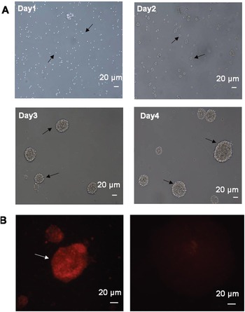
The cellular morphology of telencephalon-derived NSCs from passage 5 (P5) was analysed by microscopy during each day (D1, D2, D3 and D4). As shown in Fig. 1A, during the first 24 h, many suspended single cells were noted with clear boundaries and translucent appearances in the medium. On day 2, a few small cell clusters of the NSCs, floating in suspension, grew in size. On day 3, the number of NSCs increased rapidly and the proliferated NSCs formed neurospheres. On day 4, the neurospheres became enlarged, most reaching a size of 150–200 μm in diameter. It has been reported that the NSCs are represented by expression of nestin, the most prominent signature associated with neural precursors of the CNS (Sun et al. Reference Sun, Wang, Xie, Zhang, Wang, Li, Ma and Xin2011). Immunofluorescence staining on the isolated cell population demonstrated that the neurosphere isolates in the study were positively stained for nestin at P5 stages (Fig. 1B).
Neural stem cells were identified through morphological observation and immunocytochemical staining. (A) The morphology of the NSCs at P5 was observed by inverted light microscopy (×200) at different time points; (B) Neurospheres were positively stained for nestin (red) on day 4 (×400). (C) Negative control of immunocytochemical staining.

Toxoplasma gondii induced apoptosis of NSCs in vitro
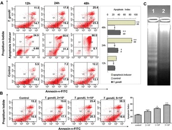
Apoptosis level of the NSCs was examined using FCM analysis with FITC-Annexin V/PI staining, and results are showed in Fig. 2. When co-cultured with 1×10⁶ RH tachyzoites for 12, 24 and 48 h, the apoptosis levels of the NSCs, including early and late apoptosis, were 26·21±0·78, 37·01±1·23 and 48·70±0·56%, respectively, whereas in the control group, the apoptosis rates were 21·12±1·34, 25·13±2·07 and 25·31±1·03%, respectively (Fig. 2A). Significant differences were found between the co-culture group and control group (P<0·05) after 24 and 48 h treatment, but not after 12 h treatment. Additionally, the apoptosis levels in the NSCs increased in a dose-dependent manner. As shown in Fig. 2B, after the treatment with different doses of tachyzoites for 48 h, the apoptotic rates were 45·73±2·42% for 1×10⁶ tachyzoites and 56·94±1·35% for 5×10⁶ tachyzoites, respectively, significantly higher than that in the control group (27·52±1·80%). No significant difference was found between 2×10⁵ tachyzoites-treated group (36·72±4·52%) and the control. In addition, a DNA laddering assay displayed an oligonucleosomal ladder of the fragmented DNA in the apoptotic NSCs co-cultured with 1×10⁶ tachyzoites for 48 h. No obvious DNA ladders were found in the control group.
Apoptosis detected by flow cytometry and DNA ladder. (A) Neural stem cells were co-cultured with 1×10⁶ T. gondii tachyzoites or Apopida (apoptosis inducer A) for different lengths of time (12, 24 and 48 h). (B) Neural stem cells were co-cultured with various doses of tachyzoites (2×10⁵ mL−1, 1×10⁶ mL−1, and 5×10⁶ mL−1) for 48 h. The cells were collected, stained with Annexin V/PI and then analysed by FCM. The presented plots are from a representative study and the graphics represent the mean and s.d. on different assays (n = 3). *P<0·05; **P<0·01 vs. control. #P<0·05; ##, P<0·01 vs. the 12-h treated group. (C) DNA ladder assay of the treated NSCs. Lane 1: Neural stem cells co-cultured with 1×10⁶ tachyzoites for 48 h; Lane 2: The untreated NSCs control.

Toxoplasma gondii induced apoptosis of NSCs via the ERS pathway
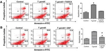
In order to identify whether the ERS signalling pathway was involved in the T. gondii-induced apoptosis of the NSCs, the NSCs were pretreated with TUDCA or Z-ATAD-FMK for 6 h before 1×10⁶ RH tachyzoites were added to the upper chamber. FCM analysis demonstrated that the apoptosis level of the NSCs pretreated with TUDCA was 28·41±0·60% after co-culture with 1×10⁶ RH tachyzoites for 48 h, significantly lower than that in the non-pretreated NSCs (52·92±0·95%) (P<0·01, Fig. 3A). When pretreated with Z-ATAD-FMK, the apoptosis level of the NSCs decreased from 49·81±0·91 to 27·63±0·82% (P<0·01) (Fig. 3B).
Effect of inhibitors on the apoptosis levels of neural stem cells co-cultured with 1×10⁶ RH tachyzoites. (A) The NSCs were pre-treated with TUDCA. (B) The NSCs were pre-treated with Z-ATAD-FMK (ZAF). The presented plots are from a representative study and the graphics represent the mean and s.d. on different assays (n = 3). *P<0·01 vs. the T. gondii-treated group; #P<0·05 vs. the control group.

Western blotting analysis showed that, as compared with the non-pretreated NSCs with co-culture of the parasites, the TUDCA pretreatment significantly down-regulated the protein level of CHOP, p-JNK and cleaved caspase-12 (Fig. 4A); however, the pretreatment of Z-ATAD-FMK significantly inhibited the cleavage of caspase-12 , but no effect was noted on the levels of CHOP and p-JNK (Fig. 4B).
The expression levels of CHOP, JNK and caspase-12 in neural stem cells. The protein levels were analysed by Western blotting, and β-actin was used as an internal loading control. (A) The NSCs were pretreated with or without TUDCA, and then co-cultured with 1×10⁶ RH tachyzoites for 48 h. (B) The NSCs were pretreated with or without Z-ATAD-FMK (ZAF), and then co-cultured with 1×10⁶ RH tachyzoites for 48 h. The presented figures are from a representative study and the graphics represent the mean and s.d. on different assays (n = 3). The NSCs without co-culture of tachyzoites were used as a control. Toxoplasma gondii+TUDCA or ZAF stands for the NSCs pretreated with TUDCA or Z-ATAD-FMK, and then co-cultured with tachyzoites. The experiments were repeated three times. *P<0·01 vs. control; #P<0·01 vs. the T. gondii-treated group.

DISCUSSION
Previous studies have reported that abnormal proliferation, differentiation or apoptosis of the NSCs induced by congenital infection of viruses can lead to brain malformations (Mutnal et al. Reference Mutnal, Cheeran, Hu and Lokensgard2011; D'Aiuto et al. Reference D'Aiuto, Di Maio, Heath, Raimondi, Milosevic, Watson, Bamne, Parks, Yang, Lin, Miki, Mich-Basso, Arav-Boger, Sibille, Sabunciyan, Yolken and Nimgaonkar2012; Sun et al. Reference Sun, Guan, Li, Li, Wang, Guan, Sheng, Yu and Liu2012). As a neurotropic parasite, the effect of T. gondii on the fate of the NSCs has not yet been fully characterized. The present study presented a facilitated effect of T. gondii on the apoptosis of the NSCs. Apoptosis is a physiological response that mediates elimination of pathogens and restores homeostasis. Toxoplasma gondii is able to promote or inhibit the cell apoptotic machinery, depending on its virulence and load, as well as on the host cell type (Nash et al. Reference Nash, Purner, Leon, Clarke, Duke and Curiel1998; Carmen and Sinai, Reference Carmen and Sinai2007; Laliberte and Carruthers, Reference Laliberte and Carruthers2008). Currently, evidence for apoptosis in brain cells infected with T. gondii is scarce and controversial (Contreras-Ochoa et al. Reference Contreras-Ochoa, Lagunas-Martinez, Belkind-Gerson, Diaz-Chavez and Correa2013). Previous studies have demonstrated that the tachyzoites of the RH strain and ME49 strain induced neuron apoptosis (Takahashi et al. Reference Takahashi, Fukuda, Tanaka, Minamitani, Onouchi and Makioka2001; el-Sagaff et al. Reference el-Sagaff, Salem, Nichols, Tonkel and Abo-Zenadah2005). However, a recent study has reported that the RH strain seemed to inhibit the apoptosis of astrocytes for up to 24 h until it replicated, egressed and generated cellular destruction (Contreras-Ochoa et al. Reference Contreras-Ochoa, Lagunas-Martinez, Belkind-Gerson, Diaz-Chavez and Correa2013). Here we found that T. gondii promoted apoptosis of the NSCs significantly, especially the late apoptosis, in a dose-dependent manner as early as 24 h (Fig. 2). The balanced regulation between cell proliferation and death is required to achieve the correct number of specific cell types within an organ and tissue, and is a crucial component in regulating organ size (Joseph and Hermanson, Reference Joseph and Hermanson2010). Therefore, the loss of the NSCs that are involved in neurogenesis of the brain in the embryonic stage may be an important cause of congenital toxoplasmic encephalopathy.
In the present study we explored a trans-well co-culture system, in which the permeable inserts with a 0·4 μm size pore only permit the passage of the excreted-secreted antigens (ESA) of T. gondii rather than of the tachyzoites. Therefore, the increased apoptosis level found in the NSCs co-cultured with T. gondii tachyzoite (Fig. 2) did not result from a direct replication of parasites in the cells but from the release of ESAs. Previous reports indicated that the major host tissue cells could act as the bystanders in acute infection, and apoptosis of those cells may result from the secretion of some soluble factors by parasite-infected cells (Mordue et al. Reference Mordue, Monroy, La Regina, Dinarello and Sibley2001; Nishikawa et al. Reference Nishikawa, Kawase, Vielemeyer, Suzuki, Joiner, Xuan and Nagasawa2007). Therefore, the trans-well co-culture system is an excellent model to identify the effect of ESA by infected cells on the bystander cells in a more natural fashion. The composition of the T. gondii ESA is surprisingly complex, and only 15 microneme proteins (MIC), 18 rhoptry proteins (ROP) and 15 dense granule proteins (GRA) have been identified. Which molecules of ESA play key roles in the apoptosis of NSCs induced by T. gondii needs to be further explored.
Apoptosis mediated by ERS plays a key role in many diseases (Amano et al. Reference Amano, Yamasaki, Yagishita, Tsuchimochi, Shin, Kawahara, Aratani, Fujita, Zhang, Ikeda, Fujii, Miura, Komiya, Nishioka, Maruyama, Fukamizu and Nakajima2003; Allen et al. Reference Allen, Nguyen, Sargent, Lipson, Hackett and Urano2004; Gao et al. Reference Gao, Lee, Chen, Zhang, Zhang, Kannan, Ortmann and Fang2008; Lei et al. Reference Lei, Zhang, Barbour, Bohrer, Ford, Koizumi, Papa and Ramanadham2010). Endoplasmic reticulum (ER) is a cellular organelle with several important functions, and various stimuli can disturb ER homeostasis, therefore resulting in the accumulation of unfolded or misfolded proteins and pathological consequences, namely ERS (Kaufman et al. Reference Kaufman, Scheuner, Schroder, Shen, Lee, Liu and Arnold2002; Nadav et al. Reference Nadav, Shmueli, Barr, Gonen, Ciechanover and Reiss2003). Prolonged and severe activation of ERS can turn on a cell death pathway through activation of C/EBP homologous protein (CHOP), caspase-12 and/or c-JUN NH2-terminal kinase (JNK) (Zinszner et al. Reference Zinszner, Kuroda, Wang, Batchvarova, Lightfoot, Remotti, Stevens and Ron1998; Nakagawa et al. Reference Nakagawa, Zhu, Morishima, Li, Xu, Yankner and Yuan2000; Urano et al. Reference Urano, Wang, Bertolotti, Zhang, Chung, Harding and Ron2000). We detected the protein expression of the three molecules involved in T. gondii ERS-mediated apoptosis pathway using Western blotting analysis, and found that caspase-12 and JNK were activated and CHOP was up-regulated in the NSCs co-cultured with T. gondii. Moreover, with pretreatment of TUDCA, a known inhibitor of ERS, the protein level of CHOP significantly decreased and the activation of caspase-12 and JNK was inhibited in the NSCs co-cultured with T. gondii (Fig. 4A), and subsequently decreased the apoptosis level of the NSCs induced by co-culture with the parasite (Fig. 3A). Additionally, pretreatment with Z-ATAD-FMK, a specific inhibitor of caspase-12, also significantly decreased the apoptosis level of the NSCs treated with the ESAs of the parasite in the co-culture system (Fig. 3B). The data from both inhibitors indicate that the T. gondii ESAs induce the apoptosis of NSCs through specific activation of CHOP, caspase-12 and JNK. As is well known, CHOP is barely detectable under physiological conditions, but is strongly induced in response to ER stress (Ron and Habener, Reference Ron and Habener1992; Wang et al. Reference Wang, Lawson, Brewer, Zinszner, Sanjay, Mi, Boorstein, Kreibich, Hendershot and Ron1996). Over-expression and targeted disruption of the CHOP gene has demonstrated that CHOP promotes apoptosis in response to ER stress (Zinszner et al. Reference Zinszner, Kuroda, Wang, Batchvarova, Lightfoot, Remotti, Stevens and Ron1998; Oyadomari et al. Reference Oyadomari, Takeda, Takiguchi, Gotoh, Matsumoto, Wada, Akira, Araki and Mori2001, Reference Oyadomari, Koizumi, Takeda, Gotoh, Akira, Araki and Mori2002). Caspase-12, an ER-localized cysteine protease, is only activated by ER stress, but not by death receptor-mediated or mitochondria-targeted apoptotic signals (Nakagawa et al. Reference Nakagawa, Zhu, Morishima, Li, Xu, Yankner and Yuan2000; Araki et al. Reference Araki, Oyadomari and Mori2003). Taken together, our results suggest that T. gondii induces apoptosis of the NSCs through the ER stress pathway via activation of caspase-12, CHOP and JNK. Whether a death receptor-mediated or mitochondria-targeted apoptotic signal pathway is involved in this pathological process remains to be elucidated.
FINANCIAL SUPPORT
This work was supported by the National Basic Research Programme of China (973 Programme) (J. S, grant no. 2010CB530001); Natural Science Foundation of China (L. Y., grant no. 30800965), (J. D. grant no.81271864); and the Outstanding Young Scholars Financial Support of Anhui Medical University (L. Y., grant no.0113014104).