Cardiovascular screening is widely recommended and performed in young competitive athletes, although the optimal strategy remains controversial nowadays. Reference Mont, Pelliccia, Sharma and Biffi1 In this regard, the inclusion of resting electrocardiogram is still a matter of debate. Despite having proved to be the most effective strategy to detect underlying causes to sudden cardiac death, Reference Harmon, Zigman and Drezner2 electrocardiogram has important limitations due to low sensitivity detecting some frequent diseases (mostly structural abnormalities). On the other hand, electrocardiogram can also generate a notable rate of false positives, leading to unnecessary additional tests or restricting from competition. Reference Maron, Friedman and Kligfield3 Point-of-care ultrasound has proven useful in some scenarios and could be a promising tool to increase detection of structural heart disease and thus improving diagnostic performance. Reference Cassels, Moulson and Regan4,Reference Moulson, Jaff and Wiltshire5
Between 2016 and 2019, 1188 young athletes [mean age 16.3 ± 4.6 years (with a range from 7 to 27)] were screened in different sports institutions (fundamentally at Villarreal CF, Spain), following the same protocol: (1) personal and family medical history 14-element checklist, according American Heart Association recommendations; Reference Maron, Friedman and Kligfield3 (2) physical examination with thorough cardiac auscultation; (3) resting 12-lead electrocardiogram, following international consensus standards for electrocardiogram interpretation in athletes; Reference Drezner, Sharma and Baggish6 and (4) point-of-care ultrasound performed by experienced sports cardiologists using a portable ultrasound (Vivid i with probe S5, GE Healthcare), consisting in visual examination in standard views (long and short parasternal, apical and subcostal), and M-mode and/or bidimensional measurement of: left ventricular diameters and wall thickness; right ventricular outflow tract; and ejection fraction by Simpson’s equation. Pulsed, continuous, and colour Doppler were performed to evaluate valvular heart disease, focussing in mitral prolapse and bicuspid aortic valve. Coronary ostia were also checked in all individuals. Left ventricular mass index was calculated according American Society of Echocardiography recommendations. Reference Lang, Badano and Mor-Avi7 All athletes signed informed consent form or their parents in case of under age.
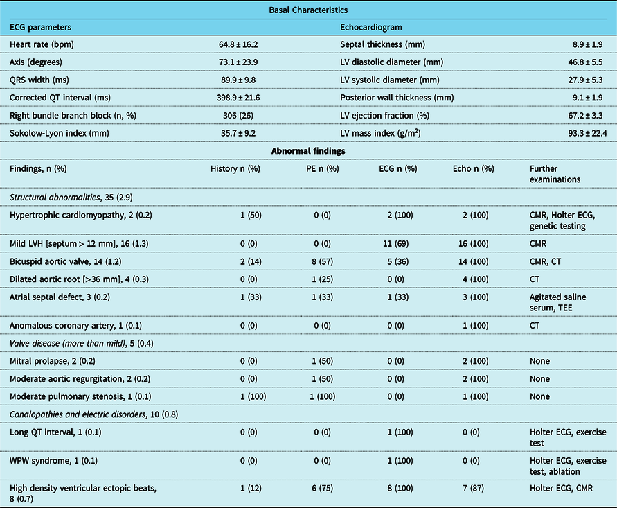
Main findings of the study are reflected in Table 1. The majority sports disciplines were: football 65.8%, tennis 21.7, and 8.2% indoor football. In total, 76.3% of athletes were male. Median time per screening was 12 minutes (with an interquartile range from 9 to 14). Of 582 athletes (48.9%) with a Sokolow-Lyon index >35 mm, only 97 (8.2%) of them had a left ventricular mass index >115 g/m2 (sensitivity 61% and specificity 44%). Normal position of both coronary ostia could not be ensured in 112 (9.4%). Abnormal findings were found in 50 athletes (4.2%), mostly structural. The most frequent finding was mild left ventricular hypertrophy [16 athletes with mean age 20.2 (with a range from 15 to 23) and mean septum thickness 13.8 mm (with a range from 13 to 16)]. Only in five individuals (0.4% of total) potential causes of sudden cardiac death were identified: a 15-year-old male handball player with hypertrophic myocardiopathy, positive family history (father with sudden death before 50 years), fibrosis in cardiac resonance, and positive genetic test, who was advised to leave sports competition; a 18-year-old male basketball player also with hypertrophic myocardiopathy, but with no family history and no fibrosis, 12-mm-thick septum, low European Society of Cardiology risk score, who was allowed to continue in competition with annual assessment; a 13-year-old male football player with anomalous right coronary artery originating from the left sinus of Valsalva (with not inter-arterial course), who was referred for clinical follow-up by cardiology; a 16-year-old male basketball player with suspected long-QT syndrome (intermediate probability according Schwartz Score), referred to study at his hospital; and a 11-year-male football player with Wolf–Parkinson–White syndrome, who was referred to consider ablation of accessory pathway.
Electrocardiogram and echocardiogram parameters, and abnormal findings of the screening

CRM: cardiac magnetic resonance. LV: left ventricular. LVH: left ventricular hypertrophy. PE: physical examination. TEE: transesophageal echocardiography.
In this study, the combined use of electrocardiogram and point-of-care ultrasound was feasible performed by experienced sports cardiologists, and allowed rule-out in situ some false positive electrocardiogram findings, mainly those derived from voltage criteria for left ventricular hypertrophy. In addition, many structural abnormalities (such bicuspid aortic valve or coronary anomalies) could be detected otherwise no diagnosed. Although these pathologies are not usually potentially fatal, their early detection allows follow-up and proper management. If major cardiac condition associated with sudden death were analysed (five athletes in our study), electrocardiogram alone was able to detect four cases (all except coronary anomaly), and the use of point-of-care ultrasound in isolation identified three cases (did not identify Wolf–Parkinson–White or long-QT). The American Heart Association’s proposed that combination of medical history and physical examination Reference Maron, Levine, Washington, Baggish, Kovacs and Maron8 would have detected only one athlete, based on a family history of early sudden death. Some previous studies have shown the poor diagnostic performance of consensus-derived history questionnaires as the primary tool for cardiovascular screening, with a very low sensitivity and positive predictive value, and advocate the reevaluation of this strategy. Reference Williams, Pelto and Toresdahl9 On the other hand, the combined use of electrocardiogram and echocardiogram has already proven effective in the diagnosis of cardiac diseases requiring sport disqualification. Reference Calo, Martino and Tranchita10 Using point-of-care ultrasound instead of standard echocardiogram facilitates the process of cardiac screening and its implantation in sports institutions with large number of young athletes could be an alternative to detect common causes of sudden death.
Limitations of the study include small sample and analysis from a single research group, the high percentage of male football players in the sample study, and the use of portable ultrasound that could limit some examinations (specially detection of coronary anomalies).
In conclusion, the combined use of electrocardiogram and point-of-care ultrasound proved to be a practical screening strategy in competitive athletes, by improving diagnostic capacity primarily with respect to structural abnormalities and also by minimising positive false cases of electrocardiogram alone, and could be a viable alternative in certain sports contexts.
Financial support
No financial or material support of any kind was received for the work described in this article.
Conflicts of interest
None.
Ethical standards
The authors assert that all procedures contributing to this work comply with the Helsinki Declaration of 1975, as revised in 2008, and has been approved by the institutional committees.