Introduction
Worldwide, a rapid loss of animal genetic resources can be observed. While many developed countries counteract this phenomenon through financial incentives to threatened breeds, it is often not a viable option in the less-developed parts of the world. This situation is addressed by a ‘proposal for a worldwide emergency programme for the creation of national gene banks of endangered breeds’ (Groeneveld, Reference Groeneveld2005). Here, the rationale is given for the initiation of gene banks based on somatic cells, which can be collected rapidly at favourable costs, and – because of this – in many cases constitute the only feasible option. A somatic cell bank consists of ear-tag tissues, which can be preserved in liquid nitrogen (LN) and subsequently used as donor cells for animal cloning. During the last decade, the technique of reproductive somatic nuclear transfer (NT) was successfully applied in livestock (sheep, cattle, goats, pigs, horses), rodents (mice, rats) and pet animals (cats, dogs), as reviewed in Kues and Niemann (Reference Kues and Niemann2004), and also in the preservation of endangered breeds (Lanza et al., Reference Lanza, Cibelli, Diaz, Moraes, Farin, Farin, Hammer, West and Damiani2000; Wells et al., Reference Wells, Misica, Tervit and Vivanco1998; White et al., Reference White, Bunch, Mitalipov and Reed1999) and wild animal species (Loi et al., Reference Loi, Ptak, Barboni, Fulka, Cappai and Clinton2001). The best success rates in terms of live offspring so far were obtained with cloned cattle and goats. The authors are aware that the NT technique needs further improvements to reach practicability for large-scale cloning. Recent developments support the notion that the need of a costly micromanipulation unit can be substantially reduced (Vajta et al., Reference Vajta, Bartels, Joubert, De la Rey, Treadwell and Callesen2004), and that NT-associated problems, such as overall low success rates, might be reduced (Andrabi and Maxwell, Reference Andrabi and Maxwell2006; Trounson, Reference Trounson2006; Wilmut, Reference Wilmut2006).
It is the objective of this paper to develop a protocol for tissue collection, treatment and storage, and to assess the costs and feasibility of the complete procedure under field conditions in a developing country, by sampling and documenting six breeds from three species, namely, pigs, sheep and goats, in the remote areas of Vietnam.
Material and methods
A functional protocol for the collection, storage and documentation of viable somatic cells needs to address the sterilisation of skin tissue, the procedures for sample collection, the cryoprotectant and storage, and their documentation.
Surface sterilisation of ear tissues
In the laboratory, ear tissues were sampled from five cattle ears, collected in the slaughterhouse, and from three mice. The ear tissues were divided into three parts, and treated with either 70% ethanol solution for 3 min or hand disinfectant (Bode Sterillium, Hamburg, Germany) for 3 min or kept in phosphate-buffered saline (PBS) solution for 3 min (control – untreated). Then the skin was removed from all tissue pieces, washed two times using PBS and cut into small pieces. These small tissue pieces were seeded in six-well culture plates using 2 ml Dulbecco’s Modified Eagle Medium (DMEM) without antibiotics and incubated at 37°C for 5 days. The cultures were then screened at 200- and 400-fold microscopic magnification for signs of bacterial or fungal growth.
Ear sample collection
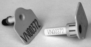
Ear tags were collected with an ear tagger (Biopsytec: http://www.biopsytec.com). The animals were fixed, one ear was cleaned and treated with 70% ethanol. The tagger is special in the sense that the tagging process integrates three actions: (1) punching a hole in the ear, (2) pushing the cut-out tissue segment into the attached vial (Figure 1) and (3) fixing the plastic identification number (ID) through the punched hole to the ear.
Ear tag with attached vial.

Assessing the optimal concentration of glycerol as cryoprotectivum and cell culture
The use of glycerol in PBS has been developed as a cryoprotectant to minimise the effects of damage by ice crystals, alternations in the concentration of electrolytes, dehydration and changes in pH (Watson et al., Reference Watson, Weigand, Clough and Hoffman1986; John, Reference John2003; Julie, 2004). An 87% glycerol stock was diluted with PBS at levels of, respectively, 100%, 90%, 50%, 30% and 10% to give concentrations of 0%, 8.7%, 26.1%, 43.5% and 78.3% of glycerol. The cattle ears were disinfected by 70% ethanol for 2 to 3 min. Five ear biopsies were taken with the ear tagger from a cattle ear (seven replications with cattle ears from a slaughterhouse) and collected in caps pre-filled with different concentrations of glycerol. The sample caps were dropped directly into LN. One day later, the sample caps were thawed, the biopsies were cut in small slices and stained with MitoTracker Red (Molecular Probes/Invitrogen, Karlsruhe, Germany). MitoTracker is a cell-permeant dye, which is selectively accumulated in viable mitochondria, allowing the live/dead staining of cells. Fluorescence was observed in a Zeiss Axiovert Microscope equipped with epifluorescence and a rhodamine filter block (excitation 520 to 550 nm, emission 580 nm). Viable cell samples were identified by Mitotracker fluorescence and the characteristic distribution of the fluorescence to mitochondria. Dead cells show no fluorescence.
In parallel, primary cell cultures were isolated from some of the samples frozen in 26.1% glycerol employing the explant culture technique (Kues et al., Reference Kues, Petersen, Mysegades, Carnwath and Niemann2005). In brief, minute tissue samples (<1 mm3) were pasted with recalcified plasma micro drops to tissue culture flasks and covered with 4 ml DMEM, containing 10% foetal calf serum, 1% non-essential amino acids, 1% vitamin solution, 2 mmol/l glutamine, 0.01 mmol/l mercaptoethanol, 100 U/ml penicillin and 100 μg/ml streptomycin (all chemicals from Sigma, Deisenhofen, Germany). After 5 to 7 days culture in a humidified incubator with 5% CO2, the explants were assessed for outgrowing cells (Kues et al., Reference Kues, Petersen, Mysegades, Carnwath and Niemann2005).
Ear biopsy collection of endemic livestock breeds in Vietnam and documentation
After the treatment of samples has been optimised and the protocol for the sample-collection process has been developed, the feasibility of the complete process has to be investigated in an environment with limited infrastructure. To this effect, collection of samples for six breeds from three species was carried out in a rural area of Vietnam.
Ideally, somatic cells would be collected from 25 males and 25 females, which are, as far as possible, unrelated. Using the expression Ne=(4NmNf)/(Nm+Nf) (Falconer and Mackay, Reference Falconer and Mackay1996), this results in an effective population size Ne = 50, a figure often considered to be the lower threshold for the long-term survival of a breeding population under human intervention, as it puts the rate of inbreeding at 1% per generation, provided the resulting population is continued with this mating structure. This threshold is, for instance, used in the National Programme of Animal Genetic Resources in Germany (DGfZ, 2004). Under practical circumstances, finding 50 unrelated animals may be a problem. Firstly, animals are often not identified individually and, furthermore, no pedigree information is available. Thus, for a group of females, it may be impossible to know their relationship. Secondly, often a very small number of adult males are required and therefore kept in breeding populations. Thus, finding 25 adult males will often be impossible. Here a pragmatic solution is proposed in that the group of candidates for sampling includes all males, adult and young. The procedure for sampling would be to start with the adult males and then continue with the males in the crop of offspring. The same procedure can be used in the less-likely case that not enough adult females can be sampled. This will clearly result in putting genetic material of possibly full sibs in store, if the population to be sampled are multiparous, as in swine. While half-sibs or even more distantly related animals are to be preferred, even with a full-sib group, still one half of the total additive genetic variation is captured.
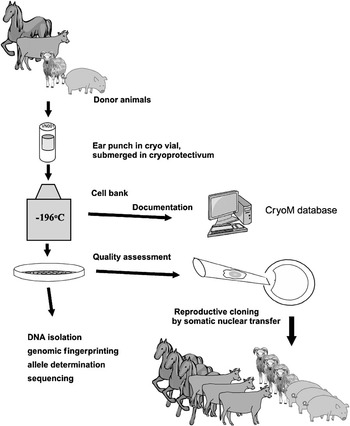
Quality assessment of samples stored should be part of the collection process, as indicated in step 3 of Figure 2. Thus, on a proportion of animals, duplicate samples should be collected, which can be used in the quality-assessment stage for testing. A figure of 10% seems useful.
Schematic outline of the creation of a somatic cell bank.

Results
Surface sterilisation of ear samples
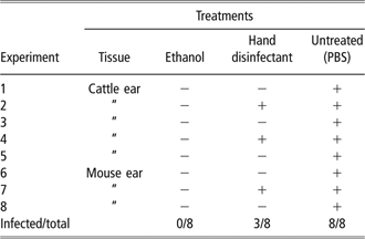
The effectiveness of 70% ethanol and a hand disinfectant for the sterilisation of ear tissue samples were tested under laboratory conditions. Therefore cattle ears collected in a slaughterhouse and mice sacrificed for other reasons were employed. A surface treatment with 70% ethanol was found to be an effective treatment for sterilisation of ear tissue, as shown in Table 1. None of the ethanol-treated samples was infected, indicating that sterile biopsies can be produced from ear tissues. In contrast, three of the samples treated with a hand disinfectant and all untreated samples resulted in bacterial contamination. The effectiveness of ethanol treatment for sterilisation was confirmed by ear sampling in a cowshed, employing the ear tagger (three replications); here too no infection was detected (results not shown).
Effect of sterilising treatmentFootnote †
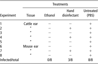
† Abbreviation is: PBS = phosphate-buffered saline; +: growth of bacteria/fungi; −: no infection.
Glycerol as cryoprotectant
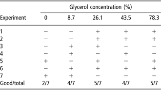
The effect of glycerol concentration on the viability of ear tags was tested. Five ear biopsies were obtained with an ear tagger from slaughterhouse cattle ears and collected in vials pre-filled with glycerol solutions ranging from 0% to 78.3% and directly frozen in LN. After thawing, viability was assessed by the live/dead stain MitoTracker; in addition, explant cultures were initiated. It seems that high viability is guaranteed over a wide range, as shown in Table 2. The best rates of outgrowing cells were obtained from tissues frozen in a 26.1% glycerol solution, which was therefore used for further experiments.
The viability of frozen tissues with different concentrations of glycerolFootnote †
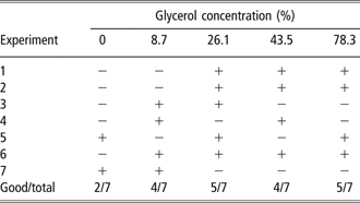
† +: good survival; −: few or no viable cells.
Sample collection: the protocol
Prerequisites and initial investment
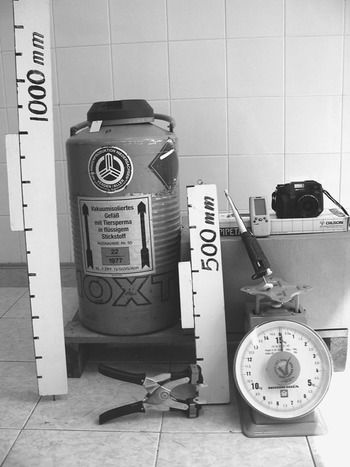
The equipment list is rather short as can be seen from Figure 3.
Equipment for collecting somatic cell samples: large ruler, transportation cryo tank, tagger, small ruler, pipette, global positioning system device, scale and digital camera (left to right).

Ear tagger: the tagger is special in the sense that the tagging process integrates three actions: (1) punching a hole in the ear, (2) pushing the cut-out tissue segment into the attached vial (Figure 1) and (3) attaching the plastic ID number through the punched hole to the ear. The tagger used here was from the Biopsytec http://www.biopsytec.com/ (Figure 3).
Ear tags and sample vials: in this pilot, a set of 400 ear tags were ordered from the supplier of the ear tagger, with an ID range of VN00001 through VN00400.
Transportation cryo tank: samples must be deep frozen right after they have been taken. This requires a cryo tank at the sampling site. A 5-l tank has proved to be adequate. For permanent storage, another stationary tank has to be available.
Scale: to keep track of the volume of LN in the tank, a metric scale can be used to determine nitrogen loss.
Photo ruler: to allow later computation of linear measurements of the animal based on the digital image.
Digital camera: to take a digital colour photo of each animal including the photo ruler being held against the animal.
GPS device: these readily available hand-held devices deliver worldwide unique coordinates of the sampling location.
Laboratory material, etc.: centrifuge tubes or vials with caps and a volume of 2 to 3 ml used for the cryoprotective medium during the period of sampling at locations, pipette or syringe to fill these tubes, gloves, ropes for restraining the animals and disinfectant spray.
Preparation for a sampling excursion
A mixture of 70% PBS and 30% of 87% glycerol giving a concentration of 26.1% is stored in small vials (2 to 3 ml) like centrifuge tubes covered with tight caps to prevent infection during use at the sampling location, sufficient for five to six tissue samples (0.3 ml per sample).
On-site sample collection
Usually, at any collection site, samples will be taken from a number of animals. The procedure for the complete collection process comprises collection of: (1) site-specific information through a global positioning system (GPS), (2) animal-specific information and finally (3) the somatic cell samples. As outlined above, to allow viability testing of the samples, a proportion of around 10% duplicate samples should be collected and used to initiate cell cultures.
Collection of samples and animal-specific data
The procedure for collecting samples and data at a site follows these steps, making use of the equipment shown in Figure 3.
1. At the sampling location, animals should be restrained by a rope.
2. Mark the photo ruler with ‘A’ or ‘Y’ to indicate if the animal is mature ‘A’ or young ‘Y’.
3. Write the number of the digital images, as they are to be stored in the camera, on the photo ruler to link the image itself to the filename, which is required for later loading into the database for documentation.
4. As soon as animals are restrained, place the photo ruler with an appropriate length close to the animal in a vertical position. Place the photo ruler perpendicular 5 to 10 cm from the ground to obtain a good reading on the basis of the digital image (see Figure 3).
5. Take a picture of the animal and the photo ruler, filling the viewfinder for maximum accuracy. To reduce the error of later measurements based on the image, the animal and the ruler should be in one plain and perpendicular to the viewing direction of the camera. A resolution of 3 megapixel has proven sufficient. To reduce distortion, a longer focal distance should be chosen.
6. Disinfect the area of ear skin from where tissue sample is to be taken by spraying both sides with 70% ethanol and wait for 1 to 3 min until dried. The ears of free-ranging animals may be very dirty, so they should be cleaned with fresh water before using 70% ethanol for disinfection.
7. While two persons are restraining the animal, taking photos (one person) and disinfecting (one person), another person should be in charge of preparing the medium, filling the sample cap with 0.3 ml cryoprotective medium and plug it into the ear tagger. To prevent the infection of tissue samples, conditions for sampling should be as clean as possible by using disposable tips, keeping hands, pipettes, medium vials and other equipment in clean conditions.
8. Take a tissue sample with the ear tagger. To be sure that the sample is safely deposited in the attached vial, it is usually necessary to squeeze the tagger twice: the first time usually only the skin is punctured, while only the second squeeze pushes the ear biopsy into the sample cap.
9. Remove the sample cap containing an ear biopsy from the tagger, turn it upside down and shake gently to make the cryoprotectant medium cover the ear biopsy inside. Finally, put the sample cap directly into the LN transport container.
10. Collect data relevant to the animals. The dataset has been reduced to a minimum that will be available everywhere around the world independent of species. The breed names are chosen in accordance with Food and Agriculture Organization of the United Nations – Domestic Animal Diversity Information Service (FAO DAD-IS), which has been redesigned in the European Union EFABIS project (Groeneveld et al., 2006; Rosati et al., 2006). This allows automatic integration of countries’ aggregate breed data on their cryo samples into the worldwide breeds databases.
Post-sampling work
After the excursion, the samples collected have to be placed in a cryo tank for permanent storage. Permanent documentation of the somatic cell samples collected is essential. Without this, the samples will be useless, as on the basis of the vial identification alone, not even the species from which the sample originated is known. The CryoM database has been developed to provide a generalised structure for storing a minimum set of data that can be collected with each somatic cell sample of any species anywhere around the world. The basis for entering data in the database is an on-site form together with the images taken by the digital camera. The form contains the GPS coordinates of the location, the species/breed, the collection date, the vial number and the number of the digital image. This information together with the location code is entered into the CryoM database.
The protocol for quality assurance
A somatic-cell bank is useful only when the cryogenic material is fit to serve as a basis for somatic cell cloning. To increase the chance of viable tissue samples in the bank, a two-step procedure was developed.
1. A simple protocol for the cryopreservation of vital ear samples was established under laboratory conditions.
2. Duplicate samples were collected from about 10% of the animals in the field study in Vietnam. These duplicate samples were successfully used to initiate growing cell cultures after 2 to 3 months of cryopreservation.
The pilot sampling collection
The pilot collection project was carried out by the staff from the Institute of Agricultural Sciences of South Vietnam (IAS) located in Ho Chi Minh City. It was the intention to test the procedures for feasibility on six breeds from swine, sheep and goats. With 50 samples per breed, this would result in 300 samples to be taken.
In Vietnam, all communication with the animal owners was routed through the Provincial Department of Agriculture and Rural Development responsible for the area where samples were to be taken. The Provincial Department then communicated with the districts and villages. The latter nominated a locally ‘respected’ person who acted as a guide and contact to the owners of the animals. To organise this process – once the list of breeds to be sampled had been established – took around 2 months under Vietnamese conditions.
The crew comprised four persons. One of them helped the owner to restrain the animal, the second applied the disinfectant and held the ruler for the photo, the third was in charge of taking the photo and keeping the records, while the last person handled the cryoprotective medium and actually took the ear samples.
Animals from the Binh Thuan and Ninh Thuan provinces were sampled. In Binh Thuan province, the tissue sample collection was conducted on the population of Co pigs named Binh Thuan-Co pigs at a mountain village. In Ninh Thuan province, the tissue sample collection was conducted on Bach Thao goat, Co goat, Phan Rang sheep and two populations of Ninh Thuan-Co pigs.
The distance from Ho Chi Minh City (central laboratory for sample storage) to the locations in Binh Thuan province is about 250 km and to the locations in Ninh Thuan it is about 400 km away.
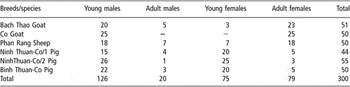
In all, 300 samples were collected as shown in Table 3. Although the two populations from the Ninh Thuan province share the same breed name, they are geographically separated with no known exchange of animals. Thus they were both sampled. The crew had to spend four trips to the locations, each trip taking 3 to 5 days.
Structure of sampled breeds

After storage periods of 2.5 to 3 months, 23 duplicate samples were taken out to be tested for their viability, comprising nine samples from pigs, 10 samples from goats and four samples from sheep. Thawed samples were transferred in cell culture and assessed for outgrowing cells. In this quality-assurance test, all of 23 tested samples were viable.
Cost appraisal
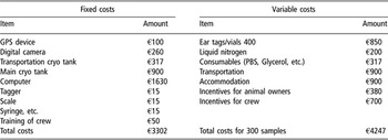
As indicated in Table 4, the total investment for setting upa somatic-cell bank is in the range of €3000, with around half of the sum having been spent on a rather high-end notebook for running the CryoM database. Not included are the costs for buildings and other infrastructure that might be used. Clearly, initial costs will also vary from country to country. The cost for local training in the use of the ear tagger and other procedures – excluding laboratory work – can be kept as low as €50.
Initial and variable cost for the collection of 300 samples in three locationsFootnote †

† Abbreviation is: PBS = phosphate-buffered saline.
Variable costs (Table 4, second part) include the well-defined costs for vials, which are around €2 per sample. Other blocks of costs are travel of the sampling crew, and incentives for both the sampling crew and the animal owners.
Discussion
The results indicate that the procedure of surface sterilisation and sample collection is simple enough to generate somatic cell samples under adverse conditions with little infrastructure that will reliably develop into cell lines as a prerequisite for NT cloning (Figure 2). Therefore, it could be argued that a post-collection test on the viability of cells collected and frozen would not be required. On the other hand, one effect of a quality-assurance procedure is that it tends to enhance careful execution of procedures, which leads to improved quality.
Even with a quality-assurance test, 100% viability of cells cannot be guaranteed, although this was the case here. However, it should be noted that it is not the individual animal that needs to be reconstituted but rather the population, which would still be possible even if some somatic cells do not lead to NT clones.
The rationale for setting up cell banks in the context of preservation of breeds hinges on the promise to reconstitute animals through NT cloning. Should this turn out to be impossible, a cell bank would have been a failure. If a breed was to be reconstituted now, both the proportion of not-normal NT clones and the high costs would currently still be a problem. As has been stated in the rationale, somatic cells are a good choice, when no other options like semen and embryos are available. Here, a quick and cheap backup can be done where other methods may not be feasible. Ideally, such a repository should later be amended by semen and embryos if time and resources permit.
The protocol presented is certainly only one option leaving it to the prospective users to modify it where required. This is particularly true for the quality-assurance test. Depending on the laboratory facilities, the outgrowing of cells described here could be extended to set up fully functional cell lines. Instead of destroying the outgrown cells, as was done here, cell lines could be refrozen for later use.
Conclusions
In the face of rapid erosion of animal genetic resources, somatic-cell banks may be the only option to create a backup repository fast enough (Ryder, Reference Ryder2002). The cell culture conditions used here favour the growth of primary fibroblast cells. Fibroblasts are one of the most used cells types for somatic nuclear transfer (Kues and Niemann, Reference Kues and Niemann2004; Andrabi and Maxwell, Reference Andrabi and Maxwell2006; Trounson, Reference Trounson2006). Cloned offspring have been obtained from long-term cell cultures and even from senescent cells. The success rates in terms of live offspring per transferred NT-embryo range from 10% to 25% for cattle, from 3% to 8% for goat and sheep and from 1% to 5% for pig cloning. It has been observed that certain cell cultures repetitively did not allow the generation of live offspring (Panarace et al., Reference Panarace, Agüero, Garrote, Jauregui, Segovia, Cane, Gutierrez, Marfil, Rigali, Pugliese, Young, Lagionia, Garnil, Forte Pontes, Ereno Junio, Mower and Medina2006). The reasons for this are unclear. It is obvious that the NT technique needs further improvements to reach practicability for large-scale cloning. However, due to the rapid loss of many endemic livestock breeds, the banking of somatic cells might be the only reasonable option to preserve these genetic resources. Future improvements of NT are likely and might circumvent the current problems associated with this technique.
The protocol developed and presented here requires far less resources than any other option like collecting semen and embryos. Gandini et al. (Reference Gandini, Pizzi, Stella and Boettcher2007) estimate the costs for establishing a gene bank, depending on the material collected, between €7000 and €33 550 per population, with only epididymical semen on the lower end, and a combination of embryos and semen on the upper. While the reconstruction of a population from semen and embryos compared with that from somatic cells is very different in terms of costs, the collection of somatic cells is an order of magnitude lower. Furthermore, the procedure is identical for all species, making sample collection much easier. Because of the limited investment in both equipment and training, somatic cell cryoconservation can be implemented rapidly in the field. This pilot study indicated that within a few months a number of breeds can be backed up in a cell bank. On the basis of 25 males and 25 females per breed, the variable costs can be kept below €1000 per breed.
The protocol has been developed with the objective to be simple, cheap and robust and to work on all species of interest. Likewise, the data to be collected can be obtained on any species, while allowing to generate more information ex post. Thus, on the basis of one scaled colour images, many linear measurements can be made on an individual animal, also making textual descriptions of coat colour and patterns obsolete. Similarly, the GPS coordinates recorded with each sample can be used ex post to obtain a whole array of information like altitude and climatic zone; soil types can be readily derived from the GPS coordinates for each sample.
As a result, large-scale creation of national somatic-cell banks as stipulated (Groeneveld, Reference Groeneveld2005) is feasible and should be started as soon as possible. The pilot study shows that the involvement of local organisations is extremely useful. On the other hand, the protocols developed and tested for sample collection have proved suitable across species and will also work for animals other than swine, sheep and goats. Also, there is no reason as to why they should not work for mammalian wildlife. The same applies to the data collected: all elements recorded are available for all species anywhere in the world. Together with a rather small initial investment on equipment and training, setting up national somatic-cell banks on the basis of somatic cells can be done quickly and efficiently. Accordingly, the procedure presented is well suited for the implementation of an international programme for a worldwide effort to establish national emergency somatic-cell banks on the basis of somatic cells perhaps under the umbrella of FAO.
Acknowledgements
The project was financially supported by the Federal Ministry of Education and Science, Germany, under VBN 03/B14, the Ministry of Science, Technology and Environment and the Ministry of Agriculture and Rural Development, Vietnam. Administrative support by Professor Pham Van Bien, Director of IAS, is gratefully acknowledged. Without the help of the Department of Agriculture and Rural Development from the Binh Thuan and Ninh Thuan provinces, locating and sampling the animals would not have been possible. In particular, their support in communication and contact to the animal owners is greatly appreciated. Finally, the staff from the Department of Animal Genetics and Breeding at IAS is recognised for their contribution to the sample collection.