Introduction
The cryotolerance of in vitro-produced bovine embryos is strongly influenced by lipid metabolism, as excessive lipid accumulation, particularly of saturated lipids, increases susceptibility to damage caused by oxidative stress, mitochondrial dysfunction and membrane rupture during freezing or vitrification (Sudano et al., Reference Sudano, Paschoal, Rascado, Magalhães, Crocomo, de Lima-Neto and Landim-Alvarenga2011). Polyunsaturated fatty acids (PUFAs), such as n-3 (e.g., α-linolenic acid) and n-6 (e.g., linoleic acid), modulate not only the fluidity of cellular and mitochondrial membranes, but also signalling pathways and gene expression involved in lipid metabolism, apoptosis and responses to oxidative stress. This modulation results in embryos with lower lipid accumulation and improved post-vitrification quality (Cavalieri et al., Reference Cavalieri, Santos, Matsushita, Petit, Rigolon, Silva, Horst, Capovilla and Ramos2005). Pantaneira, a native breed adapted to the Pantanal, shows remarkable adaptability to environmental stresses but is currently at risk of extinction. Therefore, it offers a strategic model for investigating the effects of fat supplementation on embryonic development and cryotolerance after vitrification, contributing to both genetic conservation and optimization of assisted reproduction protocols in local breeds (Juliano et al., Reference Juliano, do Egito, Araujo and Antônio2022). This study examined the effect of linseed supplementation in the diets of Pantaneira cows on the in vitro production and vitrification of bovine embryos.
Material and methods
The experiment was carried out at NUBOPAN – Pantaneira Cattle Conservation Nucleus of Aquidauana, located at 20°27′ S, 54°37′ W. All manipulations involving animals were approved by the Institutional Animal Use Ethics Committee (Protocol No. 012/2014). Pantaneira donors (n = 6), non-pregnant and non-lactating, aged 2–4 years, underwent gynaecological evaluation using transrectal ultrasound. All experimental animals were subjected to a rotational grazing regime with mombaça grass (Panicum maximum) and received mineral supplementation and water ad libitum.
The animals were distributed in a completely randomized design. Animals were assigned to two groups (n = 3 each): a CONTROL group receiving concentrate (2 kg/day) only and a LINSEED group receiving concentrate plus 0.8 kg/day of linseed grain, an amount corresponding to approximately 250 g of oil and known to produce positive effects when included in the diet (Nogueira et al., Reference Nogueira, da Silva, Amaral, Ítavo, Dias and Mingoti2012; Cardoso et al., Reference Cardoso, de Lima, Nogueira, de Oliveira and Sterza2018).
The animals underwent a 30-day dietary adaptation period, followed by 62 days of supplementation. Three ultrasound-guided follicular aspiration sessions were conducted at intervals of 31 ± 5 days during supplementation (Fialho et al., Reference Fialho, Souza-Cáceres, Silva, Arruda, Kischel, Ribeiro-Ferreira, Medeiros, Silva, Oliveira, Ferraz and Melo-Sterza2018). The COCs recovered from the CONTROL and LINSEED groups were classified as viable or nonviable. In vitro maturation and fertilization (oocytes were fertilized using frozen semen from different bulls; all bulls were obtained from a central breeding facility and tested for IVF in our own laboratory), as well as culture, were performed at 38.5°C, 5% CO2 and high humidity. At D7, embryos were assessed for blastocyst formation rate (Pereira et al., Reference Pereira, Cardoso, da Silva, Souza-Cáceres, Santos, Pöhland, Couto, Moslaves, Kadri and Sterza2020).
Blastocysts and expanded blastocysts (grades I and II, respectively) were classified as described by Stringfellow and Givens (Reference Stringfellow and Givens2010). Vitrification and warming followed the protocol described by Vajta et al. (Reference Vajta, Rindom, Peura, Holm, Greve and Callesen1999) using the Cryotop method. Embryos (CONTROL: n = 30; LINSEED: n = 52) were stored in liquid nitrogen for 20 days and then warmed in sucrose solutions (0.3 M and 0.15 M) prepared in TCM Hank’s solution (5 min each). After warming, the embryos were maintained in embryo culture (SOF) medium for re-expansion. They were re-evaluated 3 and 24 h after the warming process. After analysis (R version 3.4.0), the data were considered nonparametric based on the Shapiro–Wilk test. Subsequently, the Kruskal–Wallis test was used for oocyte quality, and the Mann–Whitney test was used for the other variables. Statistical significance was set at p < 0.05 for all analyses.
Results
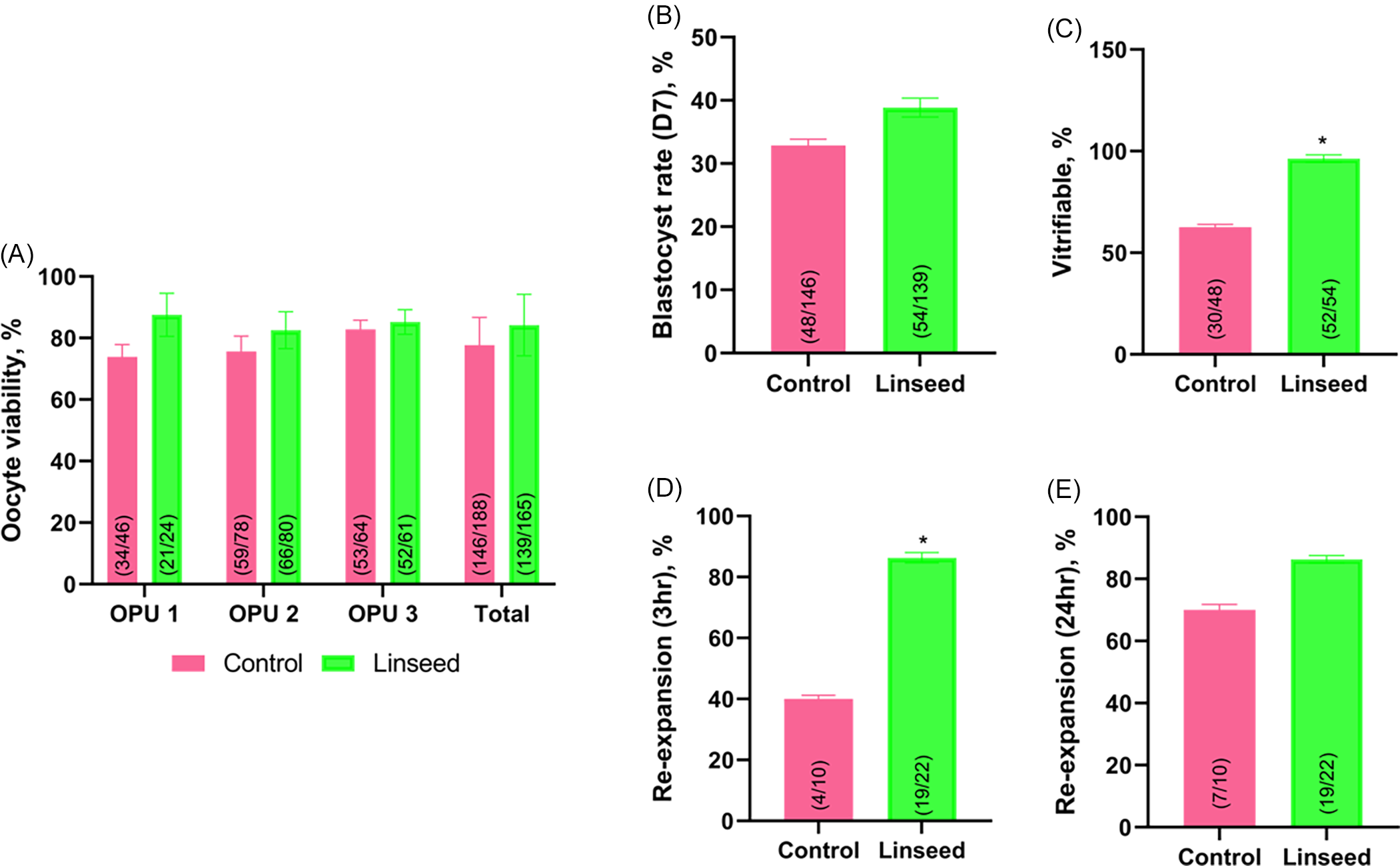
The oocyte quality rates did not vary among the OPU sessions or across the days of each session (p > 0.05). Linseed supplementation did not affect oocyte quality or D7 blastocyst rates (p > 0.05); however, the linseed group had a higher rate of vitrifiable embryos than the control group (p < 0.05). At 3 h after warming, the rate of re-expansion was higher in the linseed group (p < 0.05); however, this difference was not observed at 24 h (p > 0.05; Figure 1).
Effect of linseed supplementation in donor cows on oocyte and embryo parameters. (A) Oocyte quality (%) across three OPU sessions; (B) Blastocyst rate on day 7 (D7); (C) Percentage of vitrifiable embryos (grades I and II) (p < 0.05); (D–E) Embryo re-expansion rates after vitrification and warming, measured at 3 h (D) and 24 h (E). Data are expressed as mean ± SEM. Asterisks (*) indicate statistical significance (p < 0.05, LINSEED vs. CONTROL). Numbers within bars represent the ratio of observed outcomes to the total observations.
Discussion
Embryos of Bos taurus, including the Pantaneira breed, have a higher lipid content than those of Bos indicus, which is associated with lower cryotolerance (Sudano et al., Reference Sudano, Paschoal, Rascado, Magalhães, Crocomo, de Lima-Neto and Landim-Alvarenga2011). Therefore, specific nutritional strategies that modulate lipid metabolism can improve oocyte and embryo quality. In the present study, a 62-day supplementation period was adopted, corresponding to the estimated time required for primary follicles to reach the antral stage in large mammals (Scaramuzzi et al., Reference Scaramuzzi, Baird, Campbell, Driancourt, Dupont, Fortune, Gilchrist, Martin, McNatty, McNeilly, Monget, Monniaux, Viñoles and Webb2011), thereby ensuring adequate exposure of follicular cells to dietary effects.
These results are consistent with the findings of Ponter et al. (Reference Ponter, Guyader-Joly, Nuttinck, Grimard and Humblot2012), who observed no significant effects on oocyte characteristics or in vitro embryonic development after supplementing heifers and Holstein cows with linseed or soybean. It is important to note that these authors evaluated morphological parameters and embryo production up to D7, a stage that was also considered in this study. However, an increase in the number of grades I and II blastocysts was observed in the linseed-supplemented group, suggesting an improvement in embryo quality.
The linseed grain used in this study is less susceptible to ruminal biohydrogenation than the linseed cake used in other studies; therefore, the bioavailability of polyunsaturated fatty acids (PUFAs) may differ. However, the improvement in embryo quality was similar across studies. According to Leão et al. (Reference Leão, Rocha-Frigoni, Cabral, Coelho, Ferreira, Eberlin, Accorsi, Nogueira and Mingoti2015), an increase in PUFAs in the plasma membrane enhances membrane fluidity, which contributes to the resistance of embryonic cells to freezing damage, as described by Stubbs and Smith (Reference Stubbs and Smith1984) and Zeron et al. (Reference Zeron, Tomczak, Crowe and Arav2002). In this context, the higher re-expansion rate observed three hours after embryo warming may reflect increased membrane permeability and fluidity, facilitating intracellular exchange and the reestablishment of post-vitrification metabolism (Leão et al., Reference Leão, Rocha-Frigoni, Cabral, Coelho, Ferreira, Eberlin, Accorsi, Nogueira and Mingoti2015).
However, the lack of difference after 24 h of culture indicated that the effect on cryotolerance was transient. Therefore, the main contribution of linseed supplementation appears to be related to an increased proportion of high-quality blastocysts rather than to a sustained improvement in post-vitrification performance. The limitations of this study include the small sample size and other factors that may influence the response to supplementation. Future research should include large-scale trials, detailed analyses of embryo fatty acid composition and evaluation of additional markers of survival and cellular integrity after cryopreservation.
In summary, supplementing Pantaneira cows with 0.800 kg/animal/day of linseed resulted in a higher production of high-quality embryos, confirming the potential of this nutritional strategy to improve the efficiency of in vitro embryo production in local breeds without consistent effects on cryotolerance.
Data availability
The authors declare that the data will be available upon request.
Acknowledgements
The authors would like to thank the team of the Laboratory of Veterinary Clinical Pathology of the Federal University of Mato Grosso do Sul for analyzing serum metabolites and the team of the Nutrition laboratory of the Catholic University Dom Bosco for carrying out the bromatological analyses. This work was supported by CAPES for the scholarships and FUNDECT for supporting the project.
Author contributions
Fabiana de Andrade Melo-Sterza was responsible for the conceptualization, resources and data analysis. Marcus Vinicius Moraes de Oliveira was responsible for the animal nutritional planning and resources. Ériklis Nogueira was responsible for the statistical analysis. Christopher Junior Tavares Cardoso, Ana Caroline Bini de Lima and Mirela Brochado Souza-Cáceres was responsible for the investigation, data acquisition and performed data interpretation and wrote the manuscript draft. FAMS reviewed and edited the final manuscript version.
Competing interests
The authors have no relevant financial or non-financial interests to disclose.
Funding
This work was funding by CAPES for the scholarships awarded (n° 001) and FUNDECT for the project through the notice Biota/FUNDECT23/200.214/2014 – Statement of Animal Rights.
Declaration of animal rights
All manipulations involving the animals were carried out in accordance with the Ethical Principles in Animal Experimentation, approved by the institution”s Animal Use Ethics Committee (Protocol n° 012/2014).