Introduction
To achieve rapid growth and differentiation, an embryo must obtain sufficient energy by using products of the mitochondrial electron transport chain such as ATP, NADPH, and oxygen. Reactive oxygen species (ROS), such as superoxide, hydrogen peroxide (H2O2), and hydroxyl radical, are constant by-products of this process that are quickly eliminated by antioxidant defences (Halliwell, Reference Halliwell1991). Endogenous enzymatic antioxidants, including superoxide dismutase, catalase, and glutathione peroxidase, or non-enzymatic compounds, such as bilirubin and albumin, regulate excess ROS (Francenia Santos Sánchez et al., Reference Francenia Santos Sánchez, Salas Coronado, Villanueva Cañongo and Hernández Carlos2019). Although these defence systems reduce the amount of ROS, this can be counteracted by an imbalance in oxidizing and reducing compounds induced by oocyte ageing (Moldogazieva et al., Reference Moldogazieva, Mokhosoev, Feldman and Lutsenko2018; Wang et al., Reference Wang, Branicky, Noë and Hekimi2018). Oocyte ageing is a process normally occurring while the unfertilized oocytes pass their reproductive age and mature inside the female ovary (Němeček et al., Reference Němeček, Dvořáková, Heroutová, Chmelíková and Sedmíková2017). As a result of oocyte ageing, there is an imbalance of oxidation and reduction that results in the excess generation of ROS, which leads to developmental arrest, DNA damage, apoptosis, and lipid peroxidation (Takahashi, Reference Takahashi2012). Therefore, it is important to reduce oxidative stress from ROS using effective concentrations of antioxidant for embryonic viability.
To prevent ROS-induced oxidative stress upon ageing oocytes and to maintain quality for assisted reproduction technologies, several researchers have tested various antioxidants such as ubiquinol-10, kaempferol, melatonin, β-cryptoxanthin, hesperetin and icariin (Mehaisen et al., Reference Mehaisen, Saeed, Gad, Abass, Arafa and El-Sayed2015; Park et al., Reference Park, Lee, Son, Jeong, Shin, Kim, Kim and Park2018; Kim et al., Reference Kim, Lee, Park, Jeong, Kim and Park2019; Yao et al., Reference Yao, Jiang, NanXu, Piao, Gao and Kim2019; Niu et al., Reference Niu, Zhou, Nie, Zhou, Xu, Ock, Yan and Cui2020; Yoon et al., Reference Yoon, Lee, Park, Kim, Park, Park, Kim, Oh, Lee, Pyeon, Kim and Park2021). β-Cryptoxanthin (BCX) is a carotenoid abundant in fruits such as tangerines and peaches, and is a known precursor of vitamin A. This vitamin has several useful functions including antioxidant capacity and cell-to-cell communication (Burri et al., Reference Burri, La Frano and Zhu2016). In addition, BCX prevents the development of various cancers (e.g. lung, liver, prostate, breast, colorectal and stomach) (Lim and Wang, Reference Lim and Wang2020). Hesperetin (HES) is a flavanone class of flavonoid found in citrus fruits such as oranges and grapefruit (Kim et al., Reference Kim, Jeong, Lee, Park and Choi2003). This compound has several beneficial effects such as antioxidant and anti-inflammatory capacities (Muhammad et al., Reference Muhammad, Ikram, Ullah, Rehman and Kim2019). Icariin (ICA) is a chemical compound classified as a prenylated flavonol glycoside, a type of flavonoid. It is the 8-prenyl derivative of kaempferol 3,7-O-diglucoside. ICA can be found and isolated from several plants belonging to the genus Epimedium, which is commonly known as horny goat weed (Liu JJ et al., Reference Liu, Li and Wang2006). Flavonoid glycosides, which can be widely found in nature, are proven to have many pharmacological activities including antioxidant, anti-inflammatory, and anti-tumour effects (Liu J et al., Reference Nasheed Hamad Almohammed, Moghani-Ghoroghi, Ragerdi-Kashani, Fathi, Tahaei, Naji and Pasbakhsh2019).
Our previous studies have already revealed that BCX, HES and ICA antioxidants are respectively effective at a specific concentration, which is 1 μM BCX (BCX-1), 100 μM HES (HES-100), and 5 μM ICA (ICA-5) for in vitro maturation (IVM) of porcine oocytes (Park et al., Reference Park, Lee, Son, Jeong, Shin, Kim, Kim and Park2018; Kim et al., Reference Kim, Lee, Park, Jeong, Kim and Park2019; Yoon et al., Reference Yoon, Lee, Park, Kim, Park, Park, Kim, Oh, Lee, Pyeon, Kim and Park2021). In this study, we wanted to determine which antioxidant would be more effective against oxidative stress induced by oocyte ageing by comparing chemical and biological analyses. To compare the chemical antioxidant capacity of the three antioxidants (BCX, HES and ICA), DPPH, reducing power and H2O2 scavenging activity assays were conducted. To compare the biological antioxidant capacity of the antioxidants, oocytes were experimentally aged by 24 h of incubation in IVM medium following 44 h of IVM. A previous study has shown that 24 h of rapamycin treatment supplemented to IVM medium rescued the poor developmental capacity of aged porcine oocytes after an initial 44 h of IVM treatment (Lee et al., Reference Lee, Kim, Choi, Moon, Park, Lee, Jeong and Park2014). The levels of ROS and glutathione (GSH) were analyzed by measuring the expression of antioxidant genes (SOD1 and SOD2), anti-apoptotic genes (BCL2L1 and BIRC5), pro-apoptotic gene (CASP3), and developmental genes (POU5F1 and SOX2), as well as the apoptosis rate in aged porcine oocytes and embryos treated with or without antioxidants. The developmental integrity and quality of the embryos were compared from oocytes treated in the presence or absence of antioxidants. These results suggested that BCX-1 is a more effective antioxidant than HES-100 or ICA-5 during in vitro maturation of porcine oocytes and embryos. These findings may be applicable to in vitro fertilization to help prevent ageing of oocytes in vitro.
Materials and methods
Chemicals and reagents
All chemicals and reagents were purchased from Sigma (St. Louis, MO, USA) unless stated otherwise.
DPPH free radical-scavenging assay
The 2,2-diphenyl-1-(2,4,6-trinitrophenyl)hydrazyl (DPPH) assay, originally reported by Blois (Blois, Reference Blois1958), was used with some modification to measure the free radical-scavenging activity of the antioxidants. Stock solutions of BCX (Extrasynthese, Lyon, France), HES, and ICA were diluted in DMSO and serial dilutions were prepared (0.04, 0.08, 0.12, 0.16, and 0.20 µg/ml BCX; 1, 2, 3, 4, and 5 mg/ml HES; 10, 20, 30, 40, and 50 mg/ml ICA). Briefly, 20 µl of the samples were diluted in DMSO and added to 180 µl of a 167 µM DPPH working solution. The absorbance of the solution was measured at 517 nm with a SpectraMAX i3 (Molecular Devices, LLC; Sunnyvale, CA). DPPH free radical-scavenging activity was calculated using the following Equation (1):
 $${{\text{H}}_{\text{2}}}{{\text{O}}_{\text{2}}}{\text{DPPH}}\;{\text{scavenging}}\;{\text{effect}}\;\left( \% \right){\text{ = }}\frac{{{\text{Ac }} - {\text{ At}}}}{{{\text{Ac}}}} \times {\text{100}}$$
where Ac is the absorbance value of the control reaction (blank sample treated with no antioxidant), and At is the absorbance in the presence of the antioxidants.
Reducing power assay
The reducing power of the antioxidants at different concentrations (125, 250, 500, and 1000 µM) was determined using a method described by Gülçın et al. (Reference Gülçın, Oktay, Kıreçcı and Küfrevıoğlu2003). Briefly, 0.1 ml of sample solution was combined with 0.25 ml phosphate buffer (0.2 M, pH 6.6) and 0.25 ml potassium ferricyanide (K3[Fe(CN)6]) (1%; Poly Scientific R&D, Bay Shore, NY, USA), mixed, and then incubated at 50°C for 20 min. Trichloroacetic acid (10%, 0.25 ml) was added to the samples, which were centrifuged for 10 min at 1700 g (25°C). After centrifugation, 0.25 ml from the upper layer of the solution was transferred to a new tube, and mixed with 0.25 ml distilled water and 0.05 ml ferric chloride (FeCl3) (0.1%). The absorbance of the reaction was measured at 700 nm on a SpectraMAX i3 reader (Molecular Devices, LLC). An increased absorbance value indicated an increased reducing power.
H2O2 scavenging activity assay
The H2O2 scavenging activity of the antioxidants was determined by the method described by Ruch et al. (Reference Ruch, Cheng and Klaunig1989) with slight modifications. Briefly, a 4 mM solution of H2O2 was prepared in phosphate-buffered saline (PBS, pH 7.4) at 20°C. H2O2 concentration was determined spectrophotometrically at an absorbance of 230 nm. The three antioxidants were each dissolved in DMSO and then added to the H2O2 solution at final concentrations of 1 µM BCX, 100 µM HES, and 5 µM ICA at 20°C. A control reaction was prepared containing only 0.1% DMSO. At 10 min after incubation, the amount of H2O2 in each sample was determined using a SpectraMAX i3 reader (Molecular Devices). The H2O2 scavenging activity was calculated using the following Equation (2):
 $${{\text{H}}_{\text{2}}}{{\text{O}}_{\text{2}}}{\text{ scavenging}}\;{\text{effect}}\;\left( \% \right){\text{ = }}\frac{{{\text{Ac}} - {\text{At}}}}{{{\text{Ac}}}} \times {\text{100}}$$
where Ac is the absorbance value of the control reaction (blank sample treated with no antioxidant), and At is the absorbance in the presence of the antioxidants.
Oocyte collection and IVM
Prepubertal porcine ovaries were collected from a local slaughterhouse within 2 h of slaughter and transported to the laboratory in saline supplemented with 75 μg/ml penicillin G and 50 μg/ml streptomycin sulfate at 30–33°C. Cumulus–oocyte complexes (COCs), 2–8 mm in diameter, were aspirated from the ovarian follicle using an 18-gauge needle attached to a disposable 10-ml syringe. COCs were washed three times in tissue culture medium (TCM)-199-HEPES containing 0.1% (w/v) bovine serum albumin (BSA). Thereafter, COCs were matured in groups of 50 in 500 μl TCM-199 (Gibco, Grand Island, NY) containing Earle’s salts, 0.57 mM cysteine, 10 ng/ml epidermal growth factor, 0.5 μg/ml follicle-stimulating hormone, 0.5 μg/ml luteinizing hormone, and 10% (v/v) porcine follicular fluid. The COCs were cultured in Petri dishes under a layer of mineral oil and incubated for 44 h at 38.8°C with 5% CO2.
Antioxidant treatment during ageing of matured porcine oocytes
Oocytes were matured in a 4-well tissue culture dish containing 500 μl TCM-199, covered with a layer of mineral oil, by incubation for 68 h (with the exception of a control group for 44 h) at 38.8°C in a 5% CO2 in air atmosphere. After maturation (44 h), metaphase-II (MII)-stage oocytes were transferred into dishes containing TCM-199 supplemented with 1% DMSO, 1 μM BCX, 100 μM HES, and 5 μM ICA, for ageing BCX-1, HES-100 and ICA-5, and incubated for 24 h. After antioxidant treatment, the oocytes were collected and maturity was assessed.
Parthenogenesis and embryo culture
Following oocyte maturation, cumulus cells were isolated by repeated manual pipetting for 2–3 min in the presence of 1 mg/ml hyaluronidase. Parthenogenesis (PA) was activated by treating oocytes with porcine zygote medium (PZM)-5 containing 0.4% (w/v) BSA (IVC medium) and 5 μM Ca2+ ionomycin (Sigma) for 5 min. Following 3 h of incubation in IVC medium containing 7.5 μg/ml cytochalasin B (Sigma), embryos were washed three times with IVC medium before being cultured for 7 days at 38.8°C with 5% CO2. Oocytes and embryos were washed in Dulbecco’s PBS (DPBS) and either fixed in 3.7% (w/v) paraformaldehyde for 20 min and stored at 4°C, or snap frozen in liquid nitrogen and stored at −70°C, depending on the experiment.
Measurement of intracellular ROS and glutathione (GSH) levels
2′,7′-Dichlorofluorescin diacetate (DCFHDA) and 4-chloromethyl-6,8-difluoro-7-hydroxycoumarin (CMF2HC) were used to determine the intracellular levels of ROS and GSH, respectively, as previously described by Yang and colleagues (Yang et al., Reference Yang, Hwang, Kwon, Kim, Choi and Oh1998) and You and colleagues (You et al., Reference You, Kim, Lim and Lee2010) with slight modifications. Briefly, cumulus cells were isolated from the COCs by repeated manual pipetting in the presence of 0.1% (w/v) hyaluronidase. Denuded oocytes were incubated in DPBS containing 50 μM DCFHDA or 100 μM CMF2HC in the dark for 20 min at 38.8°C in a 5% CO2 in air atmosphere. Afterwards, oocytes were washed a minimum of five times with DPBS containing 0.1% (w/v) BSA to completely remove excess dye and then were immediately analyzed by epifluorescence microscopy (Olympus; Shinjuku, Tokyo). The ROS level was measured using excitation and emission wavelength ranges of 450–490 and 515–565 nm, respectively. The excitation and emission wavelengths of CMF2HC were 371 and 464 nm, respectively. Grey-scale images were acquired on a digital camera (Shinjuku, Tokyo) attached to the microscope, and mean grey-scale values were calculated using ImageJ software (NIH, Bethesda, MD). Background fluorescence values were subtracted from the final values before statistical analysis. In total, six or seven independent experiments were performed with 10–20 oocytes per experiment.
Terminal deoxynucleotidyl transferase dUTP nick end labelling (TUNEL) assay and Hoechst staining
At 7 days after PA, blastocysts were fixed, washed a minimum of three times with PBS containing 0.1% BSA, and then incubated with 0.1% Triton X-100 at 38.8°C in a 5% CO2 in air atmosphere for 30 min. Blastocysts were incubated with fluorescein-conjugated dUTP and terminal deoxynucleotidyl transferase (In Situ Cell Death Detection Kit; Roche, Mannheim, Germany) in the dark for 1 h at 38.8°C. The numbers of mitotic and apoptotic cells were recorded. Nuclei were stained with Hoechst 33342 (1 μg/ml) for 30 min, and embryos were washed with PBS containing 0.1% BSA. In total 5–7 blastocysts were mounted onto glass slides and examined under an inverted Olympus IX-71 fluorescence microscope. Six independent experiments were performed.
mRNA extraction and complementary DNA (cDNA) synthesis
For PCR sample preparation, mRNA was isolated from a minimum of three biological replicates, with 20 oocytes or 10 blastocysts per replicate, using a Dynabeads mRNA Direct Kit (Invitrogen, Carlsbad, CA, USA), according to the manufacturer’s instructions. The mRNA was suspended in 10-µl elution buffer that was provided with the kit. Eluted RNA was reverse transcribed into cDNA using an oligo(dT)20 primer and SuperScript II reverse transcriptase (Invitrogen), according to the manufacturer’s instructions.
Real-time RT-PCR
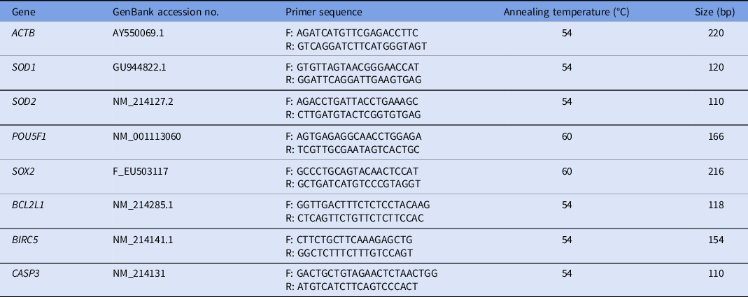
Real-time RT-PCR was performed as previously described by Lee and colleagues (Lee et al., Reference Lee, Sun, Choi, Uhm and Kim2012) using the primer sets listed in Table 1. Samples were loaded in a final reaction volume of 20 µl containing SYBR Green PCR Master Mix (Applied Biosystems, Warrington, UK). Analysis was performed using a StepOnePlus Real-time PCR System (Applied Biosystems) with the following program settings: 10 min at 95°C, followed by 39 cycles of 15 s at 95°C, and 60 s at 54 or 60°C. Samples were then cooled to 12°C. Relative gene expression levels were analyzed using the 2–ΔΔCт method (Livak and Schmittgen, Reference Livak and Schmittgen2001) after normalization against the housekeeping gene ACTB. One RT-PCR experiment was performed using three replicates of the sample. By performing statistical analysis and by repeating this experiment three times, we had finally performed nine experiments.
Primers used for real-time RT-PCR

F, forward; R, reverse.
Statistical analysis
All experimental data were analyzed using the general linear model procedure with Statistical Analysis System software (SAS User’s Guide 1985, Statistical Analysis System Inc., Cary, NC, USA). Tukey’s multiple range test was used to determine significant differences. P-values were considered as significant at *P < 0.05 and **P < 0.01.
Results
Improved DPPH scavenging activity of BCX compared with HES and ICA
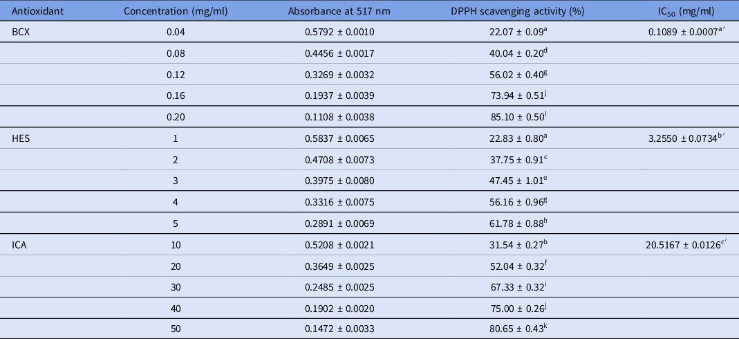
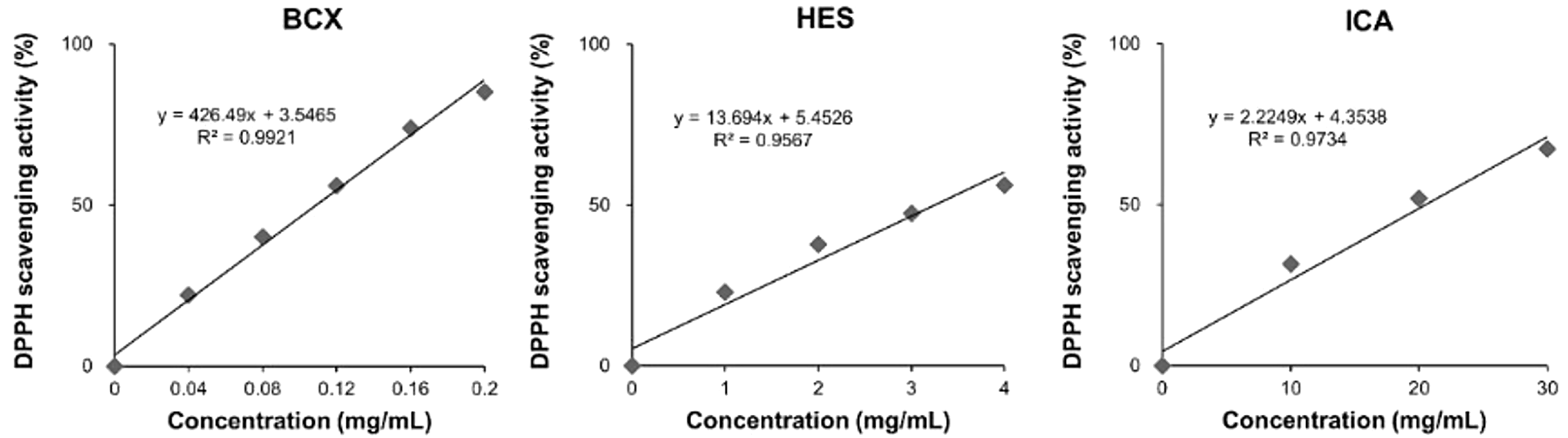
The scavenging abilities of BCX, HES, and ICA to reduce DPPH free radicals was determined by measuring the decrease in absorbance. The result showed that scavenging ability was dependent on antioxidant concentration (Table 2). To compare scavenging efficiency, an estimate for the concentration of antioxidants required to inhibit 50% of DPPH radicals (IC50) was extrapolated from a linear regression analysis (Fig. 1). The resulting IC50 values for BCX, HES, and ICA were determined to differ significantly (P < 0.001) at 0.109 mg/ml BCX, 3.255 mg/ml HES, and 20.517 mg/ml ICA (Table 2).
DPPH scavenging activity of the three antioxidants tested at different concentrations

a–l P < 0.05, a′-c′ P < 0.001.
DPPH scavenging activity of the antioxidants at different concentrations. The results are represented by concentration vs. DPPH scavenging activity (%). Data are derived from three independent experiments represented by the mean ± standard error of the mean (SEM)

Improved reducing power of BCX compared with HES and ICA
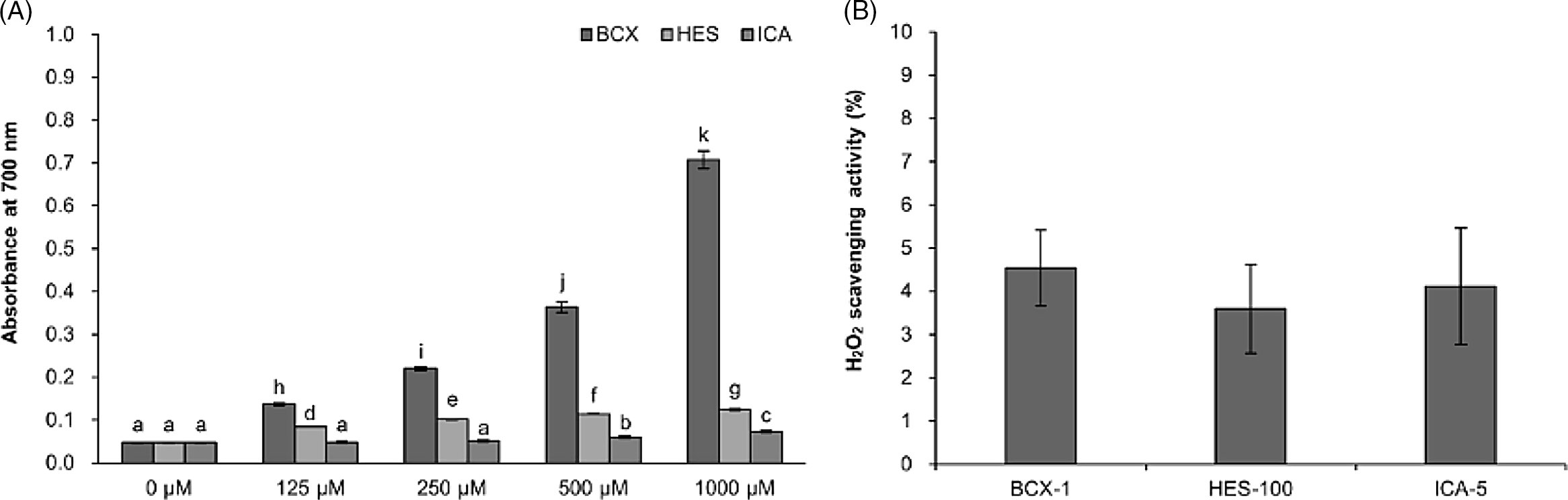
The reducing power of the antioxidants was correlated with concentration in the range 125–1000 µM. When measured at an absorbance of 700 nm, the antioxidant reducing powers were statistically different (P < 0.05, Fig. 2A). BCX had a greater reducing power than both HES and ICA at the same concentration. Compared with the controls, there were significant differences in the reducing power relative to concentration; however, BCX was the most effective even at very low concentrations (Fig. 2A). Additionally, the reducing power at the lowest concentration of HES was significantly stronger than the highest concentration of ICA (P < 0.05, Fig. 2A).
Reducing power and H2O2 scavenging activity of BCX, HES, and ICA. (A) Reducing power of the antioxidants tested at different concentrations and measured at an absorbance of 700 nm (a–k, P < 0.05). (B) H2O2 scavenging activity of the antioxidants tested at optimal concentration during in vitro maturation of porcine oocytes. Data are derived from three independent experiments represented by the mean ± standard error of the mean (SEM).

BCX, HES, and ICA demonstrate similar H2O2 scavenging activities
To compare the H2O2 scavenging activity, a H2O2 scavenging assay was performed using the optimal concentrations of each antioxidant in IVM porcine oocytes. The sample groups BCX-1, HES-100, and ICA-5 scavenged approximately 5% of H2O2 compared with the control, although this effect was not significantly different (Fig. 2B). Although the optimal concentration of BCX was lower than that for HES and ICA, BCX had an equivalent H2O2 scavenging activity compared with HES-100 or ICA-5 (Fig. 2B).
BCX reduces oxidative stress by increasing GSH in matured porcine oocytes
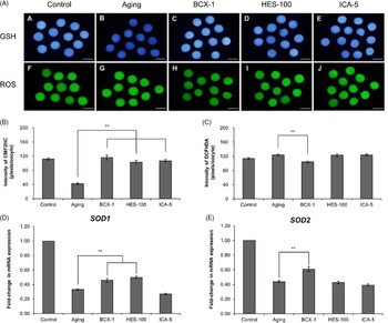
To assess oxidative stress in matured porcine oocytes, ROS and GSH levels were measured on porcine oocytes initially matured for 44 h (control) or for an additional 24 h (total of 68 h) in the presence of 1% DMSO, 1 µM BCX, 100 µM HES, and 5 µM ICA, which were then referred to as the ageing, BCX-1, HES-100, and ICA-5 groups, respectively (Fig. 3A). The GSH levels were significantly increased in the BCX-1, HES-100, and ICA-5 sample groups compared with the control (P < 0.01, Fig. 3B). Despite slightly lower GSH levels in the HES-100 (103.42 ± 4.65 pixels/oocyte) and ICA-5 (107.28 ± 3.67 pixels/oocyte) groups compared with the control group, the BCX-1 (115.97 ± 6.80 pixels/oocyte) group result was similar to the control group (111.91 ± 3.19 pixels/oocyte) but not to the ageing group (42.47 ± 3.12 pixels/oocyte) (Fig. 3B). The staining intensity of ROS was significantly lower (P < 0.01) in the control (113.73 ± 2.49 pixels/oocyte) and BCX-1 (104.13 ± 2.52 pixels/oocyte) groups compared with the ageing (124.18 ± 1.77 pixels/oocyte) group, however there was no difference between ageing, HES-100 (123.19 ± 3.87 pixels/oocyte), and ICA-5 (123.57 ± 3.07 pixels/oocyte) (Fig. 3C). Real-time RT-PCR was performed to investigate the effect of antioxidants on mRNA expression of superoxide dismutase 1 (SOD1) and superoxide dismutase 2 (SOD2) on porcine oocytes at MII (Fig. 3D,E). The BCX-1 group significantly increased the expression of SOD1 and SOD2 compared with the ageing and ICA-5 sample groups (P < 0.01). The HES-100 group increased the expression of SOD1 but not SOD2, compared with the ageing and ICA-5 sample groups (P < 0.05).
Antioxidant activity against oxidative stress during ageing of porcine oocytes. (A) Images of oocytes stained with CMF2HC (blue), DCFHDA (green) for assessing GSH, A–E, and ROS, F–J. Quantification of the fluorescence intensities of (B) CMF2HC and (C) DCFHDA (**P < 0.01). Relative expression of antioxidant genes (D) SOD1, (E) SOD2 in MII-stage porcine oocytes (**P < 0.01). Data are derived from three independent experiments represented by the mean ± standard error of the mean (SEM). Scale bars, 120 µM.

BCX enhanced developmental competence in ageing oocytes
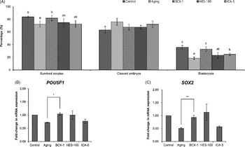
Comparing the development effect of the antioxidants, the percentages of oocytes that survived to MII stage were not significantly different between control (83.77% ± 1.58%) and treatment groups (BCX-1, 81.79% ± 4.07%; HES-100, 74.70% ± 5.78%; ICA-5, 72.77% ± 5.30%), except the ageing group (72.32% ± 4.34%; Fig. 4A). Oocyte survival rate, which means intact membrane integrity, was highest in BCX and control groups and these were similar to each other. Following PA, the percentage of oocytes that developed to the 2–4-cell cleavage stage was similar between groups (control, 63.26% ± 5.47%; ageing, 75.97% ± 5.73%; BCX-1, 67.64% ± 4.22%; HES-100, 67.57% ± 2.80%; ICA-5, 72.18% ± 5.21%; Fig. 4A). The percentage of cleavage embryos that developed into blastocysts by day 7 was significantly higher (P < 0.01) in the BCX-1 (32.87% ± 3.72%) and control (35.89% ± 2.81%) groups than in the ageing (18.46% ± 1.86%), HES-100 (23.04% ± 4.47%), and ICA-5 (24.75% ± 1.73%) groups (Fig. 4A). Additionally, the expression of important developmental genes, including POU domain, class 5, transcription factor 1 (POU5F1) and SRY (sex determining region Y)-box 2 (SOX2), were investigated on porcine blastocysts that had been cultured for 7 days (Fig. 4B,C). BCX-1 treatment significantly increased the expression of POU5F1 (P < 0.05) and SOX2 (P < 0.01), but treatment with HES-100 and ICA-5 did not increase the expression of POU5F1 or SOX2.
Effect of antioxidant treatment during ageing of porcine oocytes on subsequent embryo development and expression of developmental genes in porcine blastocysts. (A) Percentage of survived oocytes (a, b, P < 0.05), cleaved embryos, and formed blastocysts (a–c, P < 0.05) during in vitro maturation and subsequent culture for 7 days. Relative expression of the developmental genes (B) POU5F1 and (C) SOX2 in porcine blastocysts (*P < 0.05, **P < 0.01). Data are derived from three independent experiments represented by the mean ± standard error of the mean (SEM).

BCX improves the quality of blastocyst compared with HES and ICA
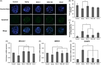
To compare whether BCX, HES, or ICA treatment during IVM of porcine oocytes influenced the subsequent embryonic development and integrity, the quality of blastocyst was assessed at 7 days post-PA by Hoechst staining (Fig. 5A). The total number of cells per blastocyst was significantly higher (P < 0.01) in the control (71.5% ± 12.2%) and BCX-1 (67.1% ± 10.0%) groups compared with the ageing, HES-100, and ICA-5 groups (ageing, 52.4% ± 7.6%; HES-100, 59.4% ± 4.0%; ICA-5, 57.8% ± 4.1%; Fig. 5B). Genomic DNA fragmentation was assessed by a TUNEL assay to detect the percentage of apoptotic cells in the blastocysts (Fig. 5A). The percentage of apoptotic cells was significantly lower (P < 0.01) in the control (1.3% ± 0.7%), BCX-1 (3.3% ± 0.6%), and HES-100 (3.1% ± 1.1%) groups compared with the ageing and ICA-5 groups (ageing, 7.7% ± 2.5%; ICA-5, 5.7% ± 1.3%; Fig. 5C). Additionally, the expression of anti-apoptotic genes including BCL2-like 1 (BCL2L1) and baculoviral IAP repeat-containing 5 (BIRC5), and pro-apoptotic gene caspase-3 (CASP3) were investigated in porcine blastocysts (Fig. 5D–F). Treatment with BCX-1 significantly increased the expression of BCL2L1 (P < 0.01, Fig. 5D) and BIRC5 (P < 0.05, Fig. 5E), and decreased the expression of CASP3 (P < 0.01, Fig. 5F). Treatment with HES-100 significantly increased the expression of BCL2L1 (P < 0.01, Fig. 5D) and decreased the expression of CASP3 (P < 0.05, Fig. 5E) and treatment with ICA-5 significantly increased the expression of BCL2L1 (P < 0.05, Fig. 5D) and BIRC5 (P < 0.05, Fig. 5E), and decreased the expression of CASP3 (P < 0.01, Fig. 5F).
Effect of the antioxidant treatment during ageing of porcine blastocysts on cell survival and apoptosis. (A) Blastocysts stain. Scale bar, 120 μm, (B) total cell number and (C) percentage of apoptotic cells in porcine blastocysts (*P < 0.05). Expression of anti-apoptotic genes (D) BCL2L1 and (E) BIRC5, and pro-apoptotic gene (F) CASP3, in porcine blastocysts (*P < 0.05, **P < 0.01). Data are derived from three independent experiments represented by the mean ± standard error of the mean (SEM).

Discussion
This study investigated whether the antioxidants BCX, HES, and ICA could effectively reduce oxidative stress induced by oocyte ageing. Although the mechanism by which oocytes are preserved from the normal ageing process in vitro is unknown, we tried to measure antioxidant activity by chemical and biological assays. First, we measured the chemical antioxidant capacity of BCX, HES, and ICA using the DPPH assay. DPPH is a stable free radical that can be used to measure the radical-scavenging activity of antioxidants. The DPPH assay is a short and simple method to measure the electron-donating ability of antioxidants during lipid oxidation (Kikuzaki et al., Reference Kikuzaki, Hisamoto, Hirose, Akiyama and Taniguchi2002). To compare chemical antioxidant activity, we calculated the IC50 value representing the concentration of the sample required to inhibit 50% of the DPPH radical. To calculate an approximate IC50 value with a 95% confidence level, a linear regression line was generated against antioxidant concentration, based on the guidelines by Sebaugh (Reference Sebaugh2011). According to the linear regression formula (Fig. 1), the IC50 values of BCX, HES, and ICA were 0.1089 mg/ml, 3.2550 mg/ml, and 20.5167 mg/ml, respectively, as shown in Table 2. As the IC50 value directly represents the sample concentration required to inhibit 50% of DPPH, we assume that the antioxidant activity of BCX was approximately 30-fold stronger than that of HES, and approximately 200-fold stronger than that of ICA, at any given concentration (mg/ml). Most previous publications did not consider the final sample concentration using the sample and DPPH reaction to calculate the IC50 value (Chen and Huang Reference Chen and Huang2019; Hara et al., Reference Hara, Someya, Sano, Sagane, Watanabe and Wijesekara2018; López-Martínez et al., Reference López-Martínez, Santacruz-Ortega, Navarro, Sotelo-Mundo and González-Aguilar2015). As the final sample concentration tested in the DPPH assay was not included in the linear regression analysis, values should be diluted 10-fold (data not shown) for application in biological experiments or as supplements to bioreagents. In this study, three antioxidants were supplemented with IVM medium; modified IC50 was referenced for biological assay. Each optimal concentration of antioxidants treated during ageing IVM was approximately 20× diluted BCX, 10× diluted HES, and 600× diluted ICA from each modified IC50 value. Although HES-100 was more concentrated than the modified IC50 value, BCX-1 treatment showed the best antioxidant activity when comparing the results from chemical and biological assays. Overall, the BCX antioxidant was more effective and efficient at scavenging ROS compared with HES and ICA.
To measure reductive ability, the Fe3+–Fe2+ transformation in the presence of different concentrations of antioxidants was investigated. Reducing power has previously been used as a good indicator of antioxidant activity as it represents the ability to reduce another substance by gaining electrons. For example, ROS can be reduced by antioxidants. Recently, the antioxidant capacities of four different kinds of microalgae were investigated using DPPH scavenging activity, chelating activity, and reducing power assays. The study revealed that polyphenol and carotenoid, but not flavonoid, contents of the microalgae were strongly correlated with antioxidant activity. Plant extracts with high amounts of carotenoid had the best antioxidant properties (Taviano et al., Reference Taviano, Filocamo, Ragusa, Cacciola, Dugo, Mondello, Paterniti Mastrazzo, De Rose, Celano, Lombardo, Melchini and Miceli2018; Haoujar et al., Reference Haoujar, Cacciola, Abrini, Mangraviti, Giuffrida, Oulad El Majdoub, Kounnoun, Miceli, Fernanda Taviano, Mondello, Rigano and Skali Senhaji2019; Miceli et al., Reference Miceli, Marino, Köroğlu, Cacciola, Dugo, Mondello and Taviano2020). The results from this study also demonstrated that BCX, a type of carotenoid, had a stronger reducing power than HES and ICA, both types of flavonoids, at the same µM concentration (Fig. 2A). This suggests that BCX has greater antioxidant activity than HES or ICA.
Although DPPH is an example of a ROS, it behaves differently to other more common free radicals in the biological environment. H2O2 is one of the most common ROS in the biological system and a major contributor to oxidative stress. Previously, the antioxidant capacity of coumarins was evaluated using a H2O2 scavenging assay (Al-Amiery et al., Reference Al-Amiery, Al-Majedy, Kadhum and Mohamad2015). Vicilin was also evaluated for its antioxidant activity using the H2O2 scavenging assay at a specific concentration (Gupta et al., Reference Gupta, Srivastava and Bhagyawant2018). Accordingly, we performed the H2O2 scavenging assay to compare the effect of three antioxidants at optimal concentration. The results indicated that approximately 5% of H2O2 was scavenged by the antioxidants compared with the control (Fig. 2B). The initial concentration of H2O2 solution was 4 mM, therefore the final concentration after antioxidant scavenging was approximately 200 µM. Most studies have previously used 200 µM H2O2 to induce ROS damage. This suggests that the optimized concentrations of the antioxidants were sufficient to reduce oxidative stress induced by 200 μM H2O2. Another study also showed that 0.1 μM kaempferol significantly increased porcine blastocyst formation rate under oxidative stress induced by 200 μM H2O2 (Yao et al., Reference Yao, Jiang, NanXu, Piao, Gao and Kim2019). In our study, the H2O2 scavenging activity was not significantly different amongst the three antioxidants; however, even at lower concentrations, BCX-1 had a similar efficiency to HES-100 and ICA-5. Therefore, we can assume that BCX had high H2O2 scavenging activities compared with HES and ICA at the same μM concentration.
Oxidative stress frequently occurs in ageing post-ovulatory oocytes in which the production of ROS is increased, and the protective abilities of antioxidants are decreased (Lord and Aitken, Reference Lord and Aitken2013). Intracellular GSH normally protects oocytes against oxidative stress induced by ROS; however, the levels of GSH decrease during the ageing process (Boerjan and De Boer, Reference Boerjan and De Boer1990). Previously, resveratrol treatment administered to goat oocytes was shown to significantly increase the levels of GSH, while allicin treatment administered to porcine oocytes significantly decreased the levels of ROS (Jeong et al., Reference Jeong, Lee, Park, Son, Shin, Kim and Park2017; Piras et al., Reference Piras, Menéndez-Blanco, Soto-Heras, Catalá, Izquierdo, Bogliolo and Paramio2019). Our previous study showed that BCX, HES, and ICA could prevent oxidative stress during IVM of porcine oocytes at concentrations of 1, 100, and 5 µM, respectively (Park et al., Reference Park, Lee, Son, Jeong, Shin, Kim, Kim and Park2018; Kim et al., Reference Kim, Lee, Park, Jeong, Kim and Park2019; Yoon et al., Reference Yoon, Lee, Park, Kim, Park, Park, Kim, Oh, Lee, Pyeon, Kim and Park2021). In this study, approximately 9% of ROS was generated by ageing oocytes, and BCX-1 was found to significantly eliminate approximately 16% of the ROS compared with HES-100 and ICA-5 (Fig. 3C). BCX-1 treatment recovered GSH to almost the same level as the controls.
SOD1 and SOD2 are genes induced by inflammatory conditions. The main function of SOD1 is to catalyze the conversion of superoxide radicals into molecular oxygen and hydrogen peroxide (Del Río et al., Reference Del Río, Sevilla, Gómez, Yañez and López1978) and SOD2 is the first line of defence against superoxide produced as a by-product of oxidative phosphorylation (Li et al., Reference Li, Huang, Carlson, Melov, Ursell, Olson, Noble, Yoshimura, Berger, Chan, Wallace and Epstein1995). Previously, SOD was expressed at a relatively high level when melatonin was given to mouse oocytes or human oocytes after the maturation phase (El Mouatassim et al., Reference El Mouatassim, Guérin and Ménézo1999; Nasheed Hamad Almohammed et al., Reference Nasheed Hamad Almohammed, Moghani-Ghoroghi, Ragerdi-Kashani, Fathi, Tahaei, Naji and Pasbakhsh2019). Also, BCX might be a most useful substance to prevent age-related oxidative damage among the various subtypes of carotenoids (Sugiura et al., Reference Sugiura, Ogawa and Yano2013). A previous study has shown that BCX may compensate for Cu/Zn-SOD and Fe-SOD in providing antioxidant protection to follow the enzyme system (Schroeder and Johnson, Reference Schroeder and Johnson1993). Therefore, investigating the expression of the antioxidant genes (SOD1 and SOD2) is a good indicator of antioxidant activity against oxidative stress. Taken together, our results suggested that BCX prevented oxidative stress in ageing oocytes by decreasing the levels of ROS, promoting GSH, and increasing the expression of SOD1 and SOD2 genes.
The most common way to evaluate oocyte quality is to assess blastocyst formation rate, cell number, apoptotic rate, and the expression of important developmental and apoptotic genes (Luna et al., Reference Luna, Copperman, Duke, Ezcurra, Sandler and Barritt2008). A recent study examined both the total cell number and proportion of apoptotic cells to check the quality of developing cat blastocysts during 6–9 days of in vitro culture (Ochota et al., Reference Ochota, Wojtasik and Niżański2016). The exact mechanism by which BCX can regulate gene expression associated with inflammation, cell proliferation, and apoptosis remains unclear. However, the recent research on the mechanism of BCX in germ cells, has shown that BCX significantly suppresses NF-κB (p65) activation as well as mitogen-activated protein kinase (MAPK) phosphorylation in mouse Sertoli cells (Liu XR et al., Reference Liu, Wang, Dan, Kumar, Ye, Yu and Yang2016). The MAPK pathway is associated with the activation of two redox-sensitive transcription factors, NF-κB and AP-1 regulate more than 400 genes related to inflammation, cell proliferation, and apoptosis (Liu C et al., 2011). Also, the MAPK pathway is an important key factor for oocyte maturation. MAPK facilitates the nuclear translocation and transcriptional activation of NFE2L (Nguyen et al., Reference Nguyen, Nioi and Pickett2009) or activated by MOS protein, an active component of cytostatic factor, which is responsible for meiotic arrest at MII (Newman and Dai, Reference Newman and Dai1996). These MAPK pathways also have a major effect on subsequent embryo development. A previous study showed that resveratrol affected the regulation of apoptosis-related genes (CASP3 and BAK) during subsequent embryonic development and leads to better monospermic fertilization, blastocyst formation, and blastomere viability (Kwak et al., Reference Kwak, Cheong, Jeon, Lee, Choi, Jeung and Hyun2012). Melatonin has also been shown to significantly increase the expression of developmental genes POU5F1 and SOX2 when administered to rabbit embryos (Mehaisen et al., Reference Mehaisen, Saeed, Gad, Abass, Arafa and El-Sayed2015). In another study, the expression of anti-apoptotic genes (BCL2L1 and BIRC5) was increased, while that of pro-apoptosis gene (CASP3) was decreased, when PC3 cells were treated with simvastatin (Goc et al., Reference Goc, Kochuparambil, Al-Husein, Al-Azayzih, Mohammad and Somanath2012). The BCL2L1 protein belongs to the BCL-family to inhibit cell death by preventing the permeabilization of the mitochondrial membrane. BIRC5, also known as survivin, controls apoptosis by inhibition of Bax and the extrinsic Fas pathways, and by binding to caspase-3 and caspase-7 (Roman et al., Reference Roman, Budziński, Suszka-Świtek, Caban, Oczkowicz, Czech, Ryszka, Wiaderkiewicz, Smorąg and Cierpka2016). The caspases are a family of protease enzymes that play essential roles in programmed cell death and inflammation. When procaspase-3 is activated by caspase 8, caspase 9, or caspase 10 (the initiators of apoptosis), caspase 3 performs mass proteolysis (Teshima et al., Reference Teshima, Ianez, Coutinho-Camillo, Tucker and Lourenço2016). Our data showed that BCX-1 treatment significantly increased blastocyst formation rate and total cell number, and decreased the number of apoptotic cells. Furthermore, BCX-1 treatment prevented apoptosis induced by IVM of ageing oocytes by increasing the expression of development genes POU5F1 and SOX2, and anti-apoptotic genes BCL2L1 and BIRC5, and by decreasing the expression of the pro-apoptotic gene CASP3, compared with the ageing control group. These results suggested that BCX treatment can improve the quality of blastocysts by preventing oxidative stress induced by oocyte ageing.
In conclusion, this study suggests that BCX is a more powerful antioxidant than HES or ICA as determined by the DPPH, reducing power, and H2O2 scavenging activity assays. BCX had the lowest IC50 value for inhibiting DPPH free radicals, and a stronger reducing power than HES or ICA at the same μM concentration. Although H2O2 scavenging activity was similar for all antioxidants, low concentrations of BCX-1 had equivalent H2O2 scavenging activity compared with HES-100 and ICA-5. BCX treatment was observed to improve porcine oocyte maturation, and enhanced blastocyst formation and development, thereby increasing the production of good-quality blastocysts for subsequent embryo development. Furthermore, BCX protected against oxidative stress induced by oocyte ageing, increased the expression of antioxidant genes (SOD1 and SOD2), developmental genes (POU5F1 and SOX2), and anti-apoptotic genes (BCL2L1 and BIRC5), and decreased the expression of pro-apoptotic gene (CASP3). Taken together, the BCX antioxidant is more effective than HES or ICA to prevent ROS-mediated oxidative stress induced during oocyte ageing. These findings could be further investigated for assisted reproductive technologies to improve embryonic viability.
Financial support
This work was supported by a grant (715003–07) from the Research Center for Production Management and Technical Development for High Quality Livestock Products through Agriculture, Food and Rural Affairs Convergence Technologies Programme for Educating Creative Global Leader, Ministry of Agriculture, Food and Rural Affairs and from Jeollanam-do Science and Technology R&D Project, Development of Stem Cell-Derived New Drug, Republic of Korea.
Conflict of interest
The authors do not have any conflicts of interest to declare.
Ethical standards
Not applicable