Introduction
Intrusive thoughts, feelings and traumatic memories are among the key symptoms of post-traumatic stress disorder (PTSD) and are likely to interfere with focusing attention to one's daily activities (APA, 2000). Selective attention can be assessed in various ways and has been extensively investigated using the classic Stroop task. This task involves naming the colour of words printed in incongruent colours (e.g. the word ‘blue’ printed in red) compared with congruent colours (‘red’ printed in red). Increased Stroop ‘interference’, referring to longer reaction times due to attention distraction of incongruent stimuli, has been found in PTSD (Litz et al. Reference Litz, Weathers, Monaco, Herman, Wulfsohn, Marx and Keane1996; Kanagaratnam & Asbjornsen, Reference Kanagaratnam and Asbjornsen2007). Although not in all studies (Kimble et al. Reference Kimble, Frueh and Marks2009), PTSD patients with various types of trauma showed increased interference to trauma-relevant material (e.g. ‘rape’ printed in red) in an emotional variant of the classic Stroop task compared with non-PTSD and non-trauma-exposed controls (McNally et al. Reference McNally, Kaspi, Riemann and Zeitlin1990; Foa et al. Reference Foa, Feske, Murdock, Kozak and McCarthy1991; Bryant & Harvey, Reference Bryant and Harvey1995; Litz et al. Reference Litz, Weathers, Monaco, Herman, Wulfsohn, Marx and Keane1996; Field et al. Reference Field, Classen, Butler, Koopman, Zarcone and Spiegel2001). This indicates interference by trauma-related material, possibly triggering personal traumatic memories.
Imaging studies in healthy individuals have demonstrated activations in the anterior cingulate cortex (ACC) in the classic Stroop contrast, next to activations in the insula, left inferior frontal cortex, supplementary motor area (SMA), temporal cortex and striatum (Pardo et al. Reference Pardo, Pardo, Janer and Raichle1990; Leung et al. Reference Leung, Skudlarski, Gatenby, Peterson and Gore2000). The dorsal ACC is involved in the detection of response conflicts (Carter & van Veen, Reference Carter and van Veen2007), i.e. between the habitual responses of reading words and colour naming of words. Compared with non-PTSD controls, PTSD veterans (Shin et al. Reference Shin, Whalen, Pitman, Bush, Macklin, Lasko, Orr, McInerney and Rauch2001, Reference Shin, Bush, Whalen, Handwerger, Cannistraro, Wright, Martis, Macklin, Lasko, Orr, Pitman and Rauch2007) and women with childhood sexual abuse-related PTSD (Bremner et al. Reference Bremner, Vermetten, Vythilingam, Afzal, Schmahl, Elzinga and Charney2004) showed increased dorsal ACC activation in addition to decreased ventral ACC activation in an emotional Stroop task. A failure to activate the ventral ACC may reflect limited functional capacity relative to processing demands with compensatory dorsal ACC recruitment (Shin et al. Reference Shin, Whalen, Pitman, Bush, Macklin, Lasko, Orr, McInerney and Rauch2001). With respect to the insula, conflicting findings have been reported: whereas PTSD veterans showed increased activation of the left anterior and bilateral insula during performance of the emotional Stroop task (Shin et al. Reference Shin, Whalen, Pitman, Bush, Macklin, Lasko, Orr, McInerney and Rauch2001, Reference Shin, Bush, Whalen, Handwerger, Cannistraro, Wright, Martis, Macklin, Lasko, Orr, Pitman and Rauch2007), women with early childhood sexual abuse-related PTSD showed a relatively decreased insula activation, compared with controls (Bremner et al. Reference Bremner, Vermetten, Vythilingam, Afzal, Schmahl, Elzinga and Charney2004). The insula is involved in processing of emotions such as anger, fear and disgust (Phan et al. Reference Phan, Wager, Taylor and Liberzon2002) and it could be argued that decreased activation in childhood abuse-related PTSD indicates avoidance or dissociation of affective states.
After repeated interpersonal trauma, such as child abuse, psychiatric symptoms may extend beyond classical PTSD as defined in the Diagnostic and Statistical Manual of Mental Disorders, fourth edition (DSM-IV) (intrusions, avoidance/numbing and hyperarousal). This syndrome has been described as complex PTSD (Pelcovitz et al. Reference Pelcovitz, van der Kolk, Roth, Mandel, Kaplan and Resick1997) or, in DSM-IV terminology, ‘PTSD with associated features’ (APA, 2000). It includes problems in affect regulation, dissociative symptoms, somatization, problems in self-perception and interpersonal relations and systems of meaning (Herman, Reference Herman1992) and is associated with high co-morbidity of depressive, dissociative and personality disorders (Kessler et al. Reference Kessler, Davis and Kendler1997; Zanarini et al. Reference Zanarini, Frankenburg, Dubo, Sickel, Trikha, Levin and Reynolds1998; Zlotnick et al. Reference Zlotnick, Warshaw, Shea, Allsworth, Pearlstein and Keller1999; Molnar et al. Reference Molnar, Buka and Kessler2001). The importance of the concept of complex PTSD is its poorer response to evidence-based treatments for PTSD, such as exposure (Ford & Kidd, Reference Ford and Kidd1998). Severity of PTSD is associated with high drop-out following exposure (McDonagh et al. Reference McDonagh, Friedman, McHugo, Ford, Sengupta, Mueser, Demment, Fournier, Schnurr and Descamps2005). Furthermore, exposure is less effective in angry patients (Foa et al. Reference Foa, Keane and Friedman2000) and eye movement desensitization reprocessing is less effective after early trauma (van der Kolk et al. Reference van der Kolk, Spinazzola, Blaustein, Hopper, Hopper, Korn and Simpson2007). There is evidence that complex PTSD improves with psycho-educational and cognitive behavioural stabilizing group treatment (Zlotnick et al. Reference Zlotnick, Shea, Rosen, Simpson, Mulrenin, Begin and Pearlstein1997; Dorrepaal et al. in press) which may also increase success rates of subsequent exposure (Cloitre et al. Reference Cloitre, Koenen, Cohen and Han2002, Reference Cloitre, Stovall-McClough, Nooner, Zorbas, Cherry, Jackson, Gan and Petkova2010).
Treatment studies in PTSD have shown some evidence for normalization of aberrant activation patterns following either medication or psychotherapy. Increased activity of the left prefrontal and inferior frontal cortex in rest and during symptom provocation (Levin et al. Reference Levin, Lazrove and van der Kolk1999; Fernandez et al. Reference Fernandez, Pissiota, Frans, von Knorring, Fischer and Fredrikson2001; Seedat et al. Reference Seedat, Warwick, van Heerden, Hugo, Zungu-Dirwayi, Van Kradenburg and Stein2004; Lansing et al. Reference Lansing, Amen, Hanks and Rudy2005; Peres et al. Reference Peres, Newberg, Mercante, Simao, Albuquerque, Peres and Nasello2007) has been found in – mainly adult trauma-related – PTSD patients after treatment. Furthermore, PTSD patients showed increased ventral ACC activation during trauma recall after treatment (Levin et al. Reference Levin, Lazrove and van der Kolk1999; Felmingham et al. Reference Felmingham, Kemp, Williams, Das, Hughes, Peduto and Bryant2007; Peres et al. Reference Peres, Newberg, Mercante, Simao, Albuquerque, Peres and Nasello2007). Altered activations following PTSD treatment have also been found in the middle frontal (Lindauer et al. Reference Lindauer, Booij, Habraken, van Meijel, Uylings, Olff, Carlier, Den Heeten, van Eck-Smit and Gersons2008) and medial temporal cortex (Seedat et al. Reference Seedat, Warwick, van Heerden, Hugo, Zungu-Dirwayi, Van Kradenburg and Stein2004; Farrow et al. Reference Farrow, Hunter, Wilkinson, Gouneea, Fawbert, Smith, Lee, Mason, Spence and Woodruff2005), left hippocampus (Carey et al. Reference Carey, Warwick, Niehaus, van der Linden, van Heerden, Harvey, Seedat and Stein2004; Peres et al. Reference Peres, Newberg, Mercante, Simao, Albuquerque, Peres and Nasello2007) and insula (Fernandez et al. Reference Fernandez, Pissiota, Frans, von Knorring, Fischer and Fredrikson2001). However, it is not clear whether PTSD symptom reduction following successful treatment is associated with ameliorated selective attention and normalization of associated regional brain activation. Also, to our knowledge, there are no functional imaging studies measuring treatment effects in child abuse-related PTSD, except for one case report (Levin et al. Reference Levin, Lazrove and van der Kolk1999).
To study changes in brain activity in childhood abuse-related complex PTSD patients, we performed a functional magnetic resonance imaging (fMRI) study in complex PTSD patients compared with non-trauma-exposed healthy controls. Second, we studied neurobiological treatment effects and correlations between clinical improvement and changes in regional blood oxygenation level-dependent (BOLD) responses during a classic and emotional Stroop task as part of a larger randomized controlled trial (RCT) (Dorrepaal et al. in press) and compared complex PTSD patients after psycho-educational and cognitive behavioural stabilizing group treatment (experimental treatment; EXP) added to treatment as usual (TAU) with complex PTSD patients receiving TAU only.
Based on the literature, we hypothesized that complex PTSD patients would show increased interference during both classic and emotional Stroop performance together with increased inferior, middle and superior prefrontal cortex, decreased ventral ACC, increased dorsal ACC and – because of high levels of dissociation (Bremner et al. Reference Bremner, Vermetten, Vythilingam, Afzal, Schmahl, Elzinga and Charney2004) – decreased insula activation compared with non-trauma-exposed healthy controls. Furthermore, we expected complex PTSD patients to show normalization of behavioural and neurophysiological abnormalities following treatment, and to find correlations of PTSD severity with brain activity in the a priori regions of interest (ROIs).
Method
Participants and study design
Patients participating in the imaging study were recruited from a larger RCT on psychological effects of psycho-educational and cognitive behavioural stabilizing group treatment described in detail elsewhere (Dorrepaal et al. in press). Supplementary Table S1 (available online) shows the clinical status variables of the complex PTSD patients. Briefly, complex PTSD patients were randomly assigned to the EXP therapy, consisting of 20 weekly sessions (about 6 months) of psycho-educational and cognitive behavioural stabilizing group treatment next to TAU, or to TAU only, i.e. mainly supportive care and/or pharmacotherapy. Patients were scanned both at baseline and after treatment. Female controls were recruited via advertisements in local newspapers and were scanned at baseline only.
From 71 referred patients with child abuse-related complex PTSD and severe Axis I and II co-morbidity, 33 also participated in the MRI study. Child abuse was assessed by trained mental health workers with the Structured Trauma Interview (Draijer & Langeland, Reference Draijer and Langeland1999). Child sexual abuse was defined as repeated, forced sexual contact with a perpetrator in an intimate relationship before the age of 16 years. Patients who reported moderate (touching and groping) to severe (penetration) abuse were eligible for the study. Child physical abuse was defined as repeated maltreatment, from moderate (i.e. sometimes injured) to severe (frequently injured, confinement, battering). To assess the presence of ‘classic’ (i.e. according to DSM-IV-criteria) and ‘complex’ PTSD, the Clinician Administered PTSD Scale (CAPS) (Hovens et al. Reference Hovens, van der Ploeg, Klaarenbeek, Bramsen, Schreuder and Rivero1994; Blake et al. Reference Blake, Weathers, Nagy, Kaloupek, Gusman, Charney and Keane1995) and Structured Clinical Interview for Disorders of Extreme Stress Not Otherwise Specified (B. A. van der Kolk et al. unpublished observations) were administered, and for co-morbid disorders the Structured Clinical Interview for DSM-IV-TR Axis I disorders (First et al. Reference First, Spitzer, Williams and Gibbon1995) and DSM-IV Axis II personality disorders (Pfohl, Reference Pfohl, Blum and Zimmerman1997) were used. Symptom severity was measured with the CAPS (Hovens et al. Reference Hovens, van der Ploeg, Klaarenbeek, Bramsen, Schreuder and Rivero1994; Blake et al. Reference Blake, Weathers, Nagy, Kaloupek, Gusman, Charney and Keane1995), Beck Depression Inventory (BDI; Beck et al. Reference Beck, Steer and Garbin1998), Dissociative Experiences Scale (DES; Bernstein & Putnam, Reference Bernstein and Putnam1986) and Borderline Personality Disorder Severity Index (BPDSI; Arntz et al. Reference Arntz, van den Hoorn, Cornelis, Verheul, van den Bosch and de Bie2003). Patients with antisocial personality disorder, recurrent severe psychoses or dissociative identity disorder (based on the Structured Clinical Interview for Dissociative Disorders, SCID-D; Steinberg et al. Reference Steinberg, Rounsaville and Cicchetti1990) were excluded from the study. Further MRI exclusion criteria were current alcohol or drug dependence or abuse; the use of psychotropic medication other than selective serotonin reuptake inhibitors (SSRIs) in stable dosage for at least 1 month or low-dosage benzodiazepines (maximally 20 mg oxazepam or its equivalent); major neurological and/or internal disorders; retained metal (e.g. pacemaker or surgical clips), and pregnancy. The Medical Ethical Committee of the VU University Medical Center, Amsterdam, approved the study. Written informed consent was obtained from each participant.
In the baseline study, 33 female patients with complex PTSD and 30 controls participated (for a detailed description, see Thomaes et al. Reference Thomaes, Dorrepaal, Draijer, de Ruiter, van Balkom, Smit and Veltman2010). One patient panicked in the scanner, one patient and one control did not perform the Stroop task, and two controls made too many omissions. Furthermore, two scans of patients and four of controls had to be discarded due to technical problems, resulting in imaging data on 29 patients and 22 controls, on which we report in this article. Matching, and clinical and behavioural data in this final baseline sample were similar to those in the initial group. Controls matched well with regard to age [mean 33.5 (s.d.=11.6) years v. 35.2 (s.d.=9.9) years, p>0.05] and right-handedness (95% v. 97%, p>0.05). Controls had significantly more years of education [mean 11.7 (s.d.=2.0) years] compared with patients [mean 10.7 (s.d.=2.2) years, Z=−2.1, p=0.04], but this difference was small. Besides PTSD and complex PTSD, patients had multiple current co-morbid Axis I diagnoses [mainly other anxiety (76%) and depressive (62%) disorders], and the majority had a personality disorder (75%). Patients' symptom ratings were indicative of severe PTSD [mean CAPS=86.9 (s.d.=14.6)] and depressive symptoms [mean BDI=30.5 (s.d.=9.7)], as well as moderate to severe dissociative [mean DES=22.1 (s.d.=13.9), 24% ‘highly dissociative’, i.e. DES >35] and borderline symptoms [mean BPDSI=22.2 (s.d.=7.9)]. Stable-dose SSRIs were used in 19 patients (66%) and low-dose benzodiazepines in 15 patients (52%). Controls did not meet criteria for any current Axis I diagnosis.
Treatment, behavioural and imaging data were completed in 16 of 29 complex PTSD patients: four patients refused a second MRI, two stopped participating in the study after baseline assessments and one dropped out early in EXP treatment, three started using antipsychotics or mood stabilizers, two were pregnant and one was unreachable at the time of the repeat MRI. This final treatment sample of patients showed similar matching, and clinical and behavioural data as the baseline patient group. Of these 16 patients, nine were randomly assigned to the EXP condition (stabilizing group therapy next to TAU) and seven to TAU only. EXP and TAU patients did not differ in years of education (t 14=0.3, p=0.76), medication use (Pearson χ2=2.1, degrees of freedom=1, p=0.15) or co-morbidity (BDI: t 14=−0.8, p=0.46; DES: t 13=0.2, p=0.84; BPDSI: t 14=−1.2, p=0.23). Number of TAU sessions did not differ between EXP and TAU-only condition [28 (s.d.=9) sessions per year in EXP v. 34 (s.d.=15) sessions per year in TAU; t 18=0.4, p=0.72].
In the overall group (n=16) CAPS total scores were significantly reduced after 6 months of treatment from 88.5 (s.d.=13.9) to 66.2 (s.d.=22.0) (main effect of time: F 1,14=20.6, p<0.001). There was no significant effect for treatment type (F 1,14=0.004, p=0.95). However, a trend for a time×treatment type (EXP v. TAU) interaction (F 1,14=3.8, p=0.07) suggested differential change over time in the EXP group [n=9; CAPS pre–post from 92.7 (s.d.=9.5) to 62.4 (s.d.=27.4)] v. the TAU group [n=7; CAPS from 83.1 (s.d.=17.5) to 71.0 (s.d.=12.9)].
Stimuli and activation paradigm
The Stroop task consisted of 3×6 randomized blocks of 16 words (total 288 words) per word category: (1) congruent words (e.g. ‘red’ written in red); (2) incongruent words (e.g. ‘red’ written in blue); (3) trauma words (e.g. ‘rape’ written in red); (4) general negative words (e.g. ‘illness’ in red); (5) neutral words (e.g. ‘table’ in red); and (6) a second series of neutral words, comparable in length and frequency in the Dutch language. All words were presented for 2000 ms, i.e. above perception threshold. Stimuli were projected on a screen at the end of the scanner table, which was seen through a mirror mounted above the person's head. Two magnet-compatible four-key response button boxes were used to record the participants' accuracy and reaction times. Patients were asked to respond as fast as possible using their left middle and index finger and right index- and middle finger, for ‘yellow’, ‘green’, ‘blue’ and ‘red’, respectively. Immediately before the scan session, patients practised a Stroop task with congruent and neutral words only (block of 44 words) outside and inside the scanner. Data from any respondent were discarded in case of accuracy below 0.50 (outliers).
MRI acquisition and data pre-processing
Functional and structural MR imaging was performed on a 1.5 T Sonata MR system (Siemens, Germany) with a standard circularly polarized head coil at the VU University Medical Center. To reduce motion artefacts, the participant's head was immobilized using foam pads. During each scanning session, whole-brain T2*-weighted echo-planar images sensitive to BOLD contrast [flip angle=90°, repetition time (TR)=2920 ms, echo time (TE)=45 ms, matrix 64×64, voxel size 3×3×3 mm, 35 coronal slices, slice order ascending] were acquired while participants performed each task. In addition, a coronal three-dimensional gradient-echo T1-weighted MR image [flip angle=8°, TR=2700 ms, TE=4 ms, inversion time (TI)=950 ms, band width=190 Hz/pixel, matrix=256×256, voxel size=1×1×1.5 mm, 160 slices] was acquired. The same scanner and sequences were used pre- and post-treatment. Each MRI session lasted about 45 min. Prior to the Stroop task, respondents performed an emotional memory task, results of which are reported elsewhere (Thomaes et al. Reference Thomaes, Dorrepaal, Draijer, de Ruiter, Elzinga, Sjoerds, van Balkom, Smit and Veltman2011).
Image pre-processing was performed using SPM5 software (Wellcome Department of Imaging Neuroscience, www.fil.ion.ucl.ac.uk/spm, running in MATLAB 7.0). DICOM images were converted to Analyze format, followed by manual reorienting to the anterior commissure. Functional images were realigned, and the mean image was co-registered to the structural (T1-weighted) MR image. Spatial normalization into approximate Talairach and Tournoux space was performed using a standard SPM T1 template. For spatial smoothing a 6 mm FWHM (full-width at half maximum) filter was used.
Statistical analysis: baseline comparison and treatment effects
Demographic data were analysed with Mann–Whitney tests (age and educational years: non-normal distribution) and a χ2 test (handedness: ordinal data).
Task performance (accuracy and reaction times) – for the classic Stroop interference (i.e. difference of incongruent minus congruent words) and emotional Stroop (general negative minus neutral, and trauma minus a second series of neutral words) separately – was assessed by one-way analyses of variance (ANOVA) with the factor ‘group’ (patients v. controls) with a two-sided threshold of p<0.05 for the baseline comparison. Covariance analyses for years of education were performed post hoc and comparisons of medicated patients versus non-medicated patients and co-morbid major depressive disorder (MDD) v. no co-morbid MDD were made for main results. Treatment effects were analysed by repeated-measures ANOVA with factors ‘time’ (pre- v. post-treatment) and ‘treatment type’ (EXP v. TAU) with a two-sided threshold of p<0.05. In the case of trends or effects on treatment type or treatment type×time interactions, post hoc tests (t test, or, when normal distribution assumptions were violated, Mann–Whitney test) were performed.
fMRI data were analysed in the context of the general linear model, using delta functions modulated by block length and convolved with a canonical haemodynamic response to model responses during each condition. The resulting contrast images containing parameter estimates for main effects were entered into a second-level (random-effects) analysis, using one-way ANOVA for group effects in the baseline comparison (patients versus controls) and repeated-measures ANOVA to investigate time effects (pre–post comparison), treatment type effects (differences between EXP and TAU) and time×treatment type interactions. In addition, we performed regression analyses using clinical improvement ratings (CAPS pre–post).
For examination of the main effects of the task, we adopted a threshold of p<0.05 whole-brain corrected for multiple comparisons according to the false discovery rate (FDR) method. For examination of group effects and time or treatment type effects and interactions, we adopted a threshold of uncorrected p<0.001 (Z>3.09), with additional small volume FDR correction (SVC) as implemented in SPM5 for our a priori ROIs, with 5 ml (r=10.6) for dorsal ACC (unilateral) and 4 ml (r=9.85) for the anterior insula (unilateral) and 80 ml (r=26.7) for the superior frontal cortex, based on previous research (Worsley et al. Reference Worsley, Marrett, Neelin, Vandal, Friston and Evans1996; Shad et al. Reference Shad, Muddasani and Keshavan2006; Kasai et al. Reference Kasai, Yamasue, Gilbertson, Shenton, Rauch and Pitman2008).
Results
Baseline task performance
Table 1 shows classic and emotional Stroop performance in complex PTSD patients and controls. On reaction times, group differences in the classic Stroop task failed to reach significance (F 1,50=2.7, p=0.107); also, in the general negative–neutral Stroop no group effects were found (F 1,50=0.7, p=0.41). There was a statistically significant group difference in the trauma–neutral Stroop (F 1,50=7.9, p=0.007), indicating a larger difference in reaction time between trauma and neutral words in patients compared with controls, which was even stronger when covaried with years of education (F 1,49=11.7, p=0.001). There were no group differences between medicated and non-medicated patients (t 27=0.6, p=0.55), or between patients with or without co-morbid MDD (t 27=−0.8, p=0.41).
Classic and emotional Stroop performance in complex post-traumatic stress disorder patients (n=29) and non-trauma-exposed healthy controls (n=22): absolute values per word category and difference values

Data are given as mean (standard deviation).
a Neutral words are to compare with general negative words. b Neutral words are to compare with trauma words.
* Mean value was significantly different from that of the control group (group effect: patients >controls) (p<0.05).
† Mean value was marginally significantly different from that of the control group (group effect: patients >controls) (accuracy: p=0.101; reaction time: p=0.107).
On accuracy, group difference approached trend level in the classic Stroop task (F 1,50=2.8, p=0.101), but not in the emotional Stroop task (F 1,50=0.5, p=0.47; F 1,50=0.3, p=0.57).
Baseline imaging data
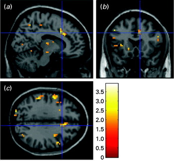
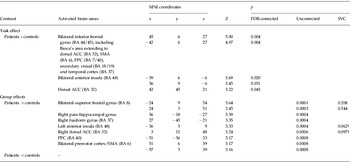
Table 2 lists BOLD activations for complex PTSD patients (n=29) and controls (n=22). For the classic Stroop contrast, both groups activated mainly a large bilateral area in the inferior frontal gyrus – including Broca's area, the dorsal ACC, SMA, posterior parietal cortex (PPC), secondary visual cortex and inferior temporal cortex – and a smaller bilateral area in the insula. In the classic Stroop task, patients showed a trend for increased left anterior insula and dorsal ACC activation compared with controls (Table 2, Fig. 1). Adjusting for years of education increased the Z value from the left anterior insula from 3.33 to 3.42 (p SVC=0.047) and decreased the Z value from the dorsal ACC from 3.24 to 3.19 (p SVC=0.116). There were no significant BOLD response differences between medicated and non-medicated patients. Patients with co-morbid MDD had decreased dorsal ACC activation (0, 27, 39, Z=3.49, p SVC=0.010) compared with non-MDD patients, whereas insular activation was similar.
Trends for increased blood oxygenation level-dependent response in the dorsal anterior cingulate cortex [Montreal Neurological Institute (MNI) coordinates 3, 12, 48, Z=3.24, p uncorrected=0.0006, p small volume correction (SVC)=0.097] and left anterior insula (MNI coordinates −36, 3, 9, Z=3.33, p uncorrected=0.0004, p SVC=0.062) in complex post-traumatic stress disorder patients (n=29) compared with non-trauma-exposed healthy controls (n=22) in the classic Stroop contrast. The cross hair is positioned at MNI coordinates 12, 18, 33 on a sagittal (a), coronal (b) and transversal (c) plane and data are thresholded at p<0.05 with an extent threshold of 10 voxels for illustrative purposes.

Activated brain areas (BOLD responses with peak MNI coordinates) in complex post-traumatic stress disorder patients (n=29) and non-trauma-exposed healthy controls (n=22) in classic Stroop contrasts (incongruent versus congruent words)

BOLD, Blood oxygenation level-dependent; MNI, Montreal Neurological Institute; FDR, false discovery rate; SVC, small volume correction; BA, Brodmann area; ACC, anterior cingulate cortex; SMA, supplementary motor area; PPC, posterior parietal cortex.
† p SVC <0.10.
In the emotional Stroop contrasts there was overall less activation across groups than in the classic Stroop task. In the general negative versus neutral words contrast, both groups activated mainly the left inferior frontal cortex, including the orbitofrontal cortex/left ventrolateral prefrontal cortex and Broca's area. In the trauma compared with neutral words contrast, both groups activated mainly the inferior frontal gyrus, including Broca's area. There were no significant group differences between complex PTSD patients and controls in these emotional Stroop contrasts.
Treatment effects: task performance
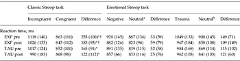
Table 3 shows reaction times in EXP and TAU patients. The overall group had shorter reaction times on the classic Stroop task after treatment (effect for time: F 1,14=4.8, p=0.046). Reaction time differences of the classic Stroop task tended to differ by treatment type (F 1,14=3.1, p=0.098). A post hoc comparison of post- versus pre-scores in EXP patients showed that classic Stroop interference significantly decreased after treatment (t 8=−2.6, p=0.034). No such decrease was found for TAU patients (t 6=−0.9, p=0.40). Reaction time differences of the emotional Stroop task did not change over time (F<1).
Reaction times in classic and emotional Stroop tasks in EXP (n=9) and TAU patients (n=7) with complex post-traumatic stress disorder pre- and post-treatment: absolute values per word category and difference values

Data are given as mean (standard deviation).
EXP, Experimental treatment (psycho-educational and cognitive behavioural stabilizing group treatment added to treatment as usual); TAU, treatment as usual.
a Neutral words are to compare with general negative words.
b Neutral words are to compare with trauma words.
* Across the groups (EXP+TAU patients) a significant effect of time (p<0.05) and a trend for treatment type (p<0.10) were found.
† In the EXP patients only, there were significantly shorter reaction times post v. pre (p<0.05; post hoc paired t test).
Accuracy improved after treatment across groups. There was an effect for time in the classic Stroop task (F 1,14=5.5, p=0.035) and a trend for time in the trauma v. neutral Stroop task (F 1,14=4.0, p=0.066), but there were no trends or effects for treatment type (data not shown).
Treatment effects: imaging data
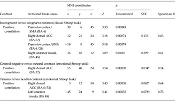
Across groups, the classic Stroop contrast showed an effect for time revealing decreased activation in the premotor cortex/SMA (6, 0, 57, Z=4.00, p uncorrected=0.000032 and −6, 12, 57, Z=3.34, p uncorrected=0.00041) and left inferior frontal cortex (−57, 6, 6, Z=3.19, p uncorrected=0.00084) after treatment (data not shown). CAPS improvement correlated not significantly with a decrease in right dorsal ACC or insula activation in the classic Stroop task (Table 4).
Positive correlation of improvement on the CAPS with decreased activation in brain areas post-treatment for classic and emotional Stroop contrasts across groups: EXP and TAU complex post-traumatic stress disorder patients together (n=16)

CAPS, Clinician Administered Post-Traumatic Stress Disorder Scale; EXP, experimental treatment (psycho-educational and cognitive behavioural stabilizing group treatment added to treatment as usual); TAU, treatment as usual; MNI, Montreal Neurological Institute; SVC, small volume correction; SMA, supplementary motor area; BA, Brodmann area; ACC, anterior cingulate cortex.
* p SVC <0.05.
† p SVC <0.10.
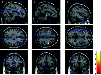
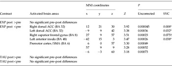
There was a treatment type×time interaction in the dorsal ACC (12, 21, 27, Z=3.04, p uncorrected=0.001) in the classic Stroop task. Post hoc tests (see Table 5, Fig. 2) revealed statistically significant decreased bilateral dorsal ACC and left anterior insula BOLD responses, and a trendwise decrease in the superior prefrontal cortex, post- compared with pre-treatment in EXP patients. This was not found in TAU patients.
Treatment effects for the classic Stroop contrast in complex post-traumatic stress disorder patients (n=9) given the experimental treatment (psycho-educational and cognitive behavioural stabilizing group treatment added to treatment as usual). There were significantly decreased blood oxygenation level-dependent responses in (a) the right and (b) left dorsal anterior cingulate cortex [Montreal Neurological Institute (MNI) coordinates: 12, 21, 30, Z=3.92, p small volume correction false discovery rate (SVC FDR)-corrected=0.008; MNI coordinates: −9, 9, 42, Z=3.38, p SVC=0.032, respectively] and (c) the left anterior insula (MNI coordinates: −42, 15, 3, Z=3.47, p SVC FDR-corrected=0.038) post- compared with pre-treatment, and a trend for the right superior frontal gyrus [MNI coordinates: 27, 9, 57, Z=3.51, p SVC=0.075, see (b) transversal and coronal plane]. Data are thresholded at p<0.05 with an extent threshold of 10 voxels for illustrative purposes.

Decreased activity post-treatment in complex post-traumatic stress disorder patients from the EXP (n=9) and not in the TAU condition (n=7): activated brain areas (BA and peak MNI coordinates) after treatment in the classic Stroop contrast

EXP, Experimental treatment (psycho-educational and cognitive behavioural stabilizing group treatment added to treatment as usual); TAU, treatment as usual; BA, Brodmann area; MNI, Montreal Neurological Institute; SVC, small volume correction; ACC, anterior cingulate cortex; SMA, supplementary motor area.
* p SVC <0.05.
† p SVC <0.10.
Across groups, there were no significant effects of time for the general negative versus neutral contrast. However, there was a significant positive correlation between CAPS improvement and decrease in right dorsal ACC activation (Table 4). In the trauma versus neutral Stroop contrast, there were no significant effects for time either, but a significant positive correlation was found between CAPS improvement and dorsal ACC and a trend for left anterior insula activation (Table 4). No significant effects for treatment type, or treatment type×time interactions, were found for the general negative and for the trauma Stroop contrasts.
Discussion
In the present fMRI study using a hybrid version of a classic and emotional Stroop task, we found performance differences and distinct cortical activation patterns as measured with fMRI between childhood abuse-related complex PTSD patients and non-trauma-exposed healthy controls. Furthermore, we found treatment effects in these patients especially with response to psycho-educational and cognitive behavioural stabilizing group treatment (EXP) next to TAU compared with TAU only. Child abuse-related complex PTSD patients with multiple current co-morbid Axis I and Axis II diagnoses and personality disorders are rarely studied and to our knowledge only a few RCTs on the neurobiological correlates of treatment outcome in PTSD following adult trauma have been published (Peres et al. Reference Peres, Newberg, Mercante, Simao, Albuquerque, Peres and Nasello2007; Lindauer et al. Reference Lindauer, Booij, Habraken, van Meijel, Uylings, Olff, Carlier, Den Heeten, van Eck-Smit and Gersons2008).
Complex PTSD patients showed increased emotional Stroop interference for trauma words compared with controls. As expected, patients showed increased activation in the dorsal ACC compared with controls in the classic Stroop contrast. Furthermore, there was increased activation in the left insula, which is not in line with another emotional Stroop fMRI study in child abuse-related PTSD (Bremner et al. Reference Bremner, Vermetten, Vythilingam, Afzal, Schmahl, Elzinga and Charney2004) showing decreased (posterior) insula activation. This could be due to a more anterior localization of the insula activation in our study and the distinct contribution of anterior and posterior insular cortices to experiencing emotions such as anger, fear and disgust (Wright et al. Reference Wright, Martis, McMullin, Shin and Rauch2003). The anterior insula is connected especially to ventral ACC regions generating emotional awareness (Taylor et al. Reference Taylor, Seminowicz and Davis2009), while the posterior insula is connected to the dorsal ACC/posterior cingulate cortex which is mainly involved in executive functions such as environmental monitoring and response selection (Taylor et al. Reference Taylor, Seminowicz and Davis2009) and detection of response conflicts (Carter & van Veen, Reference Carter and van Veen2007). This distinction has also been described as part of a ventral (arousal) and dorsal (selective attention) system (Eckert et al. Reference Eckert, Menon, Walczak, Ahlstrom, Denslow, Horwitz and Dubno2009). Increased anterior insula activation may therefore indicate a high arousal state during task performance whereas decreased posterior insular activation may be associated with impaired selective attention and poor task performance.
After 6 months of treatment, reaction times for the classic Stroop task were reduced across groups (EXP and TAU together). In an earlier study, Stroop performance was not found to be particularly sensitive to treatment effects (Devineni et al. Reference Devineni, Blanchard, Hickling and Buckley2004), but our study may provide support for the clinical utility of the classic Stroop test as a measure of treatment outcome. Furthermore, in a regression analysis we found that clinical improvement measured with the CAPS was associated with decreased activity in the dorsal ACC and anterior insula in the emotional Stroop contrasts. We suggest that these changes point to normalization of dorsal ACC and insula activity after stabilizing group therapy, which may reflect fewer response conflicts or improved selective attention, and lower emotional arousal. It has been debated if the Stroop paradigm measures automatic attention processes or rather strategic processes of effortful avoidance of threat cues (Buckley et al. Reference Buckley, Blanchard and Neill2000; Bar-Haim et al. Reference Bar-Haim, Lamy, Pergamin, Bakermans-Kranenburg and Van IJzendoorn2007), possibly indicating greater cognitive control following treatment in the current study. The treatment effect over time was mainly driven by the experimental group: only EXP patients showed decreased bilateral dorsal ACC and left anterior insula activation after treatment, while TAU patients did not. In a comparable RCT (Peres et al. Reference Peres, Newberg, Mercante, Simao, Albuquerque, Peres and Nasello2007) in adult PTSD patients with mixed trauma, significantly increased activation was observed following psychotherapy in the left anterior cingulate, but not in the insula, as well as decreased activation in the left amygdala. However, these changes are more difficult to interpret given that baseline differences between patients and controls in the latter study were absent.
Contrary to our expectations, we did not find group differences for the emotional Stroop contrasts in our neuroimaging data, at baseline or after treatment. Possibly, the classic Stroop contrast is more powerful than the emotional Stroop contrasts because of its higher cognitive demands. On the other hand, we did observe group differences with regard to the trauma Stroop reaction times, as well as a significant positive correlation between clinical improvement on the CAPS and decrease in right dorsal ACC and insula activation in the general negative and trauma Stroop tasks, respectively. While suggesting similar treatment effects as observed for the classic Stroop contrast, these latter results should be interpreted with caution due to the lack of main group effects.
A limitation of this study is the inevitable use of medication (SSRIs or benzodiazepines, no other psychoactive drugs) in a complex patient population such as the one studied. Although patients still fulfilled criteria for complex PTSD while using these medications, our main results did not differ between medicated and non-medicated patients. Recent data from our group have indicated that brain activity during emotion processing and cognitive task performance is similar in medicated and non-medicated patients with depression and/or anxiety disorders (van Tol et al. Reference van Tol, van der Wee, Demenescu, Nielen, Aleman, Renken, van Buchem, Zitman and Veltman2011); further research is needed in PTSD (Lanius et al. Reference Lanius, Brewin, Bremner, Daniels, Friedman, Liberzon, McFarlane, Schnurr, Shin, Stein and Vermetten2010). Also, because of the characteristics of our early-traumatized (female) complex PTSD population, results cannot be straightforwardly generalized to other PTSD populations. Because we chose to include a non-traumatized healthy control group as a reference for our baseline comparison we cannot fully exclude the possibility that baseline differences were related to trauma exposure rather than to PTSD diagnosis per se. Furthermore, the frequent co-morbidity of, for example, MDD makes it difficult to disentangle the effects of different Axis I diagnoses; although post hoc analyses showed that our main results were not explained by co-morbid MDD. Another limitation is the small sample size in the treatment outcome substudy, as the power was too low to reliably assess group differences outside our ROIs. Furthermore, our findings of clinical improvement on the CAPS in EXP, but also TAU patients, indicate that a fairly large sample size would be needed to detect whole-brain differences at a reasonably stringent threshold. Within our ROIs, we observed both time effects in our overall group as well as effects of time×treatment type interactions, indicating subtle differences between the treatments under study.
Concluding, compared with non-trauma-exposed healthy controls, complex PTSD patients showed greater emotional Stroop interference for trauma words and increased activation in the dorsal ACC and left anterior insula in the classic Stroop task. Post-therapy, classic Stroop interference decreased, especially in complex PTSD patients receiving psycho-educational and cognitive behavioural stabilizing group treatment next to TAU. Moreover, we found decreased – which we suggest may be interpreted as ‘normalized’ – activity in EXP patients after treatment in (bilateral) dorsal ACC and left anterior insula for the classic Stroop contrast, possibly reflecting increased selective attention and lower emotional arousal after treatment, indicating more cognitive control over PTSD symptoms.
Supplementary material
For supplementary material accompanying this paper, visit http://dx.doi.org/10.1017/S0033291712000499.
Acknowledgements
The authors thank trained psychologists N. Ran, R. J. de Vries, M. Burger-Hupkes, and psychiatry residents S. A. N. Renkema and F. E. de Vries for performing extensive assessments, Z. Sjoerds for assistance in preprocessing imaging data, and especially all patients and controls for participating in this study. This study was made possible by financing from ZonMw, the Netherlands organization for health research and innovation (grant no. 100-002-020), GGZ InGeest, the Institute for Clinical and Experimental Neurosciences (ICEN) and the VU University Medical Center, Amsterdam, The Netherlands.
Declaration of Interest
None.