Introduction
Acute, severe neck abscesses are commonly encountered. They can be caused by infections originating from the head, by primary or secondary infection of the cervical lymph nodes or thyroid gland, or by infected congenital cysts.
Congenital cervical cysts are common in adults, and are often derived from the branchiogenic cleft, thymus or thyroglossal duct. Cervical cysts of bronchogenic origin are rare.Reference Ustundag, Iseri, Keskin, Yayla and Muezzinoglu1, Reference Moz, Gamba, Pignatelli, D'Addazio, Zorzi and Fiaccavento2
A bronchogenic cyst is a malformation of the ventral foregut. The cyst is located in the thorax in more than 50 per cent of cases.Reference Cuypers, De Leyn, Cappelle, Verougstraete, Demedts and Deneffe3 Other locations include subcutaneous,Reference Zvulunov, Amichai, Grunwald, Avinoach and Halevy4, Reference Ozel, Kazez, Koseogullari and Akpolat5 subscapular,Reference Ozel, Kazez, Koseogullari and Akpolat5 abdominalReference Liang, Yee, Song and Marks6 and retroperitonealReference Sullivan, Okada, Kudo and Ebihara7 sites. Cervically located bronchogenic cysts are often asymptomatic,Reference Ustundag, Iseri, Keskin, Yayla and Muezzinoglu1, Reference Newkirk, Krowiak, Tassler and Deeb8–Reference Eng, Quraishi and Watson12 and are generally detected in childhood.Reference Ustundag, Iseri, Keskin, Yayla and Muezzinoglu1, Reference Hadi, Jammal, Hamdan, Saad and Zaatari10, Reference Touloukian13–Reference Maung, Low, Knight and Cullinane15
The symptoms caused by cervical bronchogenic cysts include stridor in neonates,Reference Touloukian13–Reference Maung, Low, Knight and Cullinane15 dyspnoea on exertion,Reference Newkirk, Krowiak, Tassler and Deeb8, Reference Park and Buford16 dysphagiaReference Newkirk, Krowiak, Tassler and Deeb8, Reference Al-kasspooles, Alberico, Douglas, Litwin, Wiseman and Rigual17 and recurrent infection.Reference Al-kasspooles, Alberico, Douglas, Litwin, Wiseman and Rigual17 One case report described a deep neck abscess due to a bronchogenic cyst.Reference McManus, Holt, Aufdemorte and Trinkle18
We describe a second case of a bronchogenic cyst presenting as a recurrent neck abscess in a middle-aged woman.
Case report
A 51-year-old woman was referred to our hospital for evaluation of a painful cervical swelling plus dysphagia of five days' duration. She had not noticed any fever or dyspnoea. The swelling had been present for as long as she could remember. Four years before admission, thyroid function tests had been performed, with normal results. She was also known to have Crohn's disease, and had bilateral hearing loss due to multiple attacks of sudden sensorineural hearing loss.
Physical examination showed a painful, tense mass located in the right supraclavicular and paratracheal area. Hypertrophic tonsils were seen in the oropharynx.
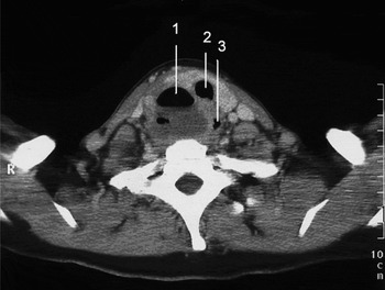
Computed tomography (CT) revealed a 4.5 cm diameter cyst with an air–fluid level located dorsal to the right thyroid lobe. The hypopharynx was compressed. No connections were seen with the lungs, trachea or thyroid gland. The most likely diagnosis was a cervical abscess, which was confirmed by surgical drainage. Rigid endoscopy of the oesophagus and the upper and lower airways did not locate any fistula. Bacterial culture of the abscess contents grew a mixture of different bacteria, but no specific pathogen was detected.
Similar complaints and signs recurred after three months. A second CT scan again showed a cyst with an air–fluid level, highly suspicious for a retrotracheal abscess. A gastrografin contrast swallow study demonstrated lateralisation of both the trachea and the oesophagus to the left. The abscess was drained surgically and a biopsy taken. Again, rigid endoscopy of the oesophagus and the upper and lower airways located no fistula. Histopathological examination of the biopsy identified pus and inflamed, ciliated, pseudo-stratified, columnar epithelium.
Three months later, when the patient had recovered completely and was free of symptoms, magnetic resonance imaging (MRI) and CT scanning of the cyst were performed (Figure 1). The cyst appeared to be filled with air, and a connection with the trachea was suspected. Ultrasonography revealed that the cyst filled with air during the Valsalva manoeuvre.
Axial computed tomography scan of the neck, showing the bronchogenic cyst filled with fluid and air (1), and displacing the trachea (2) and oesophagus (3) to the left. R = right

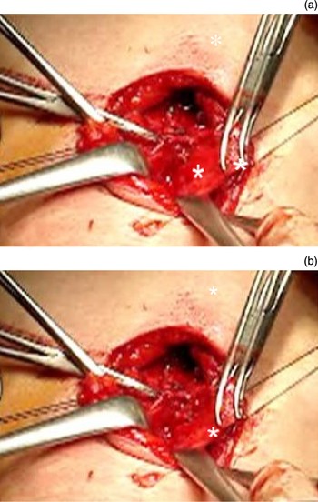
It was decided to again undertake surgery. Pre-operatively, methylene blue dye was injected into the cyst. Rigid endoscopy of the upper and lower airways again showed no fistula. The cyst was excised by a collar incision, and a thorough exposition and inspection was made of the trachea, sternocleidomastoid muscle, common carotid artery, external jugular vein, and vagal and recurrent nerves. The recurrent nerve was identified and bluntly separated from the ballooning cyst. The transparent, thin-walled cyst was seen to fill and empty synchronous with respiration (Figure 2). Because the endotracheal tube cuff pressed tightly against the trachea, a fistula had to be present somewhere between the lower airways (under the cuff) and the cyst. Upon opening of the cyst, no methylene blue dye was retrieved. Palpation of the entire trachea did not reveal any sign of a fistula. However, a strand of connective tissue connected to the upper right lobe was located and assumed to be the fistula, and was ligated. Subsequently, no air leak was seen while flooding the wound with water.
Intra-operative photographs taken during extirpation, showing the cyst (*) inflated and deflated synchronous with (a) inspiration and (b) expiration, during artificial ventilation.

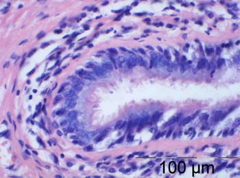
Histopathological examination of the cyst revealed ciliated, pseudo-stratified, columnar epithelium (Figure 3). No cartilage, smooth muscle or mucous glands were seen.
Photomicrograph of a section of the cyst wall, showing ciliated, pseudostratified, columnar epithelium. Note that a submucosal layer with cartilage and smooth muscle is absent. (H&E; ×400)

Post-operatively, the patient suffered a recurrent nerve palsy on the operated site, as detected by indirect laryngoscopy.
After three years' follow up, the patient was free of symptoms. Although her recurrent nerve palsy persisted, her voice improved with speech therapy.
Discussion
The symptoms of bronchogenic cysts are generally due to direct compression of surrounding structures, resulting in stridor, dyspnoea on exertion or dysphagia, or to a fistula resulting in recurrent infections or abscesses. During the Valsalva manoeuvre, bronchogenic cysts can increase in volume due to the presence of a fistula,Reference Al-kasspooles, Alberico, Douglas, Litwin, Wiseman and Rigual17 as in our case, or due to extension from an intrathoracic location into the supraclavicular area.Reference Park and Buford16 Most such fistulas are rarelyReference Al-kasspooles, Alberico, Douglas, Litwin, Wiseman and Rigual17 if everReference Eng, Quraishi and Watson12 seen on CT or MRI scanning, and are not detected until surgical extirpation. Therefore, extirpation is both diagnostic and therapeutic. A fistula should be considered in symptomatic cases, and should be searched for during surgery.
There are two embryological theories about the origin of cervical bronchogenic cysts. In the fourth embryonic week, the laryngotracheal bud sprouts as the ventral appendix of the foregut, and forms the origin of the larynx and tracheobronchial bud.Reference Sadler and Sadler19 The first theory holds that bronchogenic cysts derive from abnormal buddings of the distal part of the tracheobronchial bud. An alternative theoryReference Pujary, Pujary, Shetty, Hazarika and Rao20 hypothesises that bronchogenic cysts originate from preformed cysts of the thoracic cavity. During the development of the tracheobronchial tree and closure of the thoracic wall, this cyst pinches off and migrates to the neck region. In both theories, a connection to the thorax may persist as a fistula,Reference Al-kasspooles, Alberico, Douglas, Litwin, Wiseman and Rigual17 stalkReference Maung, Low, Knight and Cullinane15 or connective tissue strandReference Pujary, Pujary, Shetty, Hazarika and Rao20 to the mediastinum.
Diagnostic investigation includes a thorough history for cough, recurrent airway infection, dysphagia and dyspnoea. Physical and ENT examination should include the Valsalva manoeuvreReference Park and Buford16, Reference Al-kasspooles, Alberico, Douglas, Litwin, Wiseman and Rigual17 and transillumination of the cyst.Reference Gaikwad, Muthusami, Raj, Rajinikanth and John11 Radiological imaging of the cyst should comprise ultrasonography, gastrografin contrast swallow study, and CT and MRI scanning. Al-Kasspooles et al. Reference Al-kasspooles, Alberico, Douglas, Litwin, Wiseman and Rigual17 have demonstrated a fistula to the trachea using virtual bronchoscopy. Eventually, the diagnosis is made by means of rigid endoscopy (i.e. bronchoscopy, oesophagoscopy and laryngoscopy) and total extirpation of the cyst.
The differential diagnosis of a cervical neck cyst is extensive. It comprises branchial cysts, thyroglossal cysts, thymic cysts, thyroid cysts, lipomas, epidermal inclusion cysts, teratomas and cystic lymphoid vascular malformations. The location (i.e. medial or lateral) is not conclusive for the diagnosis, as has been suggested in the past.Reference Touloukian13 Branchial cysts are a commoner, better known cause of neck abscesses. Branchial cysts, being of ectodermal origin, are usually characterised by stratified, squamous epithelium, and only rarely by pseudo-stratified, columnar, ciliated epithelium.Reference Zvulunov, Amichai, Grunwald, Avinoach and Halevy4, Reference Hadi, Jammal, Hamdan, Saad and Zaatari10 Theoretically, Crohn's disease may have caused an upper digestive tract fistula in our patient.Reference Mills, Amin and Manisali21 However, her Crohn's disease had been quiescent for many years, and no ulceration of the oral cavity or oesophagus was reported. Furthermore, thorough endosopic inspection did not reveal any ulceration or fistula. This possibility was therefore excluded.
• Cervical bronchogenic cysts seldom cause symptoms after childhood
• This paper describes a neck abscess caused by a bronchogenic cyst in an adult
• The treatment of choice in this situation is total extirpation of the cyst, and its possible coexisting fistula, to avoid recurrent infection and malignant transformation
Because the diagnosis of bronchogenic cyst is made histopathologically, there is an ongoing debate regarding strict diagnostic criteria. The structures of the bronchogenic cyst are embryologically derived from endoderm, with accessory tissue of mesodermal origin. The histological criteria for a bronchogenic cyst are therefore a cyst with ciliated, pseudo-stratified, columnar epithelium and seromucinous glands, cartilage and smooth muscle. In this way the histological appearance ranges from an alveolus to a small bronchus. Although these criteria are often met in the case of intrathoracic bronchogenic cysts,Reference Cuypers, De Leyn, Cappelle, Verougstraete, Demedts and Deneffe3 in cervical bronchogenic cysts frequently only ciliated, pseudo-stratified, columnar epithelium is found.Reference Shimazu, Kuratomi and Inokuchi9, Reference Maung, Low, Knight and Cullinane15, Reference Al-kasspooles, Alberico, Douglas, Litwin, Wiseman and Rigual17 Some authors refer to such cysts as foregut cysts,Reference Liang, Yee, Song and Marks6 and consider cartilage obligatory for the diagnosis of a bronchogenic cyst.Reference Hadi, Jammal, Hamdan, Saad and Zaatari10 Others state that cartilage is seldom present.Reference Maung, Low, Knight and Cullinane15 In our case, however, the diagnosis was made at surgery, with the histopathological appearance supplying only supportive evidence.
The treatment of choice for bronchogenic cysts, irrespective of location or symptoms, is total extirpation, because of the risk of complications (e.g. abscess) and possible malignant transformation. Malignancies have been found in intrathoracicReference Cuypers, De Leyn, Cappelle, Verougstraete, Demedts and Deneffe3, Reference Sullivan, Okada, Kudo and Ebihara7 and retroperitonealReference Sullivan, Okada, Kudo and Ebihara7 bronchogenic cysts, and are known to occur in asymptomatic branchial cysts.Reference Lanzafame, Caltabiano, Puzzo and Cappellani22
In our patient, recurrent nerve palsy was seen post-operatively. This complication has also been reported by McManus et al. Reference McManus, Holt, Aufdemorte and Trinkle18 Despite such a serious complication, we still support the practice of extirpation of all cervical cysts, even asymptomatic ones.