INTRODUCTION
Physical, chemical and radioactivity characteristics of the thermal springs located around Gulf of Kuşadası
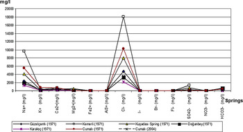
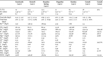
Numerous hot water springs are found around the Gulf of Kuşadası. Mineral waters have a temperature range of 19–64oC (Figure 1) (Türkiye Maden Suları 3, Ege Bölgesi, Reference Türkiye Maden and Ege1974; Barut et al., Reference Barut, Erdoğan and Başak2004). In Doğanbey Cape (Seferihisar), there are terrestrial as well as submarine springs, of which the temperatures range between 66 and 70oC. The variations observed in the concentrations of the elements are noteworthy (Figure 2). The concentration of Br is much above the average values reported for the Earth's crust (2.5 mg/l), sandstone (1 mg/l) and limestone (6.2 mg/l) (Turekian & Wedepohl, Reference Turekian and Wedepohl1961; Mason & Moore, Reference Mason and Moore1982).
Thermal mineral springs around the Gulf of Kusadası: (1) Cumalı; (2) Karakoç; (3) Doğanbey; (4) Kusadası kaynağı; (5) Kemerli; (6) Güzelçamlı.

Chemical composition of submarine spring waters in around Kusadası.

The chemical compositions show that these thermal springs are carbonated and sulphated waters with high Na, K and Mg concentration and carbonate hardness exceeds 50% (Table 1). Alkaline elements (Na, K) have higher values compared to the alkaline earth elements (Ca, Mg). Na, Ca and K are the most abundant cations, whereas the most abundant anions are Cl, HCO3 and SO4, suggesting that the waters are coming through limestones.
Physical, chemical and radioactive properties of the spring waters found around the Gulf of Kuşadası (Türkiye Maden Suları 3, Ege Bölgesi Reference Türkiye Maden and Ege1974; Barut et al., Reference Barut, Erdoğan and Başak2004).

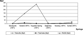
These thermal waters have radioactive characteristics (Table 1). Total alpha and total beta Rn222 values are higher in Kemerli, Kuşadası Spring and Cumalı (Figure 3). Total alpha values are ranging between 0.50 ± 5.07 and 4.57 ± 27.23 Bq/l, whereas total beta values are found to be between 2.29 ± 5.11 and 22.19 ± 26.61 Bq/l.
Radioactivity of submarine spring waters around Kusadası.

Oceanographic characteristics of the study area
Temperature of surface waters varies according to atmospheric temperature. The temperature difference between surface and 150 m varies seasonally between 0.1°C and 6.76°C. The greatest difference is observed during winter, whereas, the smallest difference is observed in summer. The temperature variations are very small in the top 50 m water column. Horizontal temperature variations observed at surface/50 m of depth are; in spring, 16.99–1.00°C/15.70–16.27°C; in summer 17.80–25.55°C/16.77–7.26°C; in autumn 21.26–22.27°C/17.52–19.88°C; and in winter 14.90–15.20°C/14.93–15.20°C (Rapor No. 239, 1989; Rapor No. 250, 1990).
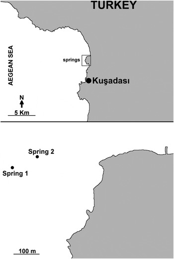
Two submarine springs (named, Kuşadası-1 and Kuşadası-2) are found 200 m off the shore in Pamucak Cove, located 50 m apart from each other (Kuşadası, Aydın), at a depth of 12.40 m (Figure 4). Kuşadası-1 was chosen for this study. The temperature of the springs is measured as 19.6oC and it is almost stable throughout the year. The foraminifer fauna around the spring and the effects of spring waters on foraminifer fauna were investigated.
Map of the study area.

MATERIALS AND METHODS
The spring was measured at its centre and four vertical lines were set on south, north, east and west directions. A total of 45 sediment samples were collected at different points on the lines. About 100 g of sediment samples were manually collected by a plastic shovel during SCUBA diving. On the south and north lines samples were collected at 5, 10, 15, 20, 25, 30, 35, 40, 45 and 50 m distances from the spring. Because of the suitable benthic structure, it was possible to collect more samples on the east line; samples were collected at 5, 10, 15, 20, 25, 30, 35, 40, 45, 50, 60, 70, 80, 90 and 100 m distances. It was not possible to take a 50 m transect on the west line and samples were taken at 5, 10, 15, 20, 25, 30, 35, 40 and 45 m distances. One sample was also taken from the spring.
Five grams of wet sediment samples were weighed and treated with 10% H2O2 for 24 hours. The samples were washed with pressurized water on 0.063 mm sieve and dried at 50°C in the oven. The dried samples were further sieved with 2.00, 1.00, 0.500, 0.250 and 0.125 mm mesh sizes. The foraminifer individuals were separated under a binocular microscope. The following literature were used for identifications: Meriç & Sakınç (Reference Meriç, Sakinç and Meriç1990); Cimerman & Langer (Reference Cimerman and Langer1991); Hatta & Ujiie (Reference Hatta and Ujiie1992); Hottinger et al. (Reference Hottinger, Halicz and Reiss1993); Sgarrella & Moncharmont-Zei (Reference Sgarrella and Moncharmont-Zei1993); Loeblich & Tappan (Reference Loeblich and Tappan1994); Meriç et al. (Reference Meriç, Yanko, Avşar and Meriç1995, Reference Meriç, Avşar, Eryilmaz and Yücesoy-Eryilmaz2001a, Reference Meriç, Avşar, Eryilmaz and Yücesoy-Eryilmazb, Reference Meriç, Avşar and Nazik2002a , Reference Meriç, Avşar and Berginb, Reference Meriç, Avşar, Görmüş and Orakc, Reference Meriç, Avşar, Bergin and Yokeşd, Reference Meriç, Avşar, Bergin and Barut2003a, Reference Meriç, Avşar, Bergin and Barutb; Reference Meriç, Avşar and Bergin2004a, Reference Meriç, Avşar and Berginb, Reference Meriç, Avşar, Nazik, Eryilmaz and Yücesoy-Eryilmazc, Reference Meriç, Avşar, Nazik, Alpar, Yokeş, Barut and Ünlu2005, Reference Meriç, Avşar, Nazik, Yokeş and Dinçer2008a, Reference Meriç, Avşar, Yokeş, Tuğrul, Bayari, Özyurt, Öncel, Barut, Uysal and Kamb, Reference Meriç, Avşar and Yokeşc, Reference Meriç, Avşar, Nazik, Tunoğlu, Yokeş, Barut, Yücesoy-Eryilmaz, Tuğrul, Görmüş, Öncel, Orak, Kam and Dinçerd); Yassini & Jones (Reference Yassini and Jones1995); Avşar & Meriç (Reference Avşar and Meriç1996, Reference Avşar and Meriç2001a, Reference Avşar and Meriçb); Avşar (Reference Avşar1997, Reference Avşar2002); Hayward et al. (Reference Hayward, Grenfell, Reid and Hayward1999); Avşar & Ergin (Reference Avşar and Ergin2001); Avşar et al. (Reference Avşar, Meriç and Ergin2001, Reference Avşar, Aksu and Dinçer2006); Meriç & Avşar (Reference Meriç and Avşar2001); Kaminski et al. (Reference Kaminski, Aksu, Box, Hiscott, Filipescu and Al-Salameen2002); Bergin et al. (Reference Bergin, Küçüksezgin, Uluturhan, Barut, Meriç, Avşar and Nazik2006). Taxonomy was based on Loeblich & Tappan (Reference Loeblich and Tappan1988, Reference Loeblich and Tappan1994).
Temperature and depth of the seawater were manually measured on the site by Casio Sea Pathfinder spf-100 diving watch. At each sampling point, we waited at least three minutes to measure a stable value. Seawater samples from both the Kuşadası-1 and Kuşadası-2 springs were manually taken from the surface for phytoplankton analysis. Cells were transferred to 1 l capacity PVC containers and fixed with borax buffered formaldehyde to a final concentration of 0.4% (Throndsen, Reference Throndsen and Sournia1978). Samples were left in the dark glass bottles to settle for a week in the laboratory and 100 ml were drawn from the bottom of the bottles to get maximum amount of cells. Phytoplankton cells were counted using a Sedgewick Rafter counting cell under an Olympus CH-2 light microscope (LM) (Guillard, Reference Guillard and Sournia1978). For qualitative analyses the cells were analysed at 40× or 100× magnification under the light microscope. For identification of species the following references were used: Cupp (Reference Cupp1943); Hendey (Reference Hendey1964); Priddle & Fryxell (Reference Priddle and Fryxell1985); and Hasle et al. (Reference Hasle, Syvertsen, Steidinger, Tangen, Throndsen, Heimdal and Tomas1997).
For heavy metal and trace element (Al, Si, Ti, Cr, Mn, Fe, Co, Ni, Cu, Zn, As, Cd, Hg and Pb) analysis a water sample was collected from each spring in a 0.5 l polyethylene bottle containing 2 ml HCl. The analyses were done at the Çukurova University, Faculty of Engineering and Architecture, Department of Geology, Geochemistry Laboratory. Since the sample was liquid, it was directly used for analysis using the Atomic Absorption Spectrophotometer 700. For As and Hg readings, the elements were reduced with sodium borohydride before measurement.
0.5 l of seawater was collected from each spring in polyethylene bottles. The water samples were frozen without adding any preservatives and sent to the TUBİTAK MAM Institute of Chemistry and Environment Laboratory as quickly as possible for nutrient analysis. N, P, Si, NH3, total nitrogen and total phosphate analysis were measured by autoanalyser.
Short term seasonal current measurements were taken with a current meter (Aanderaa RCM 4S) in two different stations: KA-01 (37°52′32″N 27°15′17″E, depth: 21 m); and KA-02 (37°58′24″N 27°14′09″E, depth: 36 m) (Figure 5). Recordings were obtained every two minutes on the surface and at 20 m of depth.
Locations of the surface current sampling stations.

RESULTS
Foraminifer assemblage
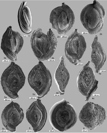
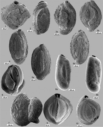
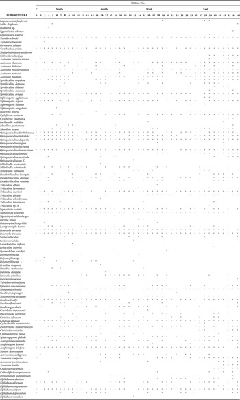
A rich foraminifer fauna was observed in the 45 sediment samples analysed. 59 genera and 103 species were identified (Table 2), which are Lagenammina fusiformis (Williamson), Iridia diaphana Heron-Allen & Earland, Haddonia sp., Eggerelloides advenus (Cushman), E. scabrus (Williamson), Textularia bocki Höglund, T. truncata Höglund, Cornuspira foliacea Philippi, Vertebralina striata d'Orbigny, Nodopthalmidium antilarum (Cushman), Nubecularia lucifuga Defrance, Adelosina carinata-striata Wiesner, A. cliarensis (Heron-Allen & Earland), A. duthiersi Schlumberger, A. mediterranensis (le Calvez J. & Y.), A. partschi (d'Orbigny), A. pulchella d'Orbigny, Spiroloculina angulosa Terquem, S. depressa d'Orbigny, S. dilatata d'Orbigny, S. excavata d'Orbigny, S. ornata d'Orbigny, Siphonaperta agglutinans (d'Orbigny), S. aspera (d'Orbigny), S. dilatata (le Calvez J. & Y.), S. irregularis (d'Orbigny), Hauerina diversa Cushman, Cycloforina contorta (d'Orbigny), C. villafranca (le Calvez J. & Y.), Lachlanella undulata (d'Orbigny), Massilina gualteriana (d'Orbigny), M. secans (d'Orbigny), Quinqueloculina berthelotiana d'Orbigny, Q. bidentata d'Orbigny, Q. disparilis d'Orbigny, Q. jugosa Cushman, Q. laevigata d'Orbigny, Q. lamarckiana d'Orbigny, Q. limbata d'Orbigny, Q. seminula (Linné), Quinqueloculina sp. C, Miliolinella semicostata (Wiesner), M. subrotunda (Montagu), M. webbiana (d'Orbigny), Pseudotriloculuina laevigata (d'Orbigny), P. oblonga (Montagu), P. rotunda (d'Orbigny), Triloculina affinis d'Orbigny, T. bermudezi Acosta, T. marioni Schlumberger, T. plicata Terquem, T. schreiberiana d'Orbigny, T. tricarinata d'Orbigny, Triloculina sp. A, Sigmoilinita costata (Schlumberger), S. edwardsi (Schlumberger), Sigmoilopsis schlumbergeri (Silvestri), Parrina bradyi (Millet), Euthymonacha polita (Chapman), Laevipeneroplis karreri (Wiesner), Peneroplis pertusus (Forskål), P. planatus (Fichtel & Moll), Sorites orbiculus Ehrenberg, S. variabilis Lacroix, Laevidentalina inflexa (Reuss), Lenticulina cultrata (Montfort), Pyramidulina catesbyi (d'Orbigny), Polymorphina sp. 1, 2, 3, Brizalina simpsoni (Heron-Allen & Earland), B. spathulata (Williamson), Bulimina elongata d'Orbigny, Reussella spinulosa (Reuss), Fursenkoina acuta (d'Orbigny), Valvulineria bradyana (Fornasini), Eponides concameratus (Williamson), Neoeponides bradyi le Calvez, Gavelinopsis praegeri (Heron-Allen & Earland), Neoconorbina terquemi (Rzehak), Rosalina bradyi Cushman, R. floridensis (Cushman), R. globularis d'Orbigny, Conorbella imperatoria (d'Orbigny), Discorbinella bertheloti (d'Orbigny), Cibicides advenum (d'Orbigny), Lobatula lobatula (Walker & Jacob), Cyclocibicides vermiculatus (d'Orbigny), Planorbulina mediterranensis d'Orbigny, Cibicidella variabilis (d'Orbigny), Cymbaloporetta plana (Cushman), Sphaerogypsina globula (Reuss), Asterigerinata mamilla (Williamson), Amphistegina lessonii d'Orbigny, A. lobifera Larsen, Nonion depressulum (Walker & Jacob), Astrononion stelligerum (d'Orbigny), Ammonia compacta Hofker, A. parkinsoniana (d'Orbigny), A. tepida Cushman, Challengerella bradyi Billman, Hottinger & Oesterle, Cribroelphidium poeyanum (d'Orbigny), Porosononion subgranosum (Egger), Elpidium aculeatum (d'Orbigny), E. advenum (Cushman), E. complanatum (d'Orbigny), E. crispum (Linné), E. depressulum Cushman and E. macellum (Fichtel & Moll) (Figures 6–11).
(1) Iridia diaphana Heron-Allen & Earland: (a and b) side views, Kusadası-45; (2) Textularia bocki Höglund: side view, Kusadası-2.; (3) abnormal individual showing Vertebralina striata d'Orbigny and Coscinospira hemprichii Ehrenberg togetherness; (a) side view; (b and c) detailed view of the Coscinospira hempriichii Ehrenberg characteristics, Kusadası-39; (4) Vertebralina striata d'Orbigny: normal individual; (a) side view; (b) detailed view of the aperture, Kusadası-16; (5) Vertebralina striata d'Orbigny: normal individual; (a) side view; (b) detailed view of the aperture, Kusadası-18; (6) Nodopthalmidium antillarum (Cushman): side view, Kusadası-4; (7) Nodopthalmidium antillarum (Cushman): side view, Kusadası-23; (8) Nubecularia lucifuga Defrance: side view, unattached side, Kusadası-40; (9) Nubecularia lucifuga Defrance: side view, attached side, Kusadası-43.

(1) Adelosina carinata-striata Wiesner: side view, Kusadası-2; (2) Adelosina duthiersi Schlumberger: (a and b) side views; (a) Kusadası-8; (b) Kusadası-36; (3) Adelosina pulchella d'Orbigny: (a b and c) side views; (a) Kusadası-27; (b) abnormal individual, Kusadası-37; (c) Kusadası-39; (4) Spiroloculina angulosa Terquem. a, b and c, side views; a, abnormal individual, Kusadası-4; (b) abnormal individual, Kusadası-4; (c) abnormal individual, Kusadası-24; (5) Spiroloculina depressa d'Orbigny: (a, b and c) side views; (a) Kusadası-8; (b) Kusadası-16; (c) Kusadası-17; (6) Spiroloculina dilatata d'Orbigny: side view, Kusadası-7; (7) Spiroloculina excavata d'Orbigny: side view, Kusadası-39; (8) Spiroloculina dilatata (le Calvez J. & Y.): side view, Kusadası-35.

(1) Siphonaperta irregularis (d'Orbigny): side view, Kusadası-6; (2) Cycloforina contorta (d'Orbigny): side view, Kusadası-2; (3) Quinqueloculina disparilis d'Orbigny: (a and b) side views, Kusadası-7; (4) Quinqueloculina jugosa Cushman: side view, Kusadası-28; (5) Quinqueloculina limbata d'Orbigny: (a and b) side views; (a) Kusadası-28; (b) Kusadası-38; (6) Quinqueloculina sp. C: side view, Kusadası-23; (7) Miliolinella webbiana (d'Orbigny): side view, abnormal individual, Kusadası-25; (8) Pseudotriloculina laevigata (d'Orbigny): (a and b) side views; (a) Kusadası-5; (b) Kusadası-37; (9) Triloculina tricarinata d'Orbigny: side view, Kusadası-2; (10) Triloculina sp. A: side view, Kusadası-39.

(1) Euthymonacha polita (Chapman): (a and b) side views; (a) Kusadası-3; (b) Kusadası-43; (2) Laevipeneroplis karreri (Wiesner): side view, Kusadası-2; (3) Peneroplis planatus (Fichtel & Moll): abnormal individual, side view, Kusadası-3; (4) Peneroplis planatus (Fichtel & Moll): (a) side view; (b) detailed view of the aperture, Kusadası-5; (5) Peneroplis planatus (Fichtel & Moll): side view, Kusadası-6; (6) Peneroplis planatus (Fichtel & Moll): (a) side view; (b) diatoms on the test, Kusadası-8; (7). Peneroplis planatus (Fichtel & Moll): abnormal individual, side view, Kusadası-23; (8) Peneroplis planatus (Fichtel & Moll): abnormal individual, side view, Kusadası-24; (9) Peneroplis planatus (Fichtel & Moll): abnormal individual, side view, Kusadası-37; (10) Sorites orbiculus Ehrenberg: side view, Kusadası-8; (11) Sorites variabilis Lacroix: side view, Kusadası-23; (12) Pyramidulina catesbyi (d'Orbigny): side view, Kuşadası-30; (13) Lenticulina cultrata (Montfort): side view, Kuşadası-40.

(1) Brizalina simpsoni (Heron-Allen & Earland): side view, Kuşadası-16; (2) Reussella spinulosa (Reuss): side view, Kuşadası-28; (3) Eponides concameratus (Williamson): side views, (a) spiral side, Kuşadası-7 and (b) umblical side, Kuşadası-5; (4) Neoeponides bradyi Le Calvez: side views, (a) spiral and (b) umbilical sides, Kuşadası-35; (5) Rosalina globularis d'Orbigny: side view, spiral side, Kuşadası-2; (6) Lobatula lobatula (Walker & Jacob): abnormal individual, (a and b) side view and spiral sides, (a) Kuşadası-12 and (b) Kuşadası-38; (7) Cyclocibicides vermiculatus (d'Orbigny): side views, (a) unattached side and (b) attached side, Kuşadası-8; (8) Planorbulina mediterranensis d'Orbigny: (a) abnormal individual, unattached side, Kuşadası-16 and (b) normal individual, unattached side, Kuşadası-43; (9) Cibicidella variabilis (d'Orbigny): abnormal individual, side view, spiral side, Kuşadası-37; (10) Cibicidella variabilis (d'Orbigny): side view, spiral side, Kuşadası-37; (11) Elphidium crispum (Linné): abnormal individual, side view, Kuşadası-31.

(1) Cibicidella variabilis (d'Orbigny): abnormal individual, side view, spiral side, Kusadası-37; (2) Cymbaloporetta plana (Cushman): side view, spiral side, Kusadası-35; (3) Cymbaloporetta plana (Cushman): edge view, Kusadası-16; (4) Cymbaloporetta plana (Cushman): side view, umbilical side, Kusadası-19; (5) Amphistegina lessonii d'Orbigny: (a) side view; (b and c) diatoms on the test, Kusadası-27; (6) Amphistegina lessonii d'Orbigny: side view, Kusadası-35; (7) Amphistegina lobifera Larsen: (a, b and c) side views, Kusadası-1; (8) Elphidium aculeatum (d'Orbigny): side view, Kusadası-35; (9) Elphidium complanatum (d'Orbigny): side view, Kusadası-30; (10) Elphidium crispum (Linné): (a and b) side views, Kusadası-28.

Distribution of benthic foraminifer species among the stations.

It was found that all the samples included Amphistegina lobifera Larsen specimens with 0.50–1.00 mm sizes. A considerable amount of foraminifer specimens were observed over the 0.250 and 0.125 mm sieves, however the amount of Amphistegina lobifera Larsen individuals was very low.
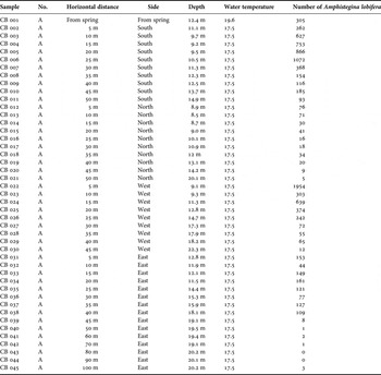
Abnormally high abundance of Amphistegina lobifera Larsen specimens observed on the south and west of the study area is noteworthy. It shows variable distribution in the sampling locations according to the direction and distance from the spring (Table 3). The diatom analysis revealed that there is diverse and rich community of diatoms around and near the springs. Many of the benthic foraminifer species feed on diatoms and it has been known that larger foraminifers, such as aphisteginids live together with endosymbiont diatoms (Lee et al., Reference Lee, McEnery, Ter Kuile, Erez, Röttger, Rockwell, Faber and Lagziel1989), suggesting that the abundance of diatoms around the spring may serve as an important food source for Amphistegina lobifera Larsen. An increase is observed in Elphidium crispum (Linné) and Ammonia compacta Hofker populations as Amphistegina lobifera Larsen populations decrease. The abundance of Coscinospira hemprichii Ehrenberg, Peneroplis planatus (Fichtel & Moll), Lobatula lobatula (Walker & Jacob), Cibicidella variabilis (d'Orbigny) with abnormal test morphologies and Spiroloculina angulosa Terquem individuals with double apertures in the study is noteworthy.
Direction, distance, depth and water temperature measurements, and the distribution of Amphistegina lobifera Larsen individuals among sediment samples.

Diatom assemblage
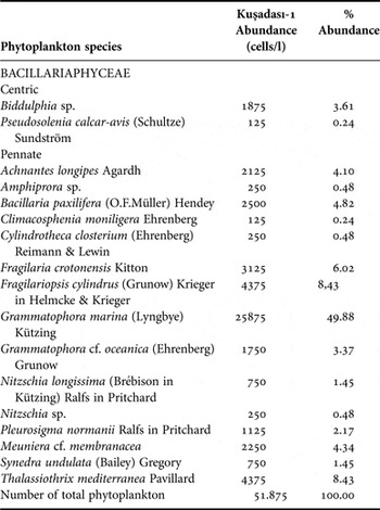
The analysis of the water which was collected from Kuşadası-1 spring on 6 June 2007 revealed that all the phytoplankton community is composed of diatoms. Centric planktic species constituted 4% of the community and the rest were pennate benthic. Grammatophora marina (Lyngbye) Kützing was found to be the dominant species, forming 50% of the community. Fragilariopsis cylindrus (Grunow) Krieger in Helmcke & Krieger and Thalassiothrix mediterranea Pavillard were the other dominant species (Table 4).
Abundance and distribution patterns of phytoplankton species, observed in the water sample collected from the submarine spring on 6 June 2007.

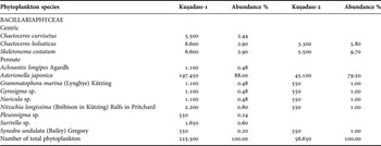
The second sampling for phytoplankton analysis was performed on 6 November 2008 and again only diatoms were found. Asterionella japonica Cleve was found to be the dominating species with 88% in Kuşadası-1 spring. 8% of the community was centric planktic and 92% was pennate benthic. A water sample from Kuşadası-2 spring was also analysed. The abundance of the diatoms was found to be 25% of the community observed around Kuşadası-1 spring. Morphological analysis showed that 12% of this community was composed of centric and 88% was pennate (Table 5). The cosmopolitan species Asterionella japonica Cleve which is known to inhabit both cold and temperate waters was also found to be the dominating species with an abundance of 80% (Cupp, Reference Cupp1943; Hasle et al., Reference Hasle, Syvertsen, Steidinger, Tangen, Throndsen, Heimdal and Tomas1997).
Abundance and distribution patterns of phytoplankton species, observed in the water sample collected from the submarine spring on 6 November 2008.

NO2 + NO3 (ppb), PO4 (ppm), P (ppb) and Si (ppm) analysis of the spring waters showed that Kuşadası-2 was more rich in nutrients; however, the diatom community was more diverse and rich around Kuşadası-1 spring.
Chemical and physical characteristics of the spring waters
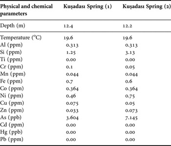
Chemical composition (Al, Si, Ti, Cr, Mn, Fe, Co, Ni, Cu, Zn, As, Cd, Hg and Pb) of the submarine springs is given in Table 6. The springs are 5 m apart and thus have similar chemical characteristics. When compared with Krauskopf (Reference Krauskopf1979), it is seen that Al, Cr, Mn, Fe, Co, Ni, Cu and As values are higher in both of the springs, whereas, Si and Zn are found to be higher only in the second spring. If the spring waters are compared, Kuşadası-1 spring has higher Cr, Fe and Cu values and lower values for Ni, Zn, Si and As. The absence of detectable amounts of Hg, Cd and Pb in both of the springs is in accordance with Krauskopf (Reference Krauskopf1979).
Physical and chemical characteristics of the submarine spring waters from Pamucak Cove.

Currents
The analysis of surface currents revealed that the current system observed in the Gulf of Kuşadası did not fit the general current system of the Aegean Sea. Regularly blowing winds produce local currents along the coasts. The local current system around the springs carries the spring water in south and south-western directions (Figures 12 and 13).
Seasonal directions and speeds of the currents observed in Station KA-01.

Seasonal directions and speeds of the currents observed in Station KA-02.

DISCUSSION
A typical Mediterranean foraminifer fauna was observed around the investigated spring. Ammonia compacta Hofker & Elphidium crispum (Linné) were found to be the highly dominating species on the eastern line. On the other hand, the observation of Indo-Pacific originated species Quinqueloculina sp. C, Triloculina sp. A, Euthymonacha polita (Chapman), Pyramidulina catesbyi (d'Orbigny), Brizalina simpsoni (Heron-Allen & Earland), Amphistegina lessonii d'Orbigny for the first time on the Turkish coastline is noteworthy. Amphistegina lessonii d'Orbigny was previously reported from north-west of Crete Island (Hollaus & Hottinger, Reference Hollaus and Hottinger1997) and Andros Island in the middle Aegean Sea (Triantaphyllou et al., Reference Triantaphyllou, Tsourou, Kouskousioura and Dermatitzakis2005). Haddonia sp. and Cymbaloporetta plana (Cushman), which are abundant on the south-western coast of Turkey, Iridia diaphana Heron-Allen & Earland which is recorded in Ayvalık (North Aegean), were represented in the sediment with few individuals. Nodopthalmidium antillarum (Cushman) which was known from the Gulf of Iskenderun and Adelosina carinata-striata Wiesner which was rarely observed on the Aegean coasts were found to be abundant in the study area.
The most interesting finding about the alien species is the distribution pattern of Amphistegina lobifera Larsen observed around the spring. It is highly abundant in the centre and in close proximity to the spring; however, the abundance decreases away from the centre (Meriç et al., Reference Meriç, Yokeş, Avşar and Bircan2010). The distribution pattern also shows differences according to the direction (Table 3). Similar abundances were reported from the south-western Mediterranean coasts of Turkey (Meriç et al., Reference Meriç, Avşar and Yokeş2008c), but not observed in Amphistegina lobifera Larsen populations found in Marmaris, Datca, Fethiye, Gokova and south-east of Gokceada on the Aegean coasts of Turkey. In a recent study (Avşar et al., Reference Avşar, Meriç, Çevik and Dinçer2009), only a single Amphistegina lobifera Larsen individual has been found in 20 sediment samples collected from south-west of Dilek Peninsula. This abnormal abundance of alien species and the presence of coloured foraminiferal tests in the sediment suggest that the submarine spring might have created special ecological conditions, such as warm temperature, high heavy metal content and low salinity.
Geochemical analysis of the recent sediments in Pamucak Cove has been previously investigated (Eyrik, Reference Eyrik2006). In the present study heavy metal and trace element contents of the 45 recent sediment samples were analysed; Al, Ti, Fe, Ca, Mg, K, Na, P, Mn, Ba, Sr, Zr, V, Cr, Rb, Ni, Zn, Cu, Ce, Y, La, Co, Li, Sc, Nb, Pb, Th, Hf, U, Sn, Ta, As, W, Cd, Ag and Au were found. Al, Ti, Fe, Ca, Mg, K, Na, P values were detected in %, and the rest of the elements were detected at ppm level. Fe (16.45%), Ti (3.08%) and Mn (6346 ppm) were found to be high; however, the values of the other elements were in accordance with their percentages observed in sedimentary rocks and in the Earth's crust. It is also noteworthy that although Ti is found in the sediments, it is not observed in the spring waters. The presence of yellow, brown and dark grey coloured foraminiferal tests around the spring suggests that heavy metals such as Fe, Mn and Ti play a role in test coloration (Eyrik, Reference Eyrik2006).
In winter, the water mass moving in south–north direction through the western coast of the Aegean Sea is blocked by Samos Island. Thus, the general current system slows down and turns to west–east direction. This water mass changes its direction when it reaches the small capes located on the north of the gulf and creates coastal currents. On the east of the gulf the current moves clockwise towards the Dilek Strait. This current pattern changes according to meteorological events. Counterclockwise currents can be formed due to strong southern winds. High evaporation rate during summer times results in the formation of considerable salinity differences between the upper and lower water layers. The density and temperature differences between layers results in the formation of local currents on the shoreline, which are more effective in the inner parts of the gulf. Besides, regularly blowing winds with west–east direction produce local currents along the coasts (Figures 12 and 13) (Rapor No. 239, 1989; Rapor No. 250, 1990; Eryılmaz et al., Reference Eryilmaz, Yücesoy-Eryilmaz, Dogan, Yüce and Bayrakatar1998; Eryılmaz & Aydın, Reference Eryilmaz and Aydin2001). The local current system around the springs carries the spring water in south and south-western directions. The distribution pattern of the Amphistegina lobifera Larsen individual can be explained by this current system and accordingly the diatom distribution patterns.
ACKNOWLEDGEMENTS
The authors thank Murat Dündar (R&D Manager, Assan Company) and Hüsnü Öztürk (Technician, Assan Company) for SEM (Jeol JSM-5600) photography, Dr Vildan Tüfekçi (TÜBİTAK MAM, Institute of Chemistry & Environment) for nutrient analysis of the spring waters and Ertuğrul Çanakçı (Çukurova University, Department of Geological Engineering) for chemical analysis of the spring waters.