Case report
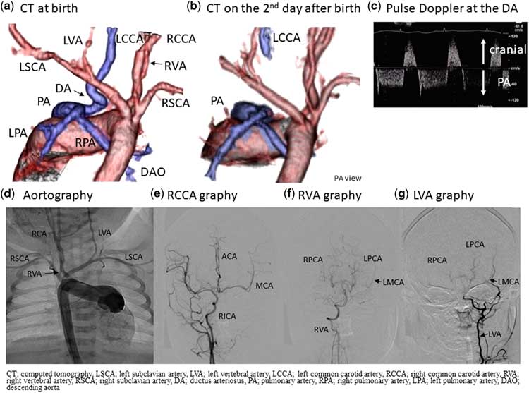
A fetus was diagnosed with a double-outlet right ventricle, pulmonary stenosis, and a right aortic arch by fetal echocardiography. A female baby was born at full term with a body weight of 3379 g and oxygen saturation of 97%. She had anovulvar fistula, without any other abnormality suspected of congenital malformation syndrome. Her echocardiography and CT (Fig 1a) on the day of birth revealed tetralogy of Fallot, crossed pulmonary arteries (Fig 1c), and infundibular and valvular pulmonary stenosis with a bicuspid pulmonary valve. The right aortic arch gave rise to the right carotid artery; second, the right subclavian artery; and third, the left subclavian artery. The left common carotid artery originated from the main pulmonary artery via a tortuous vessel, which was thought to be the ductus arteriosus with slight narrowing and bidirectional blood flow (Fig 1c). There was no ductus arteriosus present from the aortic arch. Both sides of the vertebral arteries originated from each subclavian artery. The circle of Willis was detected by cranial ultrasound and CT. Regarding the left common carotid artery circulation, we decided to observe it without surgery or prostaglandin E1 infusion. The vessel connecting the left common carotid artery and pulmonary artery was closed the next day, indicating that the vessel was ductus arteriosus tissue (Fig 1b). Cerebral angiography on the eighth day of birth (Fig 1d–g) and at an age of 9 months before surgery revealed that the left brain was perfused via the circle of Willis, and no laterality was detected. Her brain single photon emission CT image and electroencephalogram were within normal ranges during the neonatal and infantile period.
( a ) A CT on the day of birth (posteroanterior view) The left common carotid artery (LCCA) arose from the pulmonary artery (PA) via a tortuous and narrow vessel. The right aortic arch gives rise to the right common carotid artery; second, a right subclavian artery (SCA); and third, a left SCA. ( b ) A CT on the second day of birth (posteroanterior view) The tortuous vessel connecting the PA and LCCA disappeared, which was thought to be a ductus arteriosus. ( c ) Pulse Doppler at the ductus arteriosus. A pulse Doppler at the connecting vessel between the main pulmonary artery and LCCA exhibits bidirectional flow. ( d ) Aortography on the eighth day of birth The right aortic arch gives rise to the right common carotid artery, right SCA, and left SCA. Both sides of the vertebrae arteries originate from each SCA. An LCCA from the aortic arch is absent. ( e ) Frontal projection of the right common carotid arteriography The left anterior cerebral artery and middle cerebral artery (MCA) were supplied via a patent anterior communicating artery. ( f ) Frontal projection of the right vertebral arteriography The right vertebral artery supplies both the right and left posterior cerebral arteries. ( g ) Frontal projection of the left vertebral arteriography The left vertebral artery supplies both the right and left posterior cerebral arteries, and a part of the left MCA via a posterior communicating artery.

The patient underwent a tetralogy of Fallot repair at 9 months of age. During the operation, the ligamentum from the main pulmonary artery to the left common carotid artery was identified and resected. She had a good general condition and no delay in her physical growth or development was noted at 19 months of age. Her karyotype and results for fluorescence in situ hybridisation analysis for a 22q11 deletion were found to be normal.
Discussion
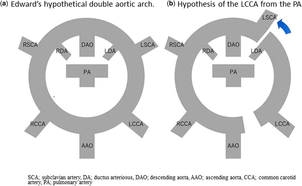
An isolated left common carotid artery from the main pulmonary artery is very rare. To the best of our knowledge, only 13 cases have been reported so far.Reference Fong and Venables 1 – Reference Osakwe, Jones and Hirsch 4 All the reported cases showed three characteristics, which are connection of the isolated left common carotid artery to the pulmonary artery, presence of right aortic arch, and presence of aberrant left subclavian artery. It was first reported by Fong and Venables in 1987.Reference Fong and Venables 1 They postulated that according to Edward’s hypothesis, the distal migration of the left subclavian artery beyond the ductus arteriosus with a subsequent disappearance of two different segments between the ascending aorta and left common carotid artery, and between the left subclavian artery and left ductus arteriosus, would produce the right aortic arch, an aberrant left subclavian artery, and an isolated left common carotid artery (Fig 2). However, there was a report that the histology of small segment of the left carotid artery resected before anastomosis showed the absence of ductal tissue; therefore, it remains controversial whether the vessel between the left common carotid artery and pulmonary artery is ductal tissue.Reference Fouilloux, Gran and Kreitmann 2 Our case was the first report tracing the closing process of the vessel between the left common carotid artery and pulmonary artery just after birth, which suggested it was ductal tissue. This finding supports the above hypothesis.
( a ) Edward’s hypothetical double aortic arch ( b ) Hypothesis of the development of the left common carotid artery (LCCA) from the pulmonary artery (PA) Interruption of the left posterior aortic arch and migration of the left subclavian artery (SCA) beyond the left ductus arteriosus results in an aberrant left SCA. Another interruption between the ascending aorta and LCCA results in the isolation of the LCCA.

Significant associated congenital cardiac defects were also often present in cases with isolated left common carotid artery from the pulmonary artery: primum atrial septal defect,Reference Gil-Jaurena, Ferreiros, Zabala and Cuenca 5 secundum atrial septal defect, tetralogy of Fallot,Reference Idhrees, Mathew, Menon, Dharan and Jayakumar 6 persistent left supra vena cava,Reference Kaushik, Saba and Rosenfeld 7 aortic atresia,Reference Ahmadi, Sabri and Dehghan 8 Ebstein anomaly,Reference Osakwe, Jones and Hirsch 4 CHARGE syndrome,Reference Fong and Venables 1 , Reference Osakwe, Jones and Hirsch 4 DiGeorge syndrome,Reference Oppido, Pace Napoleone and Gabbieri 9 or hemifacial abnormality.Reference Kaushik, Saba and Rosenfeld 7 Our case had anovulvar fistula, without any other abnormality including coloboma, genital anomaly, and ear abnormality, and she had normal karyotype and fluorescence in situ hybridisation result for 22q11 deletion. The patient had a bicuspid pulmonary valve and a crossed pulmonary artery. There was no description of bicuspid pulmonary valve in previous reports about isolated left common carotid artery from pulmonary artery. Whereas a crossed pulmonary artery is a rare form of pulmonary artery malposition, half of which is associated with genetic syndrome such as DiGeorge, Noonan, Holt-Oram, VACTERL syndrome.
It was difficult to decide the best course of treatment regarding whether the ductus arteriosus should be kept open or not, and whether an anastomosis between the left common carotid artery and aortic arch was, or was not, required during the neonatal period, or later. We were fearful of the possibility of left-brain ischemia, stenosis of the anastomosis, and steal phenomenon. We decided to close the ductus arteriosus after we evaluated the left brain and pulmonary artery circulation.
Acknowledgements
The authors would like to thank our colleagues who contributed to the care of this patient, and we would especially like to thank Dr Shigemitsu Iwai, head pediatric cardiac surgeon, who performed the surgery.
Financial Support
This case report received no specific grant from any funding agency, or from commercial or not-for-profit sectors.
Conflicts of Interest
None.