Introduction
The prostate is a fibromuscular exocrine gland, found exclusively in mammals as an accessory sex organ in the male reproductive system. In adult man, this fibromuscular exocrine gland is a small horse chestnut-shaped organ, surrounding the urethra at the base of the bladder, measuring approximately 3 cm x 4 cm and weighing approximately 18–20 g. Its growth and development are dependent on male steroid hormones, also called androgens. Prostatic secretions contribute to the ejaculate, producing a thin milky fluid, containing high levels of citrate, zinc, polyamines and a number of both anticoagulant and proteolytic enzymes, including the serine protease Kallikrein 3, more commonly known as PSA (prostate specific antigen), which plays a role in semen liquefaction. The lobular structure of the prostate, although visible in the developing prostate, is not readily apparent in the mature male. This has led to the identification of regions or ‘zones’ of the prostate based on their predisposition to various pathological processes.Reference McNeal1 These zones are demonstrable using transrectal ultrasound (TRUS), providing a more clinically useful terminology when compared with gross anatomical nomenclature. Three distinct zones and the anterior fibromuscular stroma have been identified: the central zone occupies 25% of the glandular prostate tissue and malignancies originating from this zone are rare; the transition zone can be visualized to surround the urethra, like a cuff, at the junction of the distal and proximal urethra, accounting for only 5–10% of the glandular tissue, but approximately 20% of prostate malignancies originate from this zone, and it is the principle site for benign prostatic hyperplasia; finally the peripheral zone occupies 70% of the glandular tissue and the majority of prostatic carcinomas occur in this zone.
Endocrine control of prostate growth
Prostatic growth is induced by a variety of hormones and growth factors. The most important is testosterone, which is converted into the active androgen dihydrotestosterone (DHT). Testosterone is produced in the Leydig cells of the testis from pregnenolone and converted by 5α-reductase to DHT. The hypothalamus controls androgen secretion by gonadotrophin-releasing hormone (GnRH or LHRH), which causes the pituitary to release luteinizing hormone (LH). LH acts on the Leydig cells of the testes to stimulate steroid synthesis and the production of testosterone. At least 95% of testosterone is produced by the testes; the other 5% is converted from adrenal androgens such as androstenedione peripherally. The plasma half-life of testosterone is only 10–20 minutes. DHT or testosterone binds to the androgen receptor affecting growth, differentiation and function of the prostate. DHT is 2.5 times more potent than testosterone, having greater influence on growth and secretory activities.
Carcinoma of the prostate
Prostate cancer is the most common cancer in men in the UK, accounting for nearly a quarter (22%) of all new male cancer diagnoses, and the second leading cause of cancer death in men after lung cancer (Cancer Research UK website, www.cancerresearchuk.org). In England and Wales, there were 13,481 cases registered in 1990, rising to 34,302 new cases in 2005 with 10,000 prostate cancer deaths. Whereas early preneoplastic lesions are thought to occur with a similar incidence worldwide, the global incidence of clinically detected prostate cancer varies considerably.Reference Dhom2 Geographically, the highest and lowest reported incidences are from Scandinavia (60 per 100,000 per year) and the Far East (in particular mainland China and Japan: 4 per 100,000), respectively.Reference Kirby, Christmas and Brawer3 In addition, there are wide racial and ethnic differences in incidence. The highest reported incidence of prostate cancer is amongst black men living in Atlanta, Georgia in the USA (91.2 per 100,000). African Americans tend to present at a younger age with more advanced disease. In addition, autopsy studies have revealed that this group harbours a larger volume of incidental disease and a greater number of pre-malignant foci.Reference Sakr, Grignon, Haas, Heilbrun, Pontes and Crissman4 The reason for this high incidence is not clear, though exposure to higher levels of intrauterine testosterone and a higher level of serum testosterone in adolescence may be contributory.Reference Ross5 An interesting disparity in prostate cancer incidences exists between racial groups remaining in their native countries and migrants of the same group in countries with a higher endemic rate. Indeed, incidence is increased almost threefold in Japanese immigrants in San FranciscoReference Cook, Goldoft, Schwartz and Weiss6 and HawaiiReference Akazaki and Stemmerman7 relative to their counterparts remaining in Japan.
Prostate cancer is diagnosed in very few people aged younger than 50 years (<0.1% of all patients). The mean age of patients with this disorder is 72–74 years, and about 85% of patients are diagnosed after age 65 years. Results of autopsy studies suggest that most men aged older than 85 years have histological prostate cancer. In an elegant study of 600 men in Detroit, USA, the rate of latent prostate cancer was high at all ages: 30% for men in their 30s, 50% for men in their 50s and more than 75% for men older than 85 years.Reference Sakr, Grignon, Haas, Heilbrun, Pontes and Crissman4 Due to increasing life expectancy, the incidence of prostate cancer has steadily increased over the past 50 years.
Aetiology
Prostate cancer is thought to result from a complex interplay between ageing, genetic factors, endogenous hormones, growth factors and the environment, including a growing body of evidence incriminating dietary fat.Reference Pienta and Esper8 The only consistent risk factors for prostate cancer are the life-long presence of androgens, age, race and a positive family history. The importance of androgens in prostate oncogenesis may be demonstrated by the lack of disease development in men castrated before puberty, or who are deficient in 5-α reductase.Reference Ross, Bernstein, Lobo, Shimizu, Stanczyk and Pike9 Race represents another important predisposing factor for the development of prostate cancer. Further to the evidence outlined above, North American blacks have approximately twice the lifetime risk of the disease compared with their white counterparts.Reference Pienta and Esper8 Prostate cancer can be sporadic; familial with clustering of disease within families and exposure to common risk factors; or hereditary, with typical characteristics of early age onset and an autosomal dominant inheritance pattern. The latter is likely to be triggered by a single gene passed along families, yet to be discovered and possibly located on chromosome 1.Reference Smith, Freije, Carpten, Gronberg, Xu and Isaacs10 It has been estimated that men who have three first-degree relatives diagnosed with prostate cancer have a 10.9-fold increase in risk of developing the disease.Reference Carter, Bova, Beaty, Steinberg, Childs and Isaacs11 An increased risk of prostate cancer has also been associated with familial breast cancer.Reference Pienta and Esper8 The observed differences in worldwide incidence and the increased frequency of prostate cancer in Japanese immigrants in the USA imply that a number of environmental factors may be responsible for increased prostate cancer susceptibility: large consumption of red meat has been linked to prostate cancer.Reference Hayes, Ziegler, Gridley, Swanson, Greenberg and Swanson12 One hypothesis is that cooking at high temperatures (e.g. charcoal grilling or frying) results in formation of very potent carcinogens such as heterocyclic amines, which have been correlated with colorectal, bladder and kidney cancer.Reference Augustsson, Skog, Jagerstad, Dickman and Steineck13 Another laboratory study reported that growth of tumours from prostate cancer cells in mice can be retarded by feeding them a diet containing less than 20% fat.Reference Wang, Corr, Thaler, Tao, Fair and Heston14 Men with a body mass index (BMI = weight in kg/(height in m)2) of over 26 kg/m2 are at significant increased risk of developing the disease.Reference Snowdon, Phillips and Choi15 Western countries, which have diets including higher quantities of red meat and fat intake, have a higher incidence of prostate cancer and prostate metastases than Eastern countries with low fat intake. Lycopenes (present in tomatoes) have been shown to reduce the risk of prostate cancer development in a large study involving 47,000 men.Reference Giovannucci, Ascherio, Rimm, Stampfer, Colditz and Willett16 Lycopene is a powerful naturally occurring antioxidant, which scavenges free radicals. It appears to inhibit cancer cell growth by interfering with growth factor receptor signalling and cell cycle progression.Reference Heber and Lu17 Other factors such as smoking, alcohol consumption, vasectomy and physical activity have been investigated in several studies, but the overall conclusion is that they do not affect the risk of prostate cancer.
Pathology
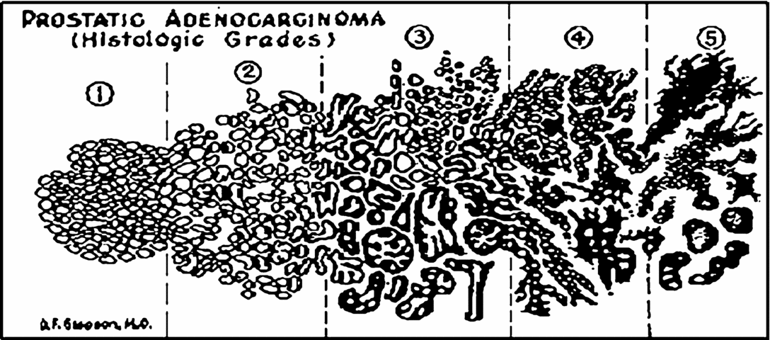
Adenocarcinoma arising from the prostatic epithelium accounts for about 95% of prostatic malignancies and is usually composed of small glandular acini that infiltrate in an irregular haphazard manner. The critical feature in prostatic adenocarcinoma is an absence of the basal cell layer, which may be detected immunohistochemically by using monoclonal antibodies against high molecular weight cytokeratin. Transition zone tumours arise in or near foci of benign prostatic hyperplasia (BPH) and are usually smaller and better differentiated. Peripheral zone cancers are often less well differentiated, larger in volume than transition zone tumours, and are frequently associated with greater stromal fibrosis, extracapsular extension, seminal vesicle invasion and lymph node metastases. The histological differentiation of prostate cancer is widely assessed using the Gleason grading score system, devised in the 1960s.Reference Gleason18 This system uses low-power architectural findings on microscopy to define the histological pattern of the tumour. Gleason grading identifies five levels of increasing disease aggressiveness, ranging from simple round glands, closely packed with well-defined margins (Grade 1) to ragged, infiltrating undifferentiated tissue with small gland size and anaplastic sheets of epithelium (Grade 5)Reference Gleason19 (Figure 1). The Gleason grading system accounts for the multifocal and heterogeneous nature of prostate cancer by assigning an overall score (2–10) based on the sum of the two most prevalent grades present within the tumour. This sum score serves as a useful prognostic indicator of disease progression, having been linked with a number of clinical end-points including clinical stage, progression to metastatic disease and survival (reviewed by Reference Humphrey20).
Original drawing of Dr Gleason's, showing the five distinct patterns of the Gleason grading system. The two most prevalent Gleason grades are added together to give the Gleason score, where 2 is the most differentiated and 10 is the least differentiated. Image available at: http://www.phoenix5.org/Infolink/GleasonGrading.html (adapted from Gleason DF. The Veteran's Administration Co-operative Urologic Research Group: histologic grading and clinical staging of prostatic carcinoma. In Tannenbaum M (ed), Urologic Pathology: The Prostate. Lea & Febiger, Philadelphia, 1997, pp. 171–198.)
Gleason grades 1 and 2: these two grades closely resemble normal prostate. They are the least important grades because they seldom occur in the general population and because they confer a prognostic benefit that is only slightly better than grade 3. In grade 2, glands are more loosely aggregated, and some glands invade the surrounding stroma.
Gleason grade 3: this is the most common grade and is also considered well differentiated (like grades 1 and 2). All 3 grades have normal glandular appearances. The lumen contains prostatic secretion like normal prostate, and each gland unit is surrounded by prostate muscle, which keeps the gland units apart. Invasion into the stroma is very prominent and is the main defining feature. The cells are dark rather than pale and the glands often have more variable shapes.
Gleason grade 4: Here there is a loss of architecture with disruption and loss of the normal gland unit. This is the main class of poorly differentiated prostate cancer, and its distinction from grade 3 is the most commonly important grading decision.
Gleason grade 5: this usually predicts another significant step towards poor prognosis. It is less common than grade 4, and it is seldom seen in men whose prostate cancer is diagnosed early in its development. This grade too shows a variety of patterns, all of which demonstrate no evidence of formed glands. This grade is often called undifferentiated, because its features are not significantly distinguishing to make it look any different from undifferentiated cancers that occur in other organs.
Diagnosis of prostate cancer
Digital rectal examination (DRE)
DRE remains the primary test for the initial clinical assessment of the prostate. It has the benefit of detecting non-PSA-secreting tumours, though DRE is a test with only fair reproducibility, even in the hands of experienced examiners. It ‘misses’ a substantial number of tumours and, compared with PSA, detects cancers at a more advanced pathological stage.
Prostate specific antigen
The measurement of serum PSA concentrations is now well established as a useful tool in the diagnosis and follow-up of patients with prostate cancer.Reference Lilja, Ulmert and Vickers21 If measured before the age of 50 years, PSA levels appear to be a strong predictor of the incidence of prostate cancer later in life.Reference Lilja, Ulmert, Bjork, Becker, Serio and Nilsson22 The greatest limitation of PSA is that it is tissue and not tumour specific in the prostate. PSA can be raised with infection, following ejaculation and with benign prostatic disease. For many years, the ‘normal’ PSA threshold below which men were not recommended prostate biopsy varied between 3 and 4 ng/ml in an age group starting at approximately 50 years. However, results from the Prostate Cancer Prevention Trial (PCPT) in men receiving the 5-α reductase inhibitor finasteride versus placebo for prostate cancer chemoprevention, showed through end-of-study biopsies that 15% of men with PSA levels less than 4 ng/ml had prostate cancer.Reference Thompson, Pauler, Goodman, Tangen, Lucia and Parnes23 There is therefore no PSA threshold below which a man can be told that he does not have prostate cancer histologically. In clinical practice, age-related PSA cut-offs are used to account for the contribution of benign prostatic hyperplasia in the elderly man. In 2002, the Prostate Cancer Risk Management Program recommended a cut-off value of 5.0 ng/ml for men over the age of 70, leading to less invasive biopsies in the elderly population. It is very important to stress that the decision to test PSA levels must be individualized following appropriate counselling, and should also include DRE. There is no place for PSA measurement as a ‘routine’ test in the elderly man, admitted to the medical ward with collapse or confusion, except in the presence of an undiagnosed cancer with painful bony metastases, or a positive bone scan.
Transrectal ultrasonography (TRUS) and biopsies
TRUS has established its place in the detection of cancer by its ability to accurately guide needle biopsies and map them to specific regions of the prostate. Patients may find the procedure uncomfortable or occasionally painful, so now local anaesthetic is used routinely, particularly with the larger number of cores now taken. It takes 5–10 minutes, allowing for 10–12 needle core biopsies to be taken. The indications for TRUS-guided biopsies include abnormal DRE, an elevated PSA or increasing PSA, and previous biopsies showing isolated prostatic intra-epithelial neoplasia (PIN) or atypical small acinar proliferation (ASAP). Exceptions include elderly or frail men with massively elevated PSA and abnormal DRE. Histological diagnosis is not always a necessity in these men. The historical sextant protocol (a para-sagittal base, mid-gland and apex from each side) has been superseded by the technique of performing 8, 10 or 12 biopsies, adding samples from the far lateral peripheral zones. Studies have shown that these extra biopsies detect up to 15% more cancers. Additional biopsies of the transition zone may be taken if a transition zone cancer is suspected or if a patient is undergoing repeat biopsies because of an increasing PSA. Complications of prostatic biopsy include urinary tract infection, requirement for appropriate antibiotics and occasional septicaemia (<1%) necessitating hospital admission. Bleeding (rectal, urinary and/or haemospermia) is usually self-limiting, but very occasionally may be severe and require hospital admission and/or transfusion. Haemospermia may persist for up to 6 weeks. Biopsy may precipitate voiding difficulty or urinary retention. It should be noted that it is not safe to biopsy patients when they are on warfarin or have coagulopathies. Also, it is important for the patient to appreciate that negative biopsies do not exclude the possibility of prostate cancer and that a positive result will not necessarily result in the recommendation of immediate treatment.
Screening for early prostate cancer
The classic triad of detection tests consists of serum PSA measurement, DRE and TRUS and biopsies, which detects up to 6% of a screened population as having prostate cancer. Based on the available evidence, there has been an apparent consensus that there is no justification in introducing population screening for prostate cancer. However, recently this consensus has been put to question, by published results from the European Randomized Study of screening for Prostate Cancer.Reference Schroder, Hugosson, Roobol, Tammela, Ciatto and Nelen24 In this large study with a median follow-up of 9 years, 72,952 men aged 55–69 years were randomized to screening and there were 89,435 age-matched controls. At follow-up to date, there was a relative risk reduction of 0.80 for dying of prostate cancer, and in absolute terms, 1410 men needed to be screened, and, of these, 48 men needed to receive treatment in order to prevent one man from dying of prostate cancer. So despite the fact that screening has now been shown to reduce prostate cancer mortality, it appears to be at a significant cost of over-detection and over-treatment. The age of the man being screened is of high improtance. At age 50, the chance of the cancer being clinically irrelevant is likely to be less than 20%, but the risk of dying prematurely (before 80 years) of prostate cancer is 60%. At age 70 years, approximately one-half of all cancers detected will be clinically irrelevant, but the risk of dying prematurely as a result of the cancer is still 38%. At the age of 75 years, two clinically irrelevant cancers will be diagnosed for every significant one, and the risk of dying prematurely as a result of the cancer is likely to have fallen to a level below 20%.
Clinical presentation
In 1530, Antonio Ferri described for the first time the role of prostate tumours in causing bladder outlet obstruction, and eight years later Vesalius provided the first anatomical drawings of the prostate. In 1649, Riolan described a prostate tumour and Billie first reported prostatic carcinoma in 1794. In 1786 the English anatomist John Hunter discovered what could be thought of as the first major breakthrough in understanding prostate cancer. He demonstrated that removing the testicles from young male animals prevented the growth of the prostate. In 1847 Benjamin Brodie first described prostatic carcinoma and metastasis to the vertebrae:
‘We found the patient complaining of excruciating pains in various parts of the body, which could be compared to nothing except the pains under which persons afflicted with carcinoma occasionally labour. He could void no urine without the assistance of a catheter. The prostate gland, examined by the rectum was found to be much enlarged and of a stony hardness. I continued to visit him in consultation for nearly a year, at the end of which time he suddenly lost the use of the muscles of his lower limbs and died a fortnight afterwards.’
Early prostate cancer is asymptomatic. The above illustrates all the relevant symptoms in advanced prostate cancer. Patients with symptomatic disease can present in a variety of ways, including bladder outflow obstruction, irritative bladder symptoms secondary to trigonal involvement and haematuria. Metastatic disease may present with skeletal pain, spinal cord compression, or with general systemic manifestations, including weight loss, weakness and anorexia. With locally advanced disease, prostate cancer may manifest itself as renal failure secondary to bilateral ureteric obstruction.
Treatment of prostate cancer
Management of prostate cancer is largely dependent on the stage and grade of the tumour, although other factors include patient's age and general health, and urologists look for a 15-year life expectancy before considering radical, curative treatments. Patient's preference also plays a significant role. For the sake of simplicity, prostate cancer can be divided into early organ-confined and advanced disease, including castrate-resistant prostate cancer.
Treatment of organ-confined disease
Organ-confined disease refers to prostate tumours in stages T1 and T2. Treatment of clinically localized prostate cancer is aimed at complete eradication of disease or minimizing tumour progression for the lifetime of the patient. A variety of treatment options are currently available for organ-confined disease, including conservative management, surgical intervention and radiotherapy.
Conservative treatment includes ‘Watchful Waiting’, where the patient is given no treatment while he remains asymptomatic, but monitoring him every 6–12 months for onset of symptoms of metastases, particularly bone pain, when palliative hormonal therapy is offered to patients. Curative treatment is not the intention. Conservative treatment also includes ‘Active Surveillance’, which involves regular 3-monthly clinical review with PSA measurement and digital rectal examination. If the PSA concentration rises repeatedly over a year, the patient can be offered radical treatment, while the disease is still potentially curable.
Radical prostatectomy involves the removal of the entire prostate together with the attached seminal vesicles, and the bladder neck is anastomosed to the remaining urethra. Laparoscopic and robot-assisted approaches have gained popularity in recent years, and this trend will certainly continue. In an observational study involving 1000 men aged around 31–81 years treated with radical prostatectomy, the 5-year cancer-free survival was 89.4% for men with Gleason scores ≤ 4, 84% for scores of 5–6, 59.9% for a score of 7 and 48.7% for scores of ≥ 8.Reference Hull, Rabbani, Abbas, Wheeler, Kattan and Scardino25 A high risk of post-surgical complications is associated with this procedure. Indeed, unwanted effects of radical prostatectomy include erectile dysfunction (in up to 60% of patients), incontinence (8–33%), severe haemorrhage and rectal injury.Reference Kaufman, McDougal, Zietman, Harisinghani and Young26
External beam radiotherapy (EBRT) is given on an out-patient basis over about 8 weeks. This form of therapy has similar progression-free and overall survival rates to those seen with surgery. In a non-randomized study of such therapy, the 5-year disease-free survival was 85% for patients with a pre-treatment PSA concentration of up to 10 ng/ml and Gleason scores of up to 6, compared with 65% for those with a higher value for one of these indicators, and 35% for those with both indicators with high values.Reference Zelefsky, Lyass, Fuks, Wolfe, Burman and Ling27 However, EBRT is associated with late chronic side-effects, such as erectile dysfunction (in up to 50% of patients), rectal bleeding (7%) and various degrees of urinary incontinence (2–3%), which limit the dose of radiation that can be administered. A recent modification of EBRT is conformal radiotherapy. In this approach the irradiated volume is matched to the tumour volume in order to reduce late chronic side-effects associated with standard radiotherapy. High levels of irradiation of the tumour tissue are thus achieved, with reported low morbidity.Reference Dearnaley, Khoo, Norman, Meyer, Nahum and Tait28
Brachytherapy (‘brachys’ being a Greek word for a ‘short distance’) involves the permanent placement of a radioactive source (most commonly iodine-125 or palladium-103 seeds) transperineally into the prostate under TRUS guidance. This treatment helps to minimize the radiation to the rectum and adjacent neurovascular structures. The risk of long-term unwanted effects is low, with proctitis in 1–2% of patients and incontinence in fewer than 2% at 12 months.Reference Henderson, Bott, Sooriakumaran, Laing and Langley29 There is as yet no conclusive evidence that brachytherapy is comparable or more effective than other radical therapies, though there is documentation from large single-institutional and multi-institutional series on patients with disease category PSA < 10, GS ≤ 6, ≤ T2b, showing that the outcome of external beam radiotherapy and brachytherapy in this group of patients is similar to those of surgery.Reference Nilsson, Norlen and Widmark30
Cryotherapy is the in situ destruction of tumours by the application of low temperatures. Initial attempts proved disappointing, but the ability of real-time TRUS to guide cryoprobe placement and accurately monitor the freezing process, in addition to the development of urethral warming devices, encouraged clinicians to attempt again the destruction of prostate cancer by freezing. High intensity focused ultrasound (HIFU) consists of delivering ultrasonic energy with resultant heat and tissue destruction to a discrete point without damaging intervening tissue. Very high temperatures are generated at the focal point using HIFU (approximately 98°C), leading to complete tumour necrosis. Currently neither cryotherapy nor HIFU are recommended as treatment for prostate cancer outside of a clinical trial.
Treatment of advanced disease
Treatment for advanced prostate cancer is aimed at temporary disease control and palliation. It focuses on prolonging life expectancy, reducing the risk of metastasis and tumour invasion in locally advanced disease and improving quality of life in patients with distant metastases by controlling or preventing symptoms of disease. Hormonal therapy is based on androgen deprivation and mediates its effects by disrupting androgen-dependent survival and proliferation of prostate tumours. Hormonal therapy is not intended to be curative and is therefore a palliative option only. Androgen deprivation can be achieved in a number of ways and these will be outlined below. Medical castration involves the use of LHRH agonists, molecules that potently mimic the effects of natural LHRH and include goserelin acetate (Zoladex®), buserelin, leuprorelin and tryptorelin. These molecules bind to the LHRH receptor, become internalized and cause the release of LH by the pituitary. Unlike natural LHRH, these agonists cause continuous super-stimulation of the receptor, preventing it from being re-externalized. This prevents further production of LH by the pituitary as it no longer has any receptors available at the cell surface and thereby prevents stimulation of testosterone production. Super-stimulation of the receptor leads to a transient increase in LH levels and testosterone production, followed by a dramatic decrease after approximately 21 days. The temporary increase in testosterone does cause increased discomfort due to bone pain in approximately 4% of patients, although this can be avoided by use of anti-androgens.Reference Kuhn, Billebaud, Navratil, Moulonguet, Fiet and Grise31 The efficacy of medical castration is comparable to orchidectomy and shares the complications of loss of libido and erectile dysfunction.Reference Seidenfeld, Samson, Hasselblad, Aronson, Albertsen and Bennett32 However, medical castration is a reversible procedure and does not have the same degree of psychological or cosmetic impact on the patient. This has resulted in a preference for medical castration in place of orchidectomy by most clinicians and, indeed, patients. Production of androgens by the adrenal gland is not affected by orchidectomy or medical castration and may account for up to 40% of DHT in the prostate. Anti-androgens are available as steroidal or non-steroidal types. Steroidal anti-androgens include cyproterone acetate and megestrol acetate. These compounds exhibit potent progestogenic activity, retarding release of LH from the pituitary gland and thereby reducing circulating testosterone levels. They also block activation of the androgen receptor, effectively reducing the impact of any residual androgen. Side-effects of lowered testosterone levels include loss of libido and may be an important consideration for sexually active patients. Non-steroidal anti-androgens include nilutamide, flutamide and bicalutamide (Casodex®). These compounds are competitive inhibitors of the androgen receptor, preventing its activation by androgens. They do not reduce testosterone production and therefore have little negative impact on libido and potency. Anti-androgens are rarely used as monotherapy but, when used in combination with LHRH agonists, these compounds serve to prevent the action of adrenal androgens.
Unfortunately, these hormone therapies have significant side-effects, and it follows that patients should be informed in detail about these before treatment is initiated. The most common side-effects are vasomotor symptoms, or hot flushes, present in about 80% of men, and still present in 40% of men after 8 years of treatment. Hot flushes can be treated symptomatically with cyproterone acetate. Awareness about the effects of androgen ablation on the skeleton has increased considerably over recent years, in particular loss of bone mineral density, which appears to occur mostly in the first 12 months of treatment, and could lead to osteopaenia and osteoporosis which may compound the effect of metastatic disease.Reference Shahinian, Kuo, Freeman and Goodwin33 Bone protection agents are therefore being tested to prevent these complications, such as bisphosphonates and RANK-ligand inhibitors (increased RANK-ligand signalling disrupts the natural balance of bone remodelling by driving osteoclast activity, resulting in bone destruction), which both act on reducing osteoclast activity. Other side-effects include depressive mood disturbance, cognitive impairment, diminution of muscular strength, increase in body fat, cardiovascular disease and gynecomastia.
Castrate-resistant prostate cancer
Hormone therapy is initially effective in up to 80% of patients, successfully reducing the size of primary and metastatic lesions and lowering serum PSA levels. However, the response is only maintained for a mean period of two years. Castrate-resistant prostate cancer (CRPC) can be defined as disease progression evidenced by a progressively rising PSA (3 consecutive rises of at least 10%) or an increase of tumour mass on bone scan, X-ray, CT scan or MRI despite a castrate level of testosterone. The development of CRPC is associated with a poor prognosis. Indeed, mean survival, measured from the date of registration of patients with clinical evidence of disease (e.g. radiographic, scintigraphic) on phase II/III trials, is estimated at 12–18 months.Reference Halabi, Small, Kantoff, Kattan, Kaplan and Dawson34–Reference Kantoff, Halabi, Conaway, Picus, Kirshner and Hars36 Furthermore, no curative therapeutic options are currently available for this stage of disease. Only palliative care (e.g. cytotoxic chemotherapy, glucocorticoids and hormonal agents), which may delay the progression of symptoms, can be provided.
Docetaxel is a member of a class of drugs known as taxanes. It works by disrupting the microtubular network that is essential for mitotic and interphase cellular functions, causing inhibition of cell division and cell death. An important study in 2004 recruited 1006 men with metastatic castrate-resistant prostate cancer. The conclusions of this study were that, when given with prednisone, treatment with docetaxel every 3 weeks led to superior survival and improved rates of response in terms of pain, serum PSA level, and quality of life, compared with mitoxantrone plus prednisone.
An important recent finding is that the high expression of enzymes involved in steroid synthesis allows CRPC to maintain high intratumoral androgen levels despite ongoing castration.Reference Stanbrough, Bubley, Ross, Golub, Rubin and Penning37 These adaptive changes by CRPC suggest that the bulk of tumour remains dependent on activation of the androgen receptor (AR) and therefore CRPC patients should benefit from further manipulations with agents targeting steroid receptor signalling. Critically, clinical data from recent phase I/II clinical trials of two novel oral agents, abiraterone acetate and MDV-3100, have reported a high level of antitumour activity in CRPC.Reference Attard, Reid, Olmos and de Bono38, Reference Tran, Ouk, Clegg, Chen, Watson and Arora39 Abiraterone acetate is a highly specific inhibitor of CYP17 and results in significant suppression of serum androgenic steroids and oestrogens.Reference Attard, Reid, Olmos and de Bono38 Declines in PSA of ≥ 50% and ≥ 90%, with abiraterone acetate in 50–60% and 20–30% of CRPC patients, respectively, have been reported.Reference Attard, Reid, A'Hern, Parker, Oommen and Folkerd40 Importantly, declines in PSA were associated with radiological tumour regression and symptomatic benefit.Reference Attard, Reid, A'Hern, Parker, Oommen and Folkerd40 MDV-3100 is a potent AR antagonist that also induces tumour responses in CRPC patients that have failed other hormonal therapies.Reference Tran, Ouk, Clegg, Chen, Watson and Arora39 This antitumour activity in heavily pretreated CRPC patients suggests that these agents are superior at inhibiting the AR signalling axis and, importantly, confirms that further hormonal manipulations in CRPC are an effective therapeutic strategy. Both of these agents are now undergoing evaluation in large, randomized, double-blind, phase III studies designed to identify a survival benefit and obtain regulatory approval to treat CRPC.
Despite these, and other encouraging research developments, there is still a need for novel therapeutic strategies to treat prostate cancer, and prevent the high morbidity and death associated with advanced disease. In order to design and develop targeted and more effective therapies, an understanding of the molecular basis of prostate cancer progression is crucial.
Androgen receptor (AR) and prostate cancer
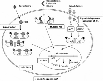
Prostate cancer and benign prostatic hyperplasia will not occur in the absence of androgen. Men castrated before puberty or those with 5α-reductase deficiency do not develop prostate cancer. Immunohistochemical studies have revealed that the AR is present in both primary and metastatic prostate cancer regardless of stage and grade, as well as in castrate-resistant tumours.Reference Hobisch, Culig, Radmayr, Bartsch, Klocker and Hittmair41 A number of different mechanisms have been described that may serve to facilitate AR transactivation in an androgen-depleted environment. These are AR amplification and over-expression, AR mutations and ligand-independent activation of AR (Figure 2).
Potential mechanisms for progression to androgen independence. Adapted from Feldman and Feldman (2001).Reference Feldman and Feldman48

AR amplification and over-expression of the AR may represent one mechanism by which prostate cancer cells proliferate in an environment with a low concentration of androgens. AR gene amplification, resulting in increased AR expression, has been reported in 25–30% of castrate-resistant tumours, but in none of the primary tumours obtained from the same patients prior to hormone therapy.Reference Koivisto, Kononen, Palmberg, Tammela, Hyytinen and Isola42 In keeping with these results, over-expression of the AR has been demonstrated at both the RNA and protein level in all castrate-resistant tumours analysed relative to androgen-dependent tumours.Reference Edwards, Krishna, Grigor and Bartlett43
Acquisition of mutations within the AR gene is thought to represent another mechanism by which prostate cancer cells may circumvent low androgen levels. The vast majority of studies have indicated that AR mutations are infrequent in untreated prostate cancers, with the exception of a subset of aggressive local or locally advanced hormone naïve tumours.Reference Marcelli, Ittmann, Mariani, Sutherland, Nigam and Murthy44, Reference Tilley, Buchanan, Hickey and Bentel45 Most AR point mutations identified in prostate cancer have been found in advanced androgen-independent carcinomas, particularly in patients treated with anti-androgens, such as flutamide and bicalutamide.Reference Taplin, Bubley, Shuster, Frantz, Spooner and Ogata46, Reference Haapala, Hyytinen, Roiha, Laurila, Rantala and Helin47 These mutations can lead to alteration of AR activity, function and ligand-binding specificity and are thought to confer growth advantage during progression to androgen-independence by allowing activation of AR-regulated genes in an androgen-depleted environment. One notable example is the Thr877Ala mutation, which enables AR activation in response to progesterone, oestradiol, cyproterone acetate, and interestingly anti-androgens, such as flutamide.
In the absence of androgen-mediated signalling, a number of alternative pathways may be utilized by the cell to compensate for the loss of androgen control of cellular growth and survival. These intracellular signalling pathways, activated by autocrine, paracrine and neuroendocrine stimulatory signals, function to enhance growth and survival by bypassing the AR or leading to transcriptional activation of the AR in the absence of its cognate ligand and include growth factor-activated pathways, receptor tyrosine kinase-activated pathways and the Akt pathway. Growth factors may act as positive or negative effectors of various cellular processes, including proliferation, differentiation and cell death. Interaction occurs with specific membrane receptors, which results in the transmission of signals through an intracellular protein cascade and the activation or the repression of a number of target genes. Aberrant signalling may result from mutation in growth factors or their downstream effector proteins leading to either loss of growth factor function (i.e. switching off the signalling pathway), or to uncontrolled expression or activation (i.e. permanently switched-on signalling pathway). Such changes are commonly associated with the malignant state and the aggressive phenotypes of cancer cells. Given the important role of growth factor receptor signalling in cell proliferation and their frequent alterations in cancer, the receptors provide attractive targets for therapeutic intervention using small molecule inhibitors and monoclonal antibodies.
An explosion of scientific discoveries in the past decade has revolutionized our understanding of cancer biology. Cytotoxic chemotherapy is here, and well established, but is inconvenient, has debilitating side-effects and often needs palliative co-medication. With the guidance of exciting research, we are moving towards targeted-therapy, with earlier diagnosis, chronic treatment of earlier disease, and fewer, more benign side-effects. Combination therapy is certainly part of the future. Lastly, perhaps we need to accept that we do not need to ‘cure’ advanced prostate cancer. Instead, we should be aiming to convert a debilitating disseminated malignancy into a controlled chronic disease with minimal symptoms and an improved quality of life.
Conflicts of interest
The authors declared no conflicts of interest.